3318
Changes in resting state functional connectivity within and between brain networks reveal effects of an injury to motor cortex in monkey brain1Institute of Imaging Science, Vanderbilt University Medical Center, Nashville, TN, United States
Synopsis
Keywords: Functional Connectivity, fMRI (resting state), brain connectivity
Motivation: To evaluate whether changes in resting state functional connectivity within and between brain functional networks reveal the effects of an injury to motor cortex in squirrel monkey brain.
Goal(s): The goal was to quantify the connectivity changes within and between functionally relevant regions of a squirrel monkey brain post a motor cortex lesion.
Approach: Resting state BOLD signals were acquired and analyzed by correlating functional MRI time courses from ICA detected networks using Pearson’s correlation.
Results: Differential changes in connectivity measures were observed both within and between functional networks post the lesion which indicates the usefulness of studying both the measures in conjunction.
Impact: The study demonstrates the utility of within-network connectivity measures in conjunction with the traditional approach of computing between-network connectivity to investigate the effects of a motor cortex injury. This has translational value in early diagnosis and treatment planning in patients.
Background and Purpose
The goal of this study was to investigate how resting state functional connectivity changes within the brain after an injury in the primary motor cortex (M1) of squirrel monkey. The M1 is a thin strip of tissue located in the frontal lobe of the brain and is responsible for initiating purposeful and intentional body movements[1,2]. Subcortical regions including the basal ganglia and thalamus are key relay and integration center responsible for transmission of motor and sensory signals to different parts of cerebral cortex including M1[3–5]. An injury to M1 alters cortical-subcortical connections[6] which then affects control of voluntary movements of specific body parts[7,8]. Previous fMRI research has studied the effects of cortical injury or neuro-disorders by quantifying the changes in connectivity between functionally relevant regions (between network connectivity or BNC) but not many have studied within network changes[9,10]. In this study we quantify the within network connectivity (WNC) changes after an M1 lesion from functionally relevant brain regions and compare it with that of BNC. In addition, animal behavior on a motor task is also being quantified to correlate with the change in connectivity measures. Our hypothesis is WNC measures are affected by injury and provides complementary information to that of BNC. We used Independent Component Analysis (ICA), a data driven method[11,12], to identify the spatial locations of functional hubs within the brain and computed resting state functional connectivity measures within and between the cortical and subcortical functional units.Methods
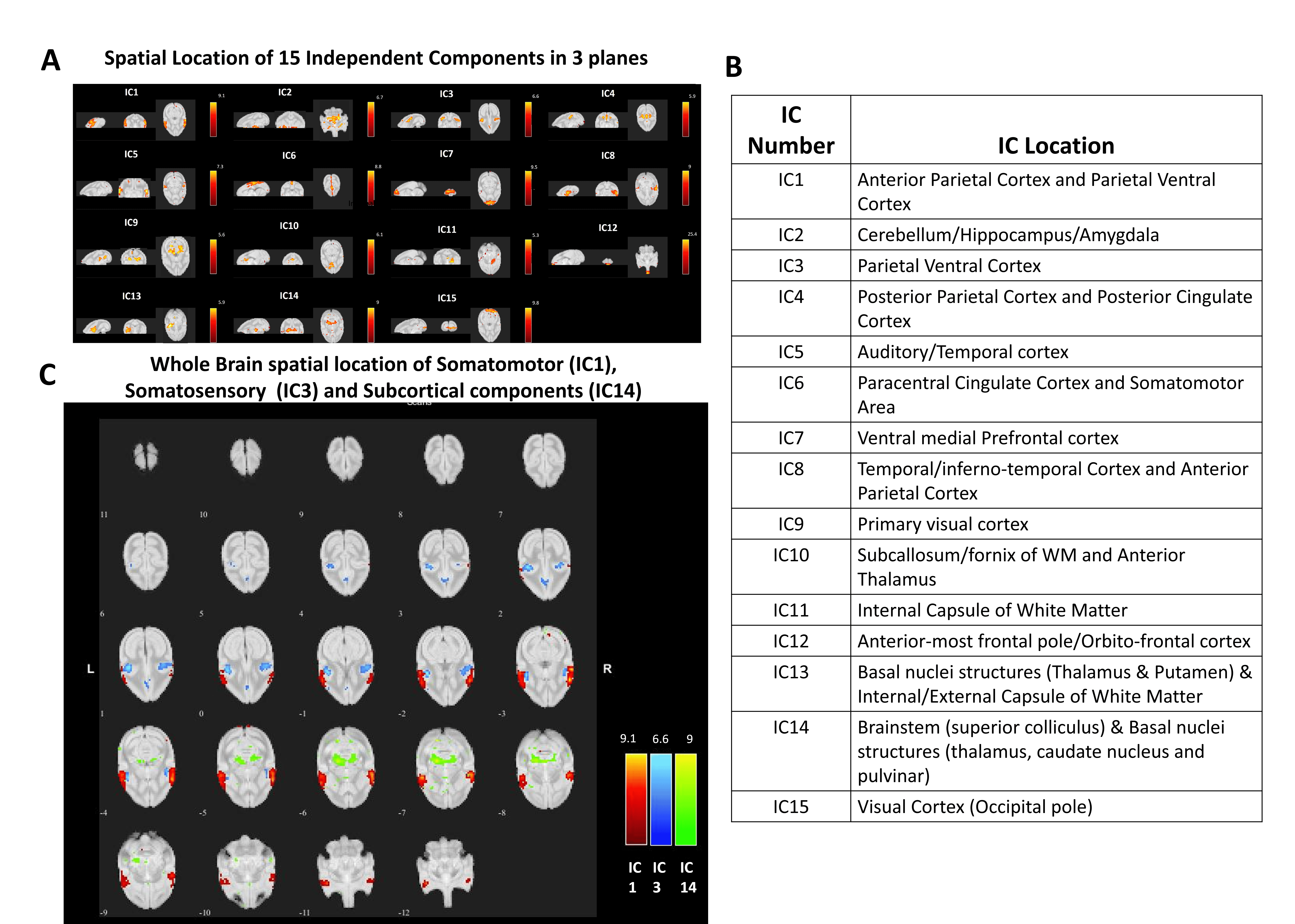
MRI scans covering the whole brain of 2 squirrel monkeys were acquired on a Bruker 9.4T scanner. High resolution (0.1 mm in-plane) T2*W anatomical images were acquired along with resting state fMRI echo-planar imaging data from the same geometry with an isotropic resolution 1x1x1 mm3 (TR=3 secs and 210 volumes each run). Functional images were acquired before and 2 weeks after a targeted M1 lesion. Ibotenic acid, which is toxic and induces neuronal cell death, was used to damage the hand region in M1[13]. Motion and physiological signal corrections and band pass filtering (0.01-0.1 Hz) were performed on the fMRI data[14,15] followed by co-registration to a monkey brain template[16] using FSL in order to facilitate template-level analyses. Functional independent components (IC) corresponding to the sensorimotor/M1 (IC1), somatosensory cortex (IC3) and subcortical region (IC14) (Figure1C) were identified from the 15 resting-state functional networks obtained from normal monkey brains using ICA (Figure1A&B) as reported in a recent study[17]. Next, WNC was computed from the functional units by correlating the fMRI time courses between the voxels within the same IC and BNC was computed by correlating the voxels between different ICs using Pearson’s correlation. The correlation values were corrected for multiple comparisons (FDR, p<0.05) and a t-test was performed to detect significantly different (p<0.05) connectivity measures before and after the M1-lesion. Each M1-injured monkey also completed a sugar pellet grasping and retrieving task and their performances were noted pre and post-injury[18]. Success rates were computed as the ratio of pellets successfully retrieved using the injured hand with the total trials.Results
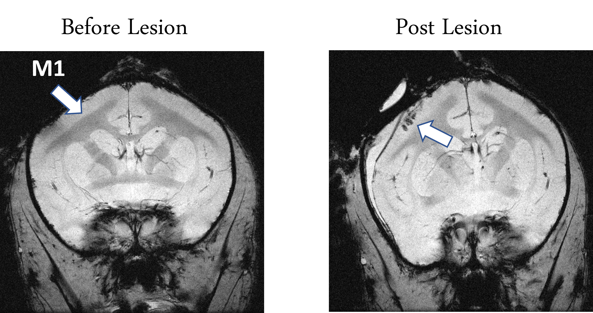
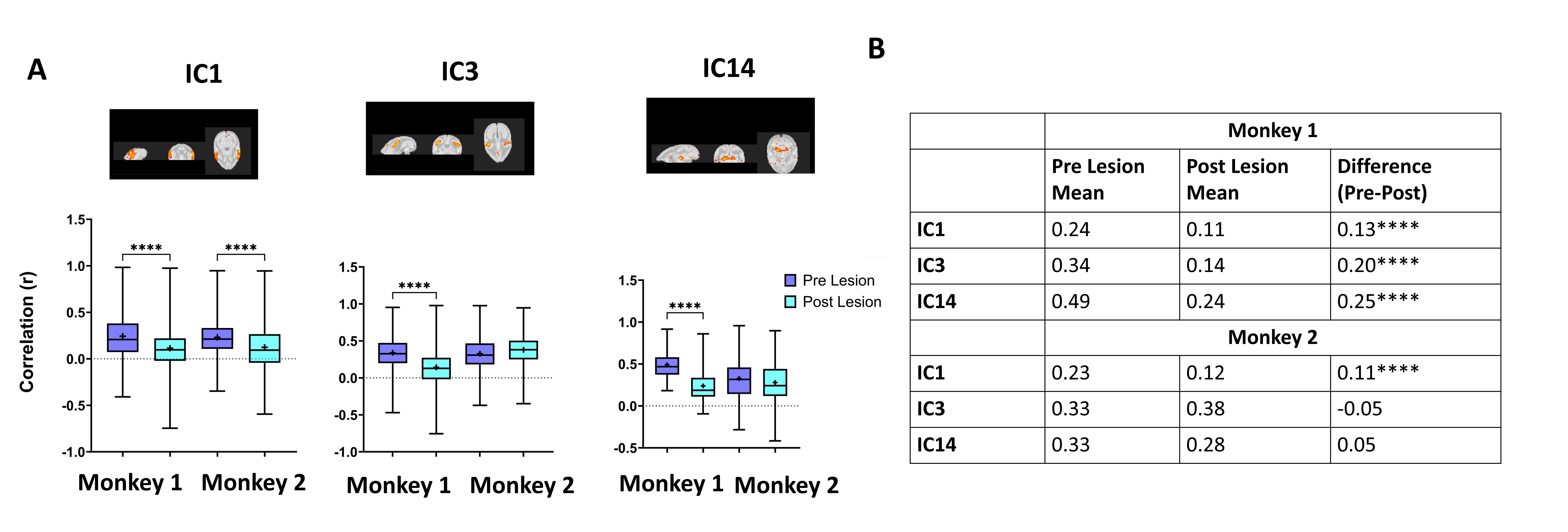
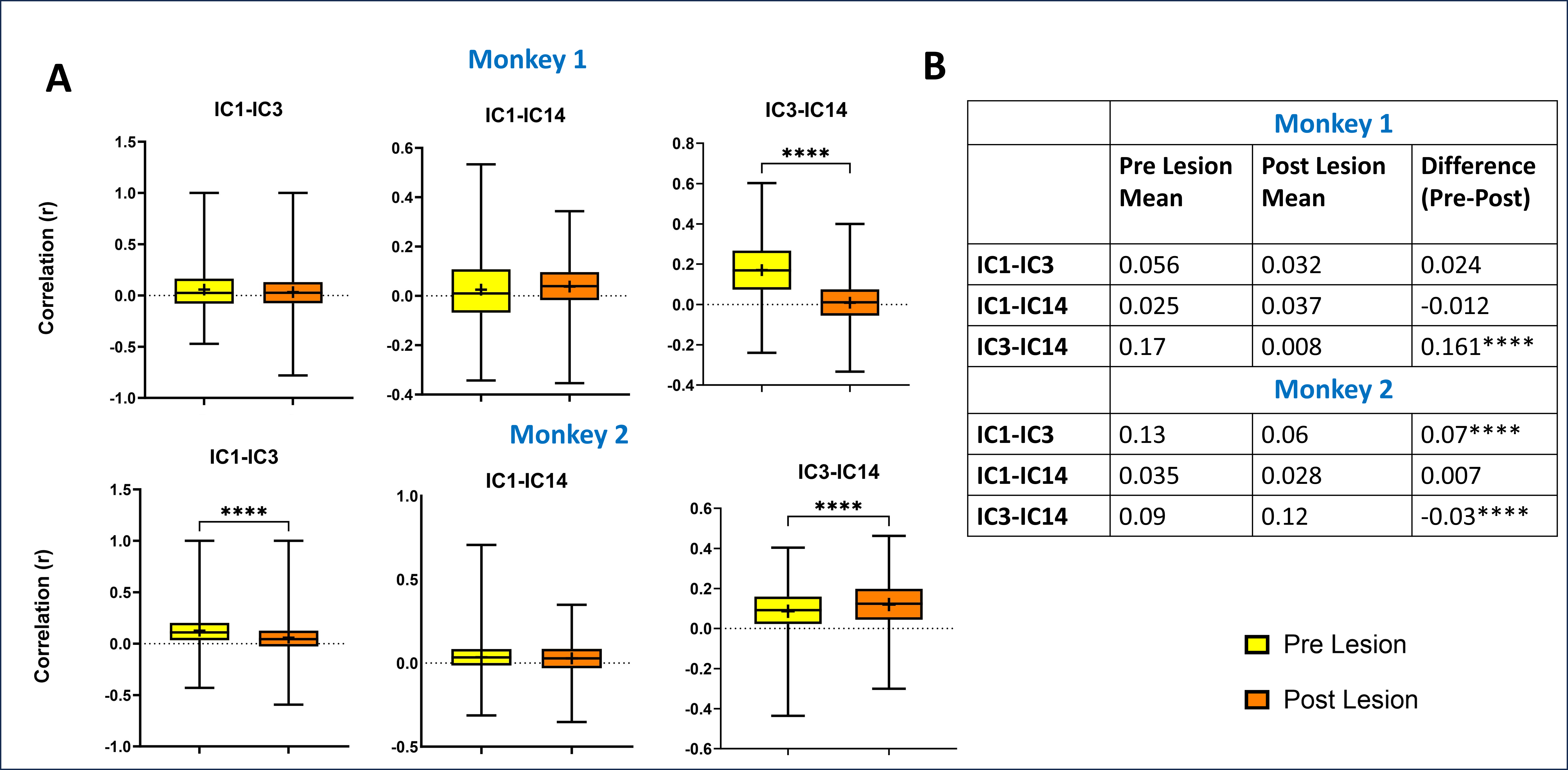
The effect of injury is visible as hypointense spots in the M1 region of monkey brain shown in Figure 2. WNC of IC1 reduced significantly for both monkeys while IC3 and IC14 reduced in the 1st monkey but remain unchanged for the 2nd monkey after M1 lesion (Figure 3). Furthermore, the BNC of IC3-IC14 was significantly reduced in the 1st monkey while in the 2nd monkey IC1-IC3 reduced and IC3-IC14 increased (Figure 4).Discussion and Conclusion
Injury to M1 produced changes both in WNC and BNC as well as behavioral deficits on the pellet retrieving task performance (task performance results not shown). The WNC changes in the three networks was not the same in both the animals possibly due to the different levels of severity of injury and recovery that each monkey experienced which was also reflected in their differential task performance. The changes in BNC measures revealed that the cortical-subcortical connection (IC3-IC14) altered significantly for both the monkeys but in opposite ways. While the reduction in connectivity indicates injury effects in the 1st monkey, an increase in cortical-subcortical connectivity in the 2nd monkey possibly indicates injury compensation. Moreover, BNC measures didn’t change significantly between the two cortical networks (IC1-IC3) in the 1st monkey, although their WNC reduced significantly. This suggests that WNC should be studied in conjunction with BNC to better understand the effects of injury. In future, we aim to establish whether the WNC and BNC measures can predict differential injury levels and recovery using the task performance measures.Acknowledgements
The authors acknowledge Chaohui Tang for animal preparation.
Funding Source: R01 NS078680 and R01NS092961
References
[1] Chen R, Cohen LG, Hallett M. Role of the ipsilateral motor cortex in voluntary movement. Can J Neurol Sci Le J Can Des Sci Neurol 1997;24:284–91. https://doi.org/10.1017/s0317167100032947.
[2] Evarts E V. Motor cortex reflexes associated with learned movement. Science 1973;179:501–3. https://doi.org/10.1126/science.179.4072.501.
[3] Basso MA, Uhlrich D, Bickford ME. Cortical function: A view from the thalamus. Neuron 2005;45:485–8. https://doi.org/10.1016/j.neuron.2005.01.035.
[4] Tang L, Ge Y, Sodickson DK, Miles L, Zhou Y, Reaume J, et al. Thalamic resting-state functional networks: Disruption in patients with mild traumatic brain injury. Radiology 2011;260:831–40. https://doi.org/10.1148/radiol.11110014.
[5] Saalmann YB. Intralaminar and medial thalamic influence on cortical synchrony, information transmission and cognition. Front Syst Neurosci 2014;8:1–8. https://doi.org/10.3389/fnsys.2014.00083.
[6] Chen J, Fan Y, Wei W, Wang L, Wang X, Fan F, et al. Repetitive transcranial magnetic stimulation modulates cortical-subcortical connectivity in sensorimotor network. Eur J Neurosci 2022;55:227–43. https://doi.org/10.1111/ejn.15571.
[7] Nudo RJ. Recovery after brain injury: Mechanisms and principles. Front Hum Neurosci 2013;7:1–14. https://doi.org/10.3389/fnhum.2013.00887.
[8] Warren G. Darling*, Marc A. Pizzimenti† and RJM. Lesions in Non-Human Primates : Experimental IMPLICATIONS FOR HUMAN STROKE PATIENTS. J Integr Neurosci 2011;10:353–84. https://doi.org/10.1142/S0219635211002737.FUNCTIONAL.
[9] Ainsworth M, Browncross H, Mitchell DJ, Mitchell AS, Passingham RE, Buckley MJ, et al. Functional reorganisation and recovery following cortical lesions: A preliminary study in macaque monkeys. Neuropsychologia 2018;119:382–91. https://doi.org/10.1016/j.neuropsychologia.2018.08.024. [10] Varangis E, Razlighi Q, Habeck CG, Fisher Z, Stern Y. Between-network Functional Connectivity Is Modified by Age and Cognitive Task Domain. J Cogn Neurosci 2019;31:607–22. https://doi.org/10.1162/jocn_a_01368.
[11] Beckmann C, Mackay C, Filippini N, Smith S. Group comparison of resting-state FMRI data using multi-subject ICA and dual regression. Neuroimage 2009;47:S148. https://doi.org/10.1016/s1053-8119(09)71511-3.
[12] Calhoun VD, Adali T, Pearlson GD, Pekar JJ. Group ICA of Functional MRI Data: Separability, Stationarity, and Inference. Proc ICA 2001 2001:155–60.
[13] Murata Y, Higo N, Oishi T, Yamashita A, Matsuda K, Hayashi M, et al. Effects of motor training on the recovery of manual dexterity after primary motor cortex lesion in macaque monkeys. J Neurophysiol 2008;99:773–86. https://doi.org/10.1152/jn.01001.2007.
[14] Jenkinson M, Smith S. A global optimisation method for robust affine registration of brain images. Med Image Anal 2001;5:143–56. https://doi.org/10.1016/S1361-8415(01)00036-6.
[15] Glover GH, Li TQ, Ress D. Image-based method for retrospective correction of physiological motion effects in fMRI: RETROICOR. Magn Reson Med 2000;44:162–7. https://doi.org/10.1002/1522-2594(200007)44:1<162::AID-MRM23>3.0.CO;2-E.
[16] Schilling KG, Gao Y, Stepniewska I, Wu TL, Wang F, Landman BA, et al. The VALiDATe29 MRI Based Multi-Channel Atlas of the Squirrel Monkey Brain. Neuroinformatics 2017;15:321–31. https://doi.org/10.1007/s12021-017-9334-0.
[17] Sengupta A, Wang F, Mishra A, Reed JL, Chen LM, Gore JC. Detection and characterization of resting state functional networks in squirrel monkey brain. Cereb Cortex Commun 2023;4:tgad018. https://doi.org/10.1093/texcom/tgad018.
[18] Duque DH, Racca JM, Manzanera Esteve I V, Yang P-F, Gore JC, Chen LM. Machine-Learning-Based Video Analysis of Grasping Behavior During Recovery from Cervical Spinal Cord Injury. Behav Brain Res 2022:114150.
Figures