3314
Investigate the correlation between resting-state functional connectivity and EEG spikes with drug-induced epilepsy1Graduate Institute of Biomedical Electronics and Bioinformatics, National Taiwan University, Taiwan, Taipei, Taiwan, 2Department of Electrical Engineering, National Taiwan University, Taiwan, Taipei, Taiwan
Synopsis
Keywords: Functional Connectivity, fMRI (resting state)
Motivation: Electroencephalography (EEG) and resting-state functional MRI (rs-fMRI) are both critical tools for investigating epilepsy, and their combination will further reveal meaningful insights.
Goal(s): This study investigates the correlation between EEG spikes and functional connectivity in a drug-induced epileptic small-animal model.
Approach: An epileptic animal model was employed by injecting pentylenetetrazol (PTZ) through intraperitoneal to trigger acute epileptic-like abnormal neuron discharges, and EEG and rs-fMRI data were acquired seperately.
Results: This study integrated longitudinal rs-fMRI monitoring and EEG recordings in the same epilepsy animal model to assess drug-induced epileptic signals, showing and increase of both EEG spike numbers and C.C. after inducing epilepsy drug.
Impact: This study demonstrate the potential of brain region-to-region connectivity becoming a new bio marker of epilepsy diagnosis. However, greater precision and repeatablility of fMRI measurements is required for future studies to accurately align fMRI data with the electrophysiological data.
Introduction
Epilepsy is a common neurological disorder characterized by abnormal discharges of brain neurons due to excessive excitation. It manifests as excessive and synchronized neural activity. Epileptic seizures can result in varying degrees of loss of consciousness, muscle spasms, sensory abnormalities, and other neurologically related symptoms. Electroencephalography (EEG) is a crucial tool for diagnosing and monitoring patients with epilepsy. During epileptic seizures, there are significant changes in the electrical activity of brain neurons, which can be observed in EEG waveform. Through the analysis of resting-state functional MRI (rs-fMRI) data, it is possible to investigate the functional connectivity between different regions within the brain, revealing the organization, networks, and functional connectivity of the brain [1]. In this study, we attempt to investigate the correlation between EEG spikes and functional connectivity in a drug-induced epileptic small-animal model.Material and Method
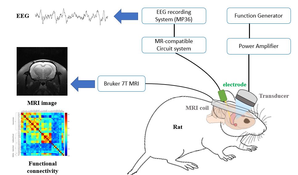
Sprague-Dawley rats were employed under the accordance of IACUC approved by National Taiwan University. Epileptic animal model was employed in this study by injecting pentylenetetrazol (PTZ) through intraperitoneal to trigger acute epileptic-like abnormal neuron discharges. MR-compatible polyimide-based microelectrodes [2] were implanted in the brain to record EEG in the Hippocampus, as shown in Figure 1. All EEG data are recorded by the 4-channel Data Acquisition System MP36(BIOPAC Systems Inc., Goleta, California, USA). For rs-fMRI, image scans was performed in a 7 Tesla Bruker BioSpec MRI scanner (Bruker Corp., Billerica, MA, USA). All fMRI data are analyzed by SPM12 and Matlab scripts RESTplus to produce correlation coefficients (C.C.) data.Results
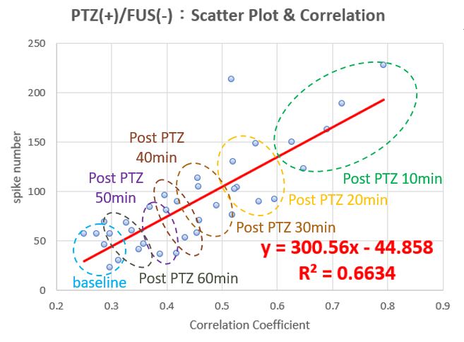
Animals injected with a seizure-inducing drug exhibited the following experimental results: After 10 minutes of PTZ injection, the spike numbers increased from 17.5 to 118.8, and the C.C. rose from 0.19 to 0.65. Both spike numbers and C.C. gradually decreased over time, reaching 75.8 for spike count and 0.29 for C.C. within 60 minutes. EEG spikes and the temporal changes in functional connectivity between brain regions were plotted as a scatter graph and their correlation was analyzed, as shown in Figure 2. It is evident that after the induction of generalized seizures by the drug, there is a strong positive correlation (R=0.66) between EEG spikes and functional connectivity.Conclusion
The results of this study demonstrate the successful integration of longitudinal rs-fMRI monitoring and EEG recordings in the same epilepsy animal model to assess drug-induced epileptic signals. The EEG spike numbers and C.C. both increase after inducing epilepsy drug, which show that brain region-to-region connectivity has potential to become a new bio marker of epilepsy diagnosis. However, subsequent research will require the accumulation of a significant number of animal experiments. In order to validate the fMRI measurements, the corresponding fMRI measurement methods must possess precision and repeatability. Each measurement needs to be precisely aligned with electrophysiological data to establish the relationship between fMRI image measurements and epileptic activity.Acknowledgements
No acknowledgement found.References
[1] [1] Vetkas, A. et al. (2022) ‘Identifying the neural network for neuromodulation in epilepsy through Connectomics and graphs’, Brain Communications, 4(3). doi:10.1093/braincomms/fcac092.[2] Chen, Y.-Y. et al. (2009) ‘Design and fabrication of a polyimide-based microelectrode array: Application in neural recording and repeatable electrolytic lesion in rat brain’, Journal of Neuroscience Methods, 182(1), pp. 6–16. doi:10.1016/j.jneumeth.2009.05.010.
Figures