3312
Evaluation of diffusion functional magnetic resonance imaging for spatial specificity of brain activation in response to visual stimulation1Center for Information and Neural Networks, National Institute of Information and Communications Technology, Suita, Japan, 2Graduate School of Frontier Biosciences, Osaka University, Suita, Japan
Synopsis
Keywords: fMRI Acquisition, fMRI (task based), diffusion
Motivation: Addressing the uncertainty of dfMRI's spatial specificity compared to BOLD-fMRI, considering the influence of perfusion and potential for more localized brain activation mapping.
Goal(s): To evaluate whether six-directional MPG dfMRI enhances the localization of brain activity in response to visual stimulation versus standard BOLD-fMRI.
Approach: Employed high-resolution 7T MRI with dfMRI and BOLD protocols, analyzing data with advanced pre-processing and statistical methods to compare spatial activation patterns.
Results: dfMRI demonstrated more confined activation regions, especially at higher b-values, suggesting improved spatial specificity, although complete isolation from BOLD-fMRI's T2* changes remains challenging.
Impact: Enhanced dfMRI specificity could refine our understanding of neural activity patterns, crucial for advancing cognitive neuroscience and developing targeted brain-based interventions.
INTRODUCTION
Diffusion functional magnetic resonance imaging (dfMRI), which employs diffusion-weighted spin-echo sequences, offers potential for accurately mapping neuronal activity. On the other hand, blood oxygenation level-dependent (BOLD) signals obtained using gradient-echo methods are thought to be significantly influenced by relatively large veins downstream of neural activity, and the use of spin-echo sequence has been suggested to minimize such effects. Consequently, dfMRI could potentially contribute to more accurate measurements of neuronal activity in principle. Moreover, because dfMRI utilizes diffusion weighting, it is capable of suppressing the influence of perfusion effects. The comparison of initial response after stimulus-onset in dfMRI with BOLD signals has been a point of debate, yet reports on its effectiveness in localizing activity regions remains insufficient1-3. Furthermore, most of previous dfMRI studies have been limited to single-direction diffusion weighting, thus not comprehensively reducing the influence of perfusion. This study investigates whether the use of dfMRI with six-directional motion-probing gradients (MPGs) can enable the detection of brain activity regions with enhanced specificity.METHODS
Nine participants volunteered using a 7T MRI scanner (Magneton Terra, Siemens Healthcare, Erlangen, Germany) equipped with a 32-channel head coil. Functional diffusion-weighted spin-echo EPI data were acquired using monopolar diffusion scheme at 2.0 mm isotropic resolution, with parameters including TR/TE = 2.0 s/60 ms, MB of 2, iPAT of 2, a 200 x 200 mm FOV, a 100 x 100 matrix, 7/8 phase partial Fourier, 14 slices with a 50% slice gap, and six orthogonal diffusion gradients. Data at b-values of 400 and 1200 s/mm2 across three runs were collected. For BOLD comparison, gradient-echo EPI data with matched parameters but TE of 20 ms were obtained4. High resolution T2 TSE image was obtained with the same slice thickness with the gap. Whole-brain anatomical T1-weighted images were collected using a MP2RAGE sequence with compressed sensing at a resolution of 0.70 mm isotropic, which is a vendor-supplied as works in progress packages. Visual stimuli were radial checkerboard patterns with an 8 Hz contrast reversal rate. The paradigm involved 10-second stimulation with 30-second intervals. Participants pressed a button upon color changes in a central fixation point, with data below 70% accuracy discarded. Pre-processing was done in FSL (FMRIB's Software Library), which included susceptibility and eddy-current distortion, motion, and slice timing. Spatial smoothing was performed on all normalized images using a 4-mm FWHM Gaussian kernel. First-level fMRI analysis employed a canonical HRF with a Z score threshold of 2.3 and P < 0.05, corrected for multiple comparisons. dfMRI data were analyzed as averaged and minimum intensity projections across six MPG datasets.RESULTS & DISCUSSION
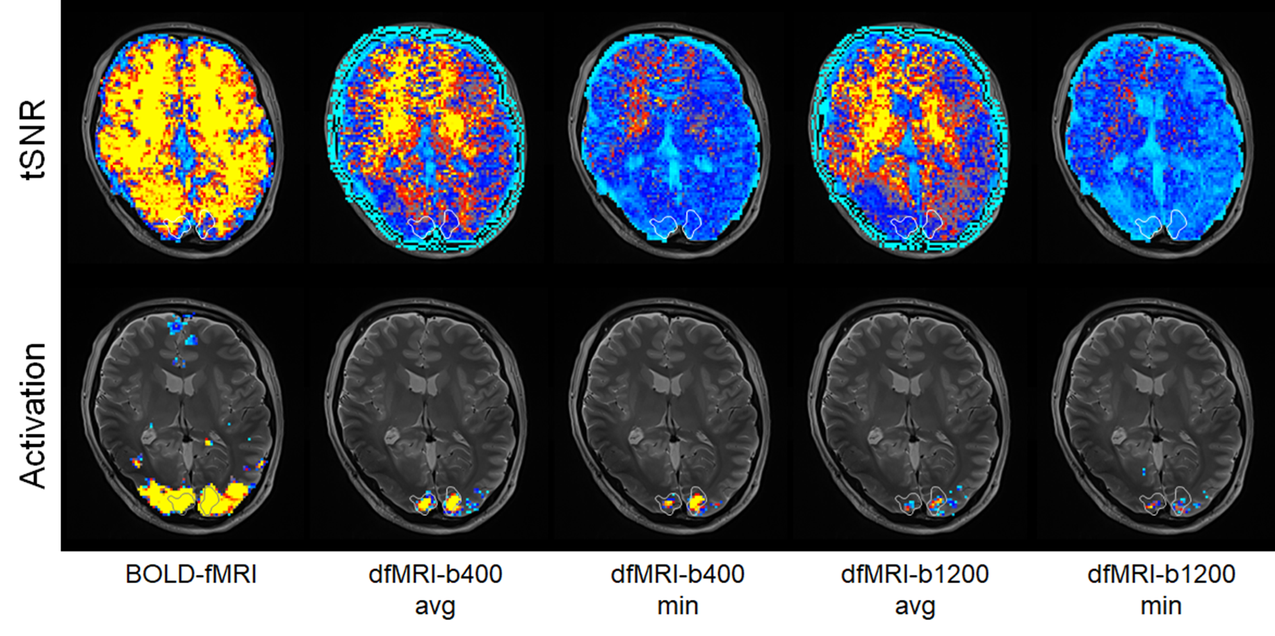
Figure 1 compares the brain activation areas elicited by visual stimulation in BOLD-fMRI and dfMRI at the same threshold (p<0.01). BOLD-fMRI shows widespread activation extending into higher-order visual areas. In contrast, dfMRI reveals activity more focused within the primary visual cortex. Notably, under the b=1200 condition, which is thought to suppress the influence of perfusion more strongly, the activation area is more localized compared to b=400. Furthermore, min-dfMRI, which uses the minimum values across six MPG directions and is considered to reduce the impact of perfusion even further, demonstrates more confined activation areas than avg-dfMRI, which simply averages across the six MPG directions. However, the extent of the activation area can expand depending on the threshold used. Meanwhile, the areas with the highest statistical values in BOLD-fMRI (enclosed in the figure) coincide with the localized activity in dfMRI. The tSNR for BOLD-fMRI is 2 to 3 times higher than avg-dfMRI, and 2 to 10 times higher than min-dfMRI. This dependency of the threshold on tSNR suggests that dfMRI activation regions might not be necessarily more localized. While high b-values and the use of minimum values in dfMRI reduce the influence of perfusion, the complete elimination of T2* changes, which are fundamental to BOLD-fMRI, is challenging as long as dfMRI employs EPI. Therefore, some overlap with BOLD-fMRI activation regions may be unavoidable. Furthermore, the current dfMRI measurements require six times longer than BOLD-fMRI, also imposing constraints on the participant's head movement.Acknowledgements
No acknowledgement found.References
1. Le Bihan et al. Direct and fast detection of neuronal activation in the human brain with diffusion MRI. Proc Natl Acad Sci USA. 103:8263-8, 2006.
2. Miller et al. Evidence for a vascular contribution to diffusion FMRI at high b value. Proc Natl Acad Sci USA. 104:20967-72, 2007.
3. Williams et al. Comparison of block and event-related experimental designs in diffusion-weighted functional MRI. J Magn Reson Imaging. 40:367-75, 2014.
4. Moeller et al. Multiband multislice GE-EPI at 7 tesla, with 16-fold acceleration using partial parallel imaging with application to high spatial and temporal whole-brain fMRI. Magn Reson Med. 63:1144-53, 2010.
Figures