3311
Microvascular specificity of spin-echo BOLD fMRI at 7T: the impact of EPI echo train length1Department of Radiology, Center for Image Sciences, University Medical Center Utrecht, Utrecht, Netherlands, 2Spinoza Centre for Neuroimaging Amsterdam, Royal Netherlands Academy of Arts and Sciences, Amsterdam, Netherlands
Synopsis
Keywords: fMRI Acquisition, Blood vessels, SE-BOLD EPI
Motivation: Macrovascular contributions to the BOLD signal reduce microvascular specificity, which can be alleviated by using SE sequences -refocusing local field inhomogeneities near large veins. Thus, the microvascular specificity of SE-EPI scan will rely on ETL duration, however, this dependence is not well-characterized in humans at 7T.
Goal(s): Determining how microvascular-specific SE-EPI BOLD responses vary with ETL in humans at 7T.
Approach: A biophysical model was developed and a validation experiment was conducted during a hyperoxic gas-challenge aiming at determining this goal.
Results: Both our simulations and measurements indicate an increase in macrovascular contamination with longer ETL-durations, leading to a decrease in microvascular specificity.
Impact: Through biophysical simulations and measurements, we show an increase in macrovascular contamination with longer ETL durations, leading to a substantial decrease in microvascular SE-BOLD specificity
INTRODUCTION
Gradient-echo (GE)-BOLD echo-planar-imaging (EPI) is a widely used functional MRI sequence for studying brain function through functional hyperemia1. However, its sensitivity to both macro- and micro-vessels affects specificity in detecting underlying neuronal activity, especially near large veins2. Spin-echo (SE)-BOLD-EPI sequences, designed to minimize macrovascular interference, aim to enhance the BOLD signal's spatial specificity3. The extended EPI-readout window (ETL-duration) reintroduces some macrovascular T2*-weighting due to full refocusing and T2-contrast occurring only at the instantaneous spin-echo timing4. Biophysical models have been pivotal in studying BOLD behavior across various field strengths and vascular properties. However, previous simulation studies often simplified BOLD signal formation by focusing solely on sequences with instantaneous readouts rather than the commonly used EPI-readouts in fMRI5,6. Previous experimental studies uncovered a positive correlation between readout duration and vessel bias, linked with macrovascular contamination. This study aimed to bridge this gap by developing a biophysical model that incorporates intrinsic biophysical MR effects induced on water molecules using realistic SE-EPI sequences. As part of this, a hyperoxic gas-challenge, inducing a global BOLD signal increase without pronounced vasoactive physiological changes, was conducted to validate our simulations at 7T. This effort sought to characterize the dependence of microvascular specificity on SE-EPI ETL-duration in humans7.METHODS
A biophysical model to simulate tissue-vascular environments within a single voxel, using infinite cylinders to represent vessels was developed. Extravascular spin diffusion was simulated via Monte Carlo methods, presuming minimal intravascular contribution at 7T. The model calculates the impact of image encoding gradients on field offset and samples voxel signals at each EPI-echo to compute the total signal energy in image space. Oxygenation levels in resting and active physiological states for GM and CSF vascular models were simulated, calculating the BOLD effect between these states. Additionally, functional SE-EPI scans on a healthy 26-year-old male at 7T were acquired. The scans involved precisely manipulation of the end-tidal O2 partial pressure using a RespirAct system. Each scan included a 60-second baseline, followed by a 180-second interval targeting specific O2 levels, conducting scans for five ETL values (with implemented SENSE) values: 33 (3.8), 41 (3.1), 51 (2.5), 63 (2.0), and 71 (1.8) -ETL durations: 23.1 - 49.7 ms. All scans used TE=55 ms, TR=4 s, and a symmetric readout window8.RESULTS
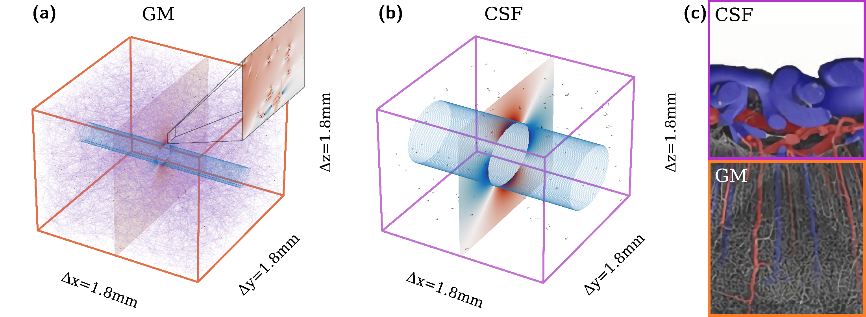
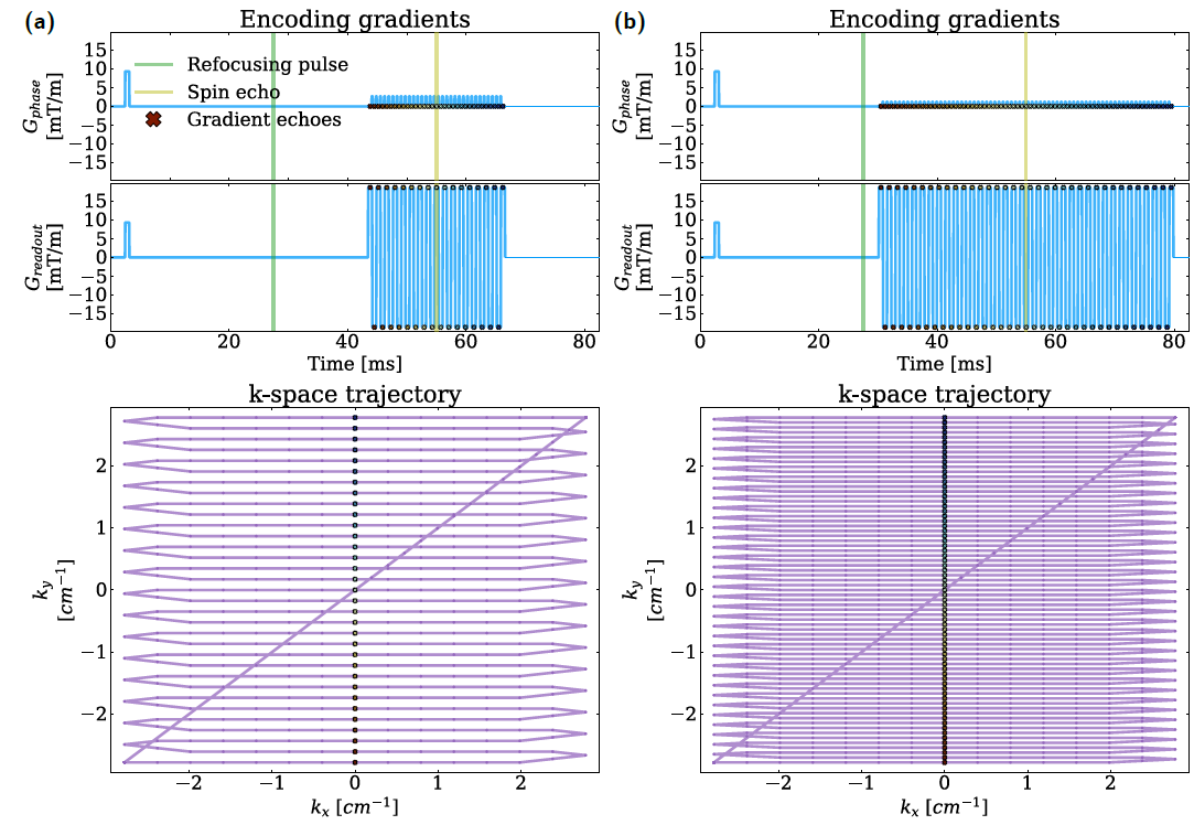
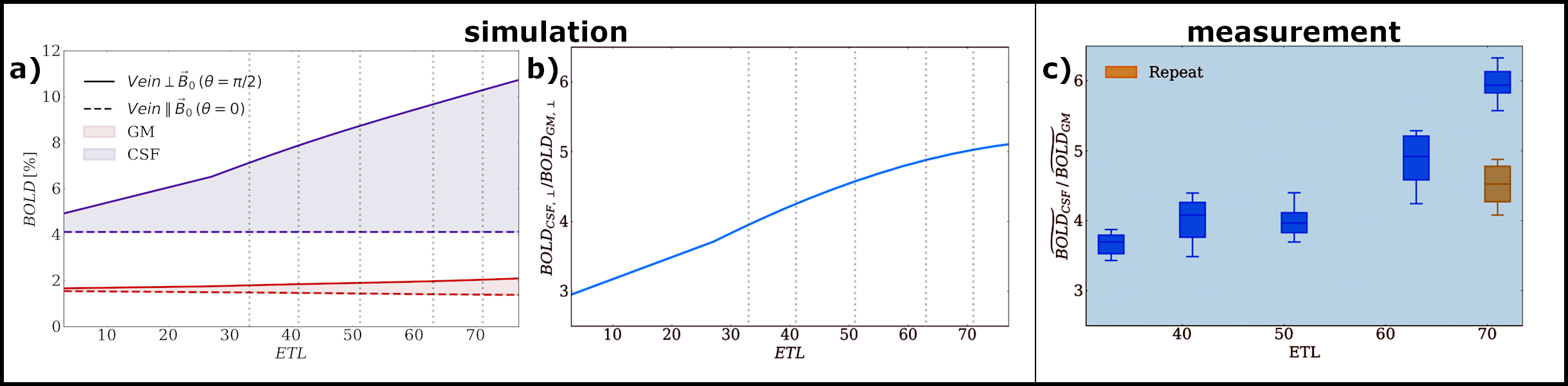
Figure 1 illustrates the biophysical model, depicting the GM and CSF voxels in panels (a) and (b) respectively. The vein in GM is significantly smaller than the pial vein in CSF, consistent with the realistic vascular morphology depicted in panel (c)9. A representative magnetic field offset distribution in a slice perpendicular to the vein is displayed. In the zoomed GM view, microvessels are observed to generate highly localized field offsets compared to the veins. Figure 2 showcases the simulated diagram of the SE-EPI sequence for ETL=33 (panel a) and ETL=71 (panel b). As the ETL increases, readout lobes and phase encoding blips are added on both sides of the spin echo, extending the ETL-duration. The occurrence of gradient echoes at kx = 0 indicates well-balanced readout gradient lobes and a suitable choice of Δt in our simulation. In Figure 3, the %BOLD signal changes are shown for both CSF and GM voxels (panel a), along with their ratio relative to ETL (panel b). The %BOLD increases proportionally with ETL in both GM and CSF. Similar to the simulation (panel b), an increase in the CSF to GM %BOLD ratio is observed with the increase in ETL. The computed increase in the CSF to GM %BOLD ratio increases by 30% within the ETL range used (ETL range = 33-71).DISCUSSION / CONCLUSION
Results reveal that SE-BOLD at 7T exhibits increased macro-vascular contamination with longer ETL durations. Macrovascular signals are notably pronounced near pial veins in CSF compared to GM microvasculature, particularly with extended ETL durations (>14 ms). Reducing the ETL duration from 49.7 ms to 23.1 ms significantly diminishes macrovascular contamination, demonstrating up to a 30% reduction in simulations and up to 60% in validation experiments for the CSF to GM %BOLD ratio. However, shortening the ETL duration through SENSE acceleration encounters limitations due to increased k-space undersampling and the necessity to mitigate GM-CSF partial volume effects, distorting GM's BOLD signal and affecting its microvascular specificity. This study introduced a versatile biophysical model accounting for EPI readouts to evaluate macrovascular contamination across diverse vascular organizations and pulse sequences. To mitigate macrovascular contamination, leveraging the shortest possible ETL duration and reducing partial volume effects are crucial. Moreover, advanced gradient insert coils with increased gradient amplitudes show potential in enhancing microvascular specificity by covering k-space faster without additional undersampling penalties.Acknowledgements
This work was supported by the Dutch Research Council under the Award Number 18969. The content is solely the responsibility of the authors.References
[1] Ogawa, S. et al. Functional brain mapping by blood oxygenation level-dependent contrast magnetic resonance imaging. A comparison of signal characteristics with a biophysical model. Biophys. J. 1993; 64:803–812
[2] Uludag K. et al. An integrative model for neuronal activity-induced signal changes for gradient and spin echo functional imaging. Neuroimage 2009; 48:150-165
[3] Weisskoff, R., et al. Microscopic susceptibility variation and transverse relaxation: Theory and experiment. Magn. Reson. Med 1994; 31:601–610
[4] Goense BM and Logothetis N. Laminar specificity in monkey V1 using high-resolution SE-fMRI. (2006) Magnetic Resonance Imaging 24.4, pp. 381–392
[5] Kiselev V.G., Posse S. Analytical model of susceptibility-induced MR signal dephasing: effect of diffusion in a microvascular network. Magn Reson Med 1999; 41:499-509
[6] Báez-Yánez MG et al. The impact of vessel size, orientation and intravascular contribution on the neurovascular fingerprint of BOLD bSSFP fMRI (2017) NeuroImage 163, pp. 13–23
[7] Schellekens W et al. The many layers of BOLD. The effect of hypercapnic and hyperoxic stimuli on macro- and micro-vascular compartments quantified by CVR, M, and CBV across cortical depth. Journal of Cerebral Blood Flow & Metabolism. 2022;0(0)
[8] van Horen TWP, Siero JCW, Bhogal AA, Petridou N, Báez-Yáñez MG. Microvascular Specificity of Spin Echo BOLD fMRI: Impact of EPI Echo Train Length. bioRxiv [Preprint]. 2023 Sep 15:2023.09.15.557938.
[9] Hirsch S et al. Topology and Hemodynamics of the Cortical Cerebrovascular System. (2012) Journal of Cerebral Blood Flow & Metabolism 32.6, pp. 952–967.
Figures