3308
Proposal of self-resonance spin-lock sequence: adaptable contrast fMRI sequences1Department of Electrical Engineering, Kyoto University, Kyoto, Japan, 2Medical Systems Research & Development Center, FUJIFILM Corporation, Minato, Japan
Synopsis
Keywords: fMRI Acquisition, fMRI, fMRI(Non-BOLD)
Motivation: To propose the novel spin-lock sequence employing phase modulation (self-resonance spin-lock: SR-SL). There is no report on self-resonance during the spin-lock pulse, and we would like to share this idea through this report.
Goal(s): To demonstrate the feasibility of SR-SL and characterize its pros and cons.
Approach: Mathematical analysis of the Bloch equation, numerical simulation, and phantom experiment.
Results: All the results of three approaches agreed. We confirmed the phase modulation could control the operating point of MR image contrast, which provides us with desired contrast change depending on the target magnetic field.
Impact: We proposed the novel spin-lock sequence named self-resonance spin-lock (SRSL), which can control the operating point of MR image contrast and provides desired contrasts. SR-SL has potential to improve sensitivity to magnetic fields and save saturation absorption rate.
INTRODUCTION
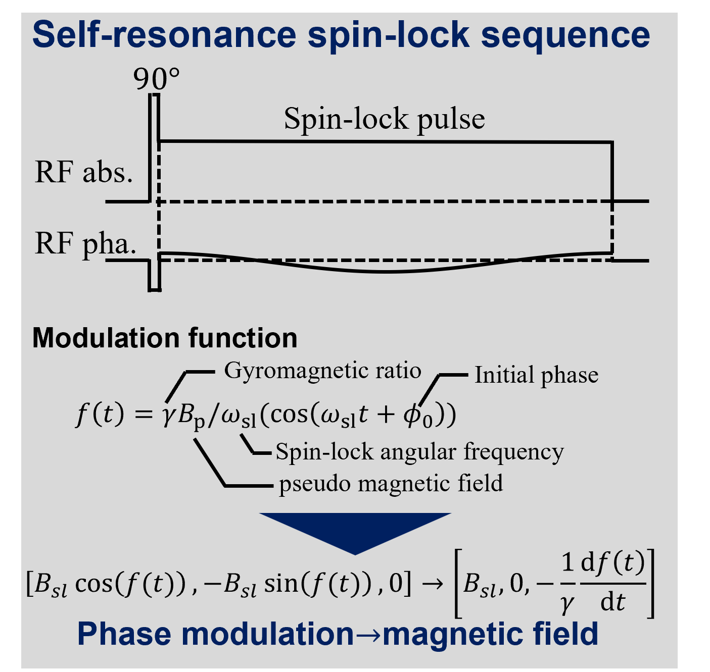
Spin-lock sequences1,2 are a kind of fMRI principles, which visualize brain activity as image contrast. The spin-lock sequences are expected to overcome the limitations of the conventional fMRI such as low temporal sampling or adaptable only to high-field MRI scanners. Among the spin-lock sequences3–5, there is no report referring to phase modulation. Thus, this study performs proof-of-principle to self-resonance spin-lock sequence (SR-SL) and reveals it feasibility using theory, simulation, and phantom experiments. The phase modulation can be interpreted as pseudo magnetic field along the static magnetic field, and magnetization can resonate with it. Thus, adjusting phase modulation, we can obtain various image contrast change with no additional RF pulse. In conclusion, SR-SL can replicate the image contrast of the conventional spin-lock sequences simply by modifying parameters without additional burden to a scanner.METHODS
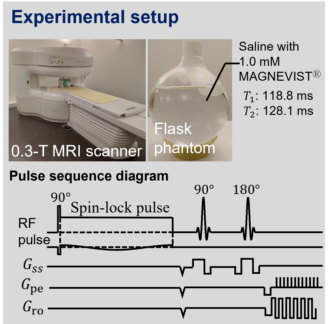
Theory: Assuming that the phase of the spin-lock pulse is modulated by function f(t), we converted the coordinate using rotation matrix whose angle is f(t). This conversion confirms that the phase modulation of the spin-lock pulse is equal to the oscillatory pseudo magnetic field Bp (= -1/γdf(t)/dt) along the static magnetic field. Therefore, even without external magnetic fields, magnetization can resonate with Bp and image contrast with this sequence changes.Simulation: To validate this concept, we performed numerical simulation using the Bloch equation. We calculated the Bloch equation during the spin-lock sequence illustrated in Figure. 1 and T1 relaxation between the end of the spin-lock pulse and the start of acquisition sequence. The spin-lock direction, frequency and its duration were x-axis, 40 Hz and 50 ms, respectively. Each T1 and T2 values of the phantom was 118.8 ms and 128.1 ms, respectively.
Phantom study: We also performed phantom experiments using 0.3-T low-field MRI scanner. We illustrated the pulse sequence diagram and overview of phantom in Figure 2. We visualized the component parallel to the static magnetic field of magnetization after the spin-lock pulse. The acquisition sequence was SE-EPI whose TR/TE = 1000ms/40.8ms. The voxel size was 3 mm isocubic and T1 and T2 values of the flask phantom is same as the simulation. The region of interest was manually selected and was 5x6 rectangular area in the center of the phantom.
RESULTS
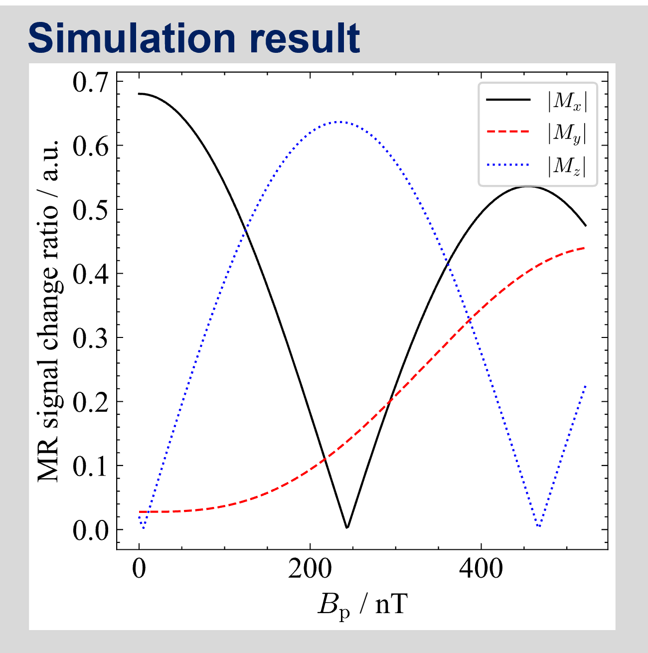
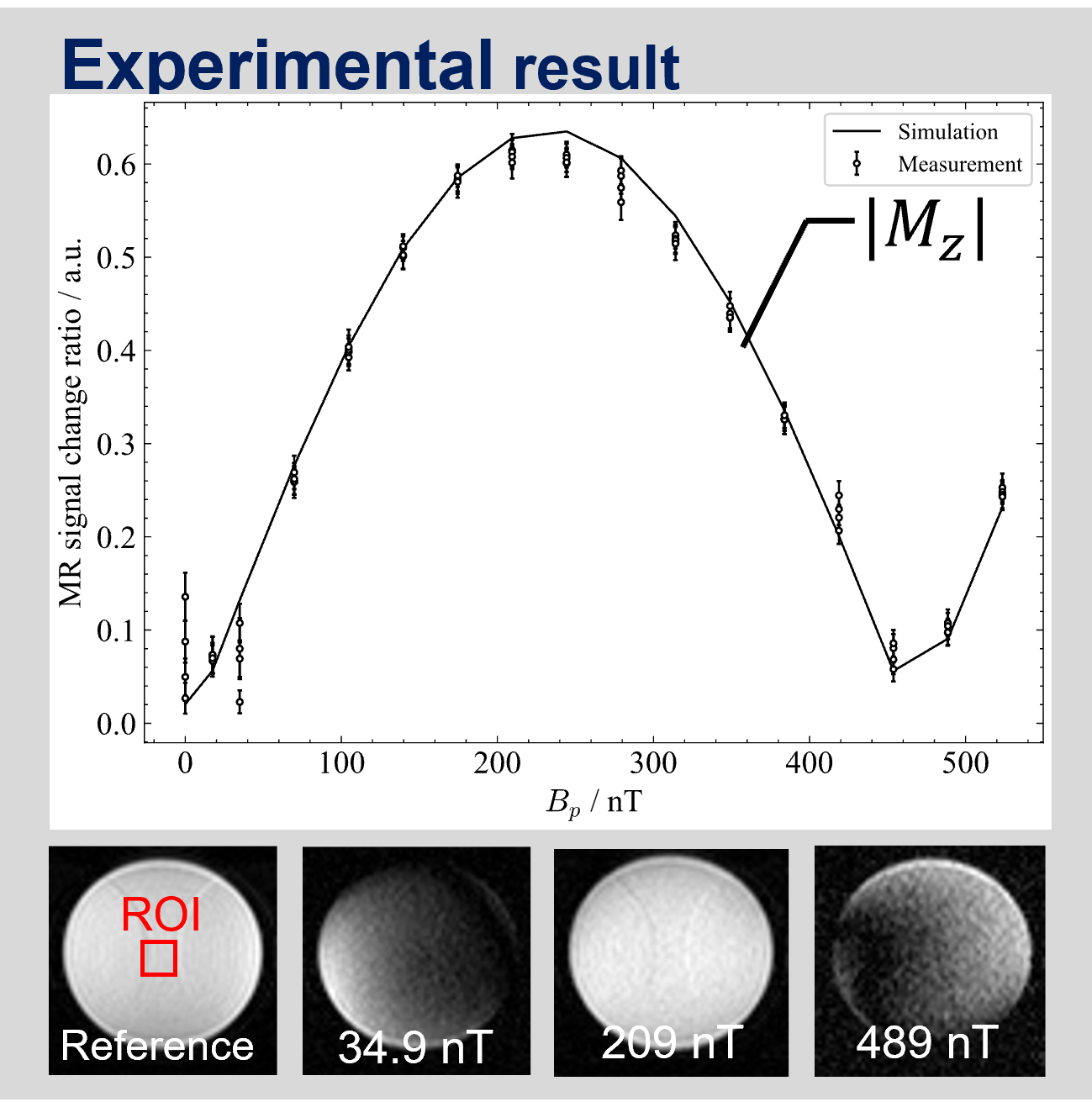
Simulation: Figure 3 illustrates each component of magnetization before the acquisition. The horizontal axis is the strength of the pseudo magnetic field, and vertical one represents the MR signal change ratio between with and without the spin-lock sequence.Phantom study: In the Figure 4, we showed the simulation and experimental result which visualized Mz in Figure3. Likewise, the horizontal axis is the strength of the pseudo magnetic field, and vertical one is the MR signal change ratio. The solid line represents simulation result with same condition as the experiments, and both agreed.
DISCUSSION
Based on the theoretical and simulation results, SR-SL can be alternative to all the other spin-lock sequences. For example, SR-SL without no modulation is same as one proposed by Truong et al., and with modulation whose Bp is about 209 nT, we can obtain MR image whose contrast is same as stimulus-induced rotary saturation (SIRS) without any additional RF pulse. In other words, we can control the operating points of image contrast depending on the external (target) magnetic field, because the Bp is controllable parameters for users.Considering the experimental results, this merits also give us another advantage such as sensitivity improvement. When Bp is lower than 30 nT, the variance of data is larger, because the signal-to-noise ratio (SNR) becomes low and the probabilistic distribution of pixel value approaches to the Rayleigh one. Therefore, to improve the sensitivity, SR-SL is one solution when we adjust Bp such that the pixel values are large enough that SNR improves, and the slope of characteristic curve is large.
CONCLUSION
This study revealed the feasibility of SR-SL based on theory, simulation, and phantom scanning. We confirmed the MR signal change depending on the depth of phase modulation with simulation and experiments. We also found that SR-SL could change the operating point of MR image contrast. This finding leads us to improvement of sensitivity and reduction in saturation absorption rate (SAR).In the future work, we plan to investigate how much the sensitivity improves and validate that phase modulation can be alternative to refocusing pulse to realize rotary-echo spin-lock sequences6,7.
Acknowledgements
This work was partially supported by Grants-in-Aid for Young Scientists (B) (JSPS KAKENHIGrant Number JP22K15621) from Japan Society for the Promotionof Science (JSPS), Japan.References
1. Witzel T, Lin F-H, Rosen BR, Wald LL. Stimulus-induced Rotary Saturation (SIRS): a potential method for the detection of neuronal currents with MRI. Neuroimage. 2008;42(4):1357-1365.2. Halpern-Manners NW, Bajaj VS, Teisseyre TZ, Pines A. Magnetic resonance imaging of oscillating electrical currents. Proc Natl Acad Sci U S A. 2010;107(19):8519-8524. doi:10.1073/pnas.1003146107
3. Jiang X, Sheng J, Li H, et al. Detection of subnanotesla oscillatory magnetic fields using MRI. Magn Reson Med. 2016;75(2):519-526. doi:10.1002/mrm.25553
4. Truong TK, Roberts KC, Woldorff MG, Song AW. Toward direct MRI of neuro-electro-magnetic oscillations in the human brain. Magn Reson Med. 2019;81(6):3462-3475. doi:10.1002/mrm.27654
5. Sveinsson B, Koonjoo N, Zhu B, Witzel T, Rosen MS. Detection of nanotesla AC magnetic fields using steady-state SIRS and ultra-low field MRI. J Neural Eng. 2020;17(3):34001. doi:10.1088/1741-2552/ab87fe
6. Gram M, Seethaler M, Gensler D, Oberberger J, Jakob PM, Nordbeck P. Balanced spin-lock preparation for B1-insensitive and B0-insensitive quantification of the rotating frame relaxation time T1ρ. Magn Reson Med. 2021;85(5):2771-2780. doi:10.1002/mrm.28585
7. Capiglioni M, Turco F, Wiest R, Kiefer C. Analysis of the robustness and dynamics of spin-locking preparations for the detection of oscillatory magnetic fields. Sci Rep. 2022;12(1):1-10. doi:10.1038/s41598-022-21232-1
Figures