3306
Silent resting-state fMRI using Looping Star Multi-echo acquisition in a 3T high-performance gradient (300 mT/m & 750 T/m/s) MRI system (MAGNUS)1Technology & Innovation Center, GE HealthCare, Niskayuna, NY, United States, 2GE HealthCare, Munich, Germany, 3GE HealthCare, Buc, France, 4Uniformed Services University of the Health Sciences, Bethesda, MD, United States, 5Walter Reed National Military Medical Center, Bethesda, MD, United States
Synopsis
Keywords: Neurotransmission, fMRI (resting state)
Motivation: To leverage the intrinsic multi-echo capabilities of Looping Star (ME-LS) for fMRI applications to minimize susceptibility-related signal loss to enhance sensitivity to underlying neurovascular changes.
Goal(s): Our objective is to demonstrate feasibility of detecting resting state networks using a silent multi-echo fMRI approach enabled by Looping Star
Approach: Resting-state Multi-echo Looping Star fMRI was implemented on MAGNUS and scanned in healthy volunteers.
Results: This feasibility study shows established resting-state networks and connectome matrixes for multi-echo Looping Star fMRI and highlights the impact of the broad parameter space accessible with MAGNUS for Looping Star.
Impact: Silent ME-fMRI has a multitude of advantages, ranging from increased sensitivity to neuro-correlates of sleep dysregulation, auditory-sensory studies, neurological disorders and to provide a more immersive experience by combining sound with visual stimuli in the MR environment.
Introduction
A common patient complaint of MR scanning is the acoustic discomfort during scanning. For high performance gradient systems[1-3] operating at maximum slew rates, scanner noise levels easily approach human auditory pain levels(>120 dB). Looping star was introduced as a novel acoustically silent, 3D radial, multi-gradient echo MRI pulse sequence[4]. It allows for whole-brain coverage and BOLD sensitivity at just a few dB above ambient noise, with the utility of the sequence previously demonstrated in block design, event-related paradigms[5]. The intrinsic multi-echo capabilities of Looping Star (ME-LS) for fMRI applications offer a practical solution to minimize susceptibility-related signal loss by leveraging echo combination which enhances sensitivity to underlying neurovascular changes by broadened optimal T2* coverage than single-echo fMRI acquisitions.In this study MAGNUS[1] was utilized to evaluate the performance of Looping Star by extending the implementation to silent multi-echo resting-state fMRI. The MAGNUS platform delivers 300 mT/m and 750 T/m/s using standard clinical 3.0-T power electronics (GE HealthCare SIGNA Premier), allowing for substantially shorter encoding pulse-widths, TEs, with reduced distortion and blurring. In addition, MAGNUS is integrated with a local transmit ‘body’ coil resulting in higher peak B1+ max which permits use of higher flip angle excitation[6] for a given minimum RF pulse length(8μs) which can be used to achieve significant improvements in SNR efficiency as the Ernst angle can be better approached. These benefits further lead to more uniform chemical shift effects and minimization of slab selection artifacts. Furthermore, due to the higher encoding efficiency, higher bandwidths can be explored allowing for even shorter repetition times. These properties make MAGNUS ideally suited for demanding acquisition sequences such as Looping Star and ME-fMRI.
Methods
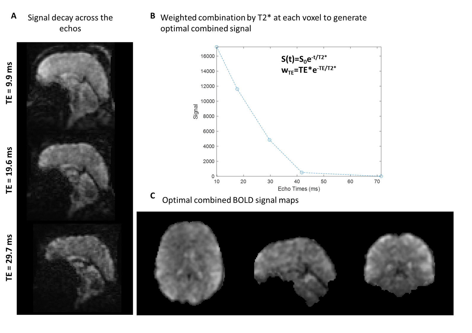
Acquisition: Three healthy volunteers were(40±5y) scanned under IRB-approved protocols with MAGNUS. A 32-channel phased array head coil (NOVA Medical, Wilmington, MA, USA) was used for all scans. In addition, the fBIRN phantom was scanned with both ME-LS and ME-EPI for static quality comparisons. To match TEs in a standard protocol for ME-fMRI using EPI on MAGNUS, the following parameters for ME-LS were used: TR 2.98s, sub-Nyquist sampling factor-per-volume of 0.4, flip-angle 3°, 3.0-mm isotropic resolution, BW=±62.5kHz, and a total of 220 volumes. These acquisition parameters provided echo-times TE1/TE2/TE3= 9.9/19.6/29.7 ms in addition to the first FID (ZTE). The third echo was assumed to be equivalent to conventional single-echo fMRI. As an anatomical reference, a 1-mm isotropic T1-weighted image was acquired. Looping Star image reconstruction was implemented offline[7].Signal processing ME-rs-fMRI: The pre-processing of resting state data was performed using a custom-built pipeline[8]. For the ME-LS, a T2*-weighted echo combination with ICA denoising (OC-ICA) was performed between the time-series alignment (Figure 2), detrending and the regressions using tedana[9]. Motion correction parameters from first-echo processing were used for correcting the later echoes and OC-ICA data. Similarly, co-registration to T1-weighted image and normalization to MNI atlas were performed on first-echo data, and the same parameters were used for processing the later echoes and OC-ICA. For functional connectivity estimation, artifact-reduced time series were bandpass-filtered with a frequency range of 0.01 to 0.1 Hz and smoothed by a Gaussian kernel with a 3-mm FWHM. Pearson correlation coefficient between the seed region mean time-course and the voxel time-course for every voxel in the brain was computed. Correlation-coefficient was converted to functional-connectivity maps using Fisher z-transform.
Results & Discussion
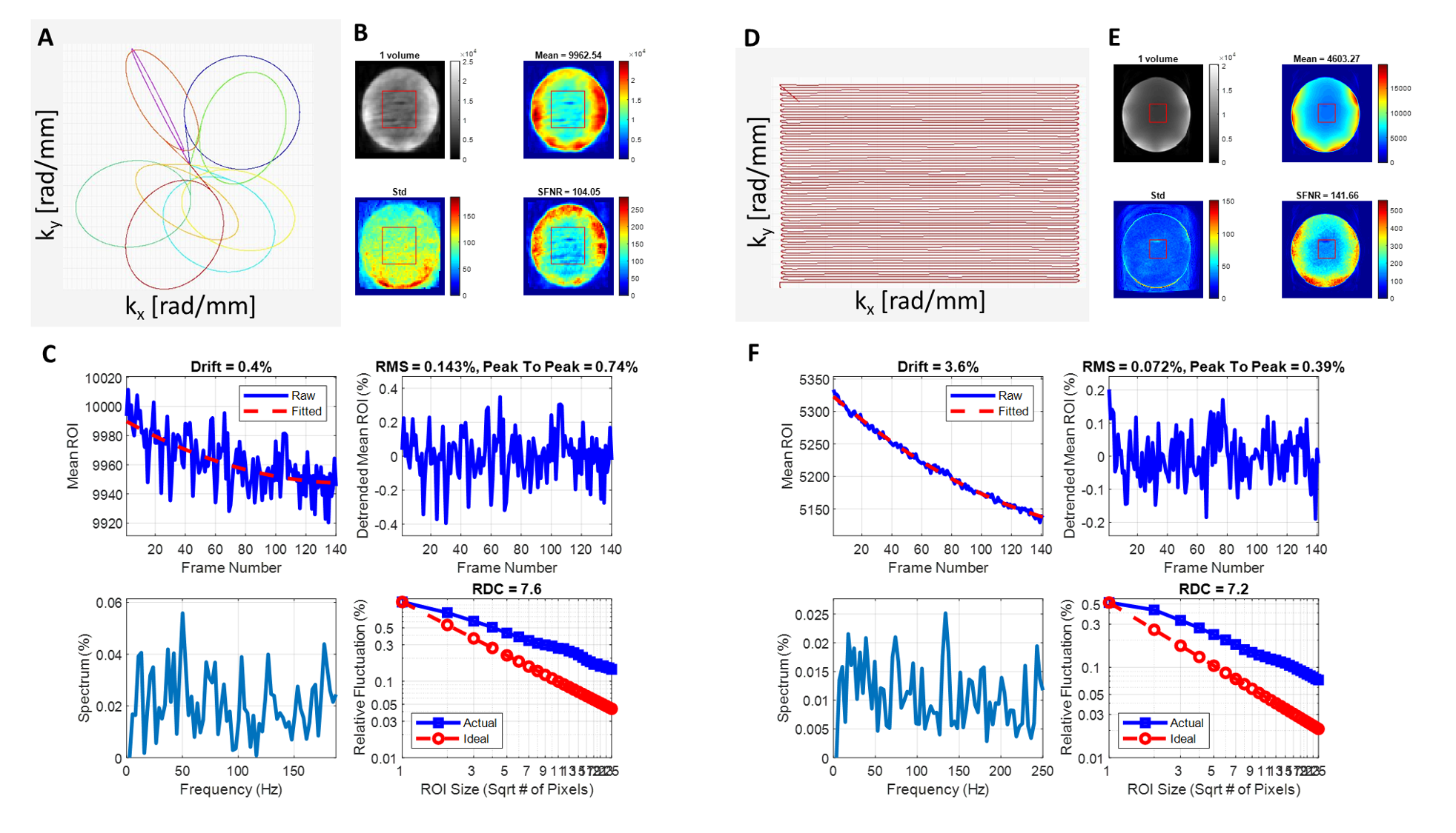
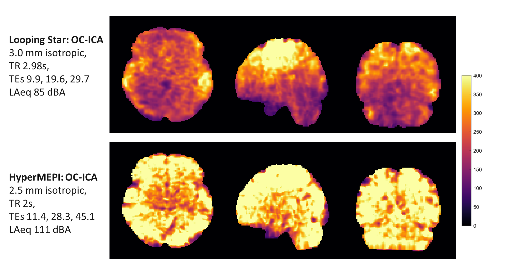
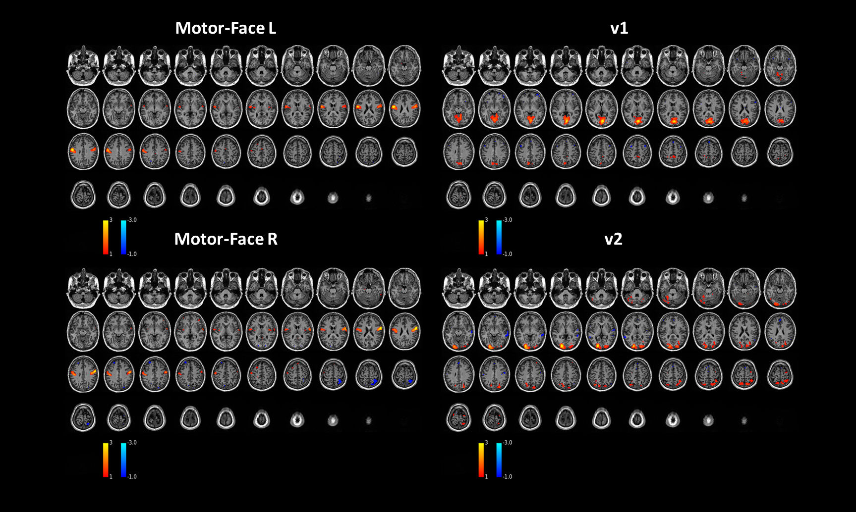
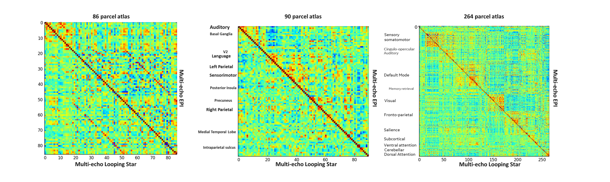
Results from the fBIRN phantom highlight comparable temporal stability between both ME-LS and ME-EPI(Figure 1b). Peak-to-peak percent fluctuations measured in the phantom indicated typical stable values (0.74% for Looping Star and 0.39% for GE-EPI), with a smaller drift (0.4% for Looping Star), and comparable RDC values. Figure 2. shows the optimal combined ME-LS image. ME-LS had lower tSNR for OC-ICA signal compared to ME-EPI potentially due to off-resonance characteristics of 3D radial acquisitions and the difference in parameter space(Figure 3). Representative seed-based network maps for ME-LS are highlighted broad agreement in localization with conventional ME-EPI(Figure 4). Functional connectomes highlight discretization of brain-based parcels and coherent identification of the networks as parcel size is decreased(Figure 5). On average a lower activation extent was noted between ME-EPI and ME-LS. However, any comparisons are significantly confounded by the difference in acquisition space (spatial resolution, TR), image-reconstruction, acceleration and the sampling depth required for stable repeatability of FC.Conclusion
This feasibility study highlights the impact of the broad parameter space accessible with MAGNUS for multi-echo fMRI with Looping Star. Further optimization is planned to encompass a wide range of paradigms and acquisition parameters to identify the limits of sensitivity and effect size needed to reveal additional or complementary information regarding cognitive processes.Acknowledgements
Grant funding from CDMRP W81XWH-22-2-0038
The opinions or assertions contained herein are the views of the authors and are not to be construed as the views of the U.S. Department of Defense, Walter Reed National Military Medical Center, or the Uniformed Services University.
References
1. Foo, T.K.F., et al., Highly efficient head-only magnetic field insert gradient coil for achieving simultaneous high gradient amplitude and slew rate at 3.0T (MAGNUS) for brain microstructure imaging. Magnetic Resonance in Medicine, 2020. 83(6): p. 2356-2369.
2. Huang, S.Y., et al., Connectome 2.0: Developing the next-generation ultra-high gradient strength human MRI scanner for bridging studies of the micro-, meso- and macro-connectome. Neuroimage, 2021. 243: p. 118530.
3. Weiger, M., et al., A high-performance gradient insert for rapid and short-T(2) imaging at full duty cycle. Magn Reson Med, 2018. 79(6): p. 3256-3266.
4. Wiesinger, F., A. Menini, and A.B. Solana, Looping Star. Magn Reson Med, 2019. 81(1): p. 57-68.
5. Dionisio-Parra, B., et al., Looping Star fMRI in Cognitive Tasks and Resting State. J Magn Reson Imaging, 2020. 52(3): p. 739-751.
6. Tarasek, M.R., et al., Average SAR prediction, validation, and evaluation for a compact MR scanner head-sized RF coil. Magn Reson Imaging, 2022. 85: p. 168-176.
7. Pruessmann, K.P., et al., Advances in sensitivity encoding with arbitrary k-space trajectories. Magn Reson Med, 2001. 46(4): p. 638-51.
8. Madhavan, R., et al., Longitudinal Resting State Functional Connectivity Predicts Clinical Outcome in Mild Traumatic Brain Injury. J Neurotrauma, 2019. 36(5): p. 650-660.
9. Kundu, P., et al., Differentiating BOLD and non-BOLD signals in fMRI time series using multi-echo EPI. Neuroimage, 2012. 60(3): p. 1759-70.
Figures