3305
Silent Looping Star fMRI with enhanced Encoding and Reconstruction Performance1GE HealthCare, Munich, Germany, 2King's College London, London, United Kingdom, 3GE HealthCare, Buc, France
Synopsis
Keywords: fMRI Acquisition, fMRI, silent, Looping Star, pulse sequence design, non-cartesian reconstruction
Motivation: Silent Looping Star fMRI offers unique advantages for neuroscience investigation, but image quality is affected by undersampling artifacts.
Goal(s): Here, we describe novel methods for improving the spatiotemporal encoding efficiency of Looping Star and thereby further advance its utility for silent fMRI.
Approach: A new Looping Star encoding scheme which adds extra out-of-plane oscillations and thereby improves overall encoding efficiency is introduced and combined with auto-calibrated cgSENSE parallel imaging reconstruction.
Results: The sharpest, most intensity uniform images and lowest background noise are demonstrated by the combination of both enhancements while maintaining BOLD sensitivity in a simultaneous, combined visual and auditory fMRI task.
Impact: The combination of a new trajectory and auto-calibrated parallel imaging leads to sharper and more uniform silent fMRI images with reduced streaking artifacts as demonstrated visually and on a visual-auditory fMRI task.
Introduction
Looping Star is a novel multi gradient echo (GRE) acquisition scheme1. Its singular features include quiet scanning (≤ 15dBA within scanner ambient acoustic noise), 3D isotropic image encoding, high sampling efficiency, and acquisition of a free-induction decay (FID) image. So far, Looping Star has been demonstrated for quiet T2* BOLD functional MRI (fMRI) and high-resolution susceptibility weighted structural MR imaging1-4. In this work, we describe further enhancements of Looping Star in terms of 1) image encoding efficiency and 2) auto-calibrated parallel imaging.Methods
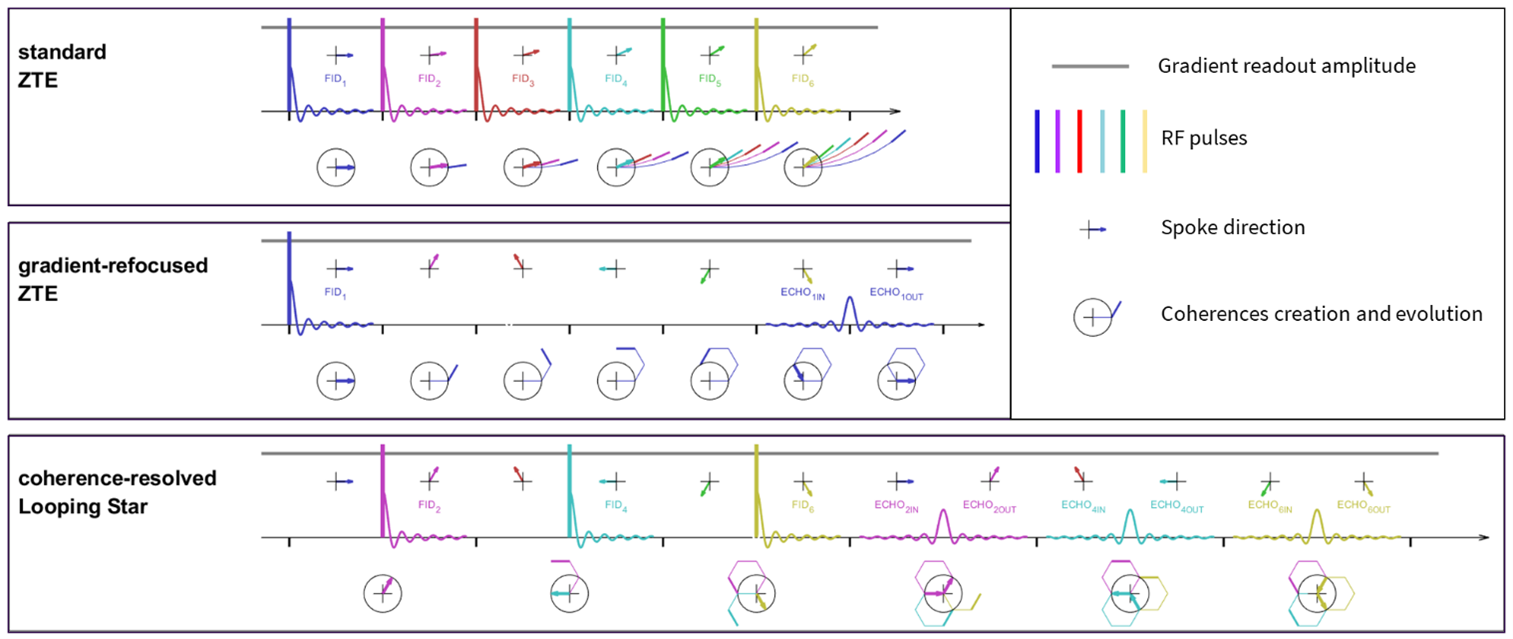
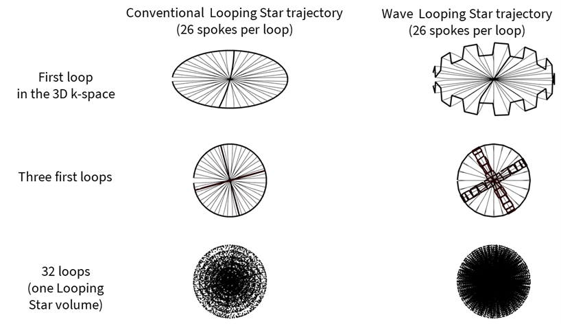
Figure 1 explains Looping Star (bottom row) as a modification of standard ZTE (top row) for time-multiplexed gradient-echo refocusing (middle row). Two enhancements are presented in this work.Image encoding: Gradient-refocusing is achieved by choosing spokes so that their cumulative trajectory rewinds back into the center of k-space. Figure 2 shows a conventional polygonal Looping Star k-space trajectory and a more efficient encoding trajectory, named wave trajectory, which includes an extra out-of-plane oscillation.
Image reconstruction: Conventional Looping Star image reconstruction is based on 3D nearest-neighbor gridding (nnGRID), followed by Fourier transformation and root-sum-of-square coil combination1. To enhance spatiotemporal encoding performance, parallel imaging in form of conjugate-gradient coil sensitivity encoding (cgSENSE)5,6 including coil compression and noise pre-whitening was implemented. The single coil FID images were used as pseudo coil sensitivities maps thereby providing intrinsic normalization of the reconstructed images (i.e., normalized relative to the complex FID image) such that the obtained GRE images can be used directly for quantitative T2* and/or susceptibility mapping. Since Looping Star acquires FID and GRE data simultaneously and with identical spatial encoding, the FID pseudo coil sensitivity maps perfectly match the GRE images without being affected by spatiotemporal incongruity due to, e.g., motion, geometric distortions, or resampling errors.
Acquisition: Healthy volunteers were scanned on a 3T SIGNATM Premier scanner (GE HealthCare, Chicago, IL) using a 48-channel head coil. Single-echo (FID+GRE) and dual-echo (FID+2GRE, parameters listed in brackets) fMRI Looping Star scans were acquired with the following parameters: 24(16) spokes per loop, BW=±31.25kHz(±41.625kHz), FOV=(19.2cm)^3, resolution=(3mm)^3, FA=3º(2)º, TEs=[0,26.88]ms([0,14.3,28.6])ms. A silent sub-millimeter T1-weighted ZTE scan was also obtained as an anatomical reference.
Visual-auditory task: A combined asynchronous visual and auditory fMRI paradigm was applied involving an 8Hz visual checkerboard with 30s duration and 30s break between blocks and a variable speed English words recording (30 to 120 words per minute) with 24s duration and 24s break between blocks. Single-echo Looping Star fMRI (FID+GRE) was run for 4:38 min, and the same data was reconstructed with nnGRID and with auto-calibrated cgSENSE.
fMRI Pre-processing & Analysis: Steady-state signal stabilization was corrected for by removing the first 4 volumes from the fMRI datasets. Pre-processing included motion correction using McFLIRT7, smoothing with 6mm FWHM kernel and registration to the T1w silent structural scan using FLIRT8. First level GLM with the regressors of interest being the hemodynamic response function convolved with the interleaved auditory and visual paradigm block designs was used to obtain two activation maps, one for each task from the same acquisition. Activation maps were considered statistically significant using clusters determined by Z>3 and a (corrected) cluster significance threshold of P=0.059.
Results
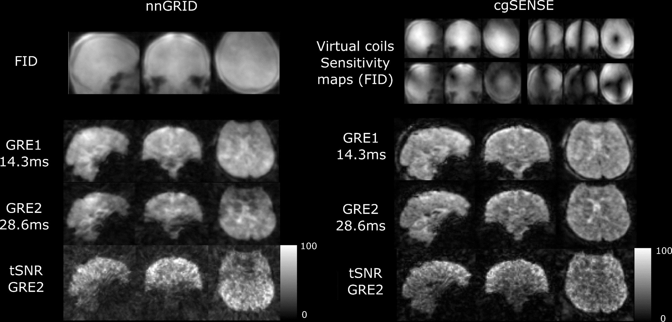
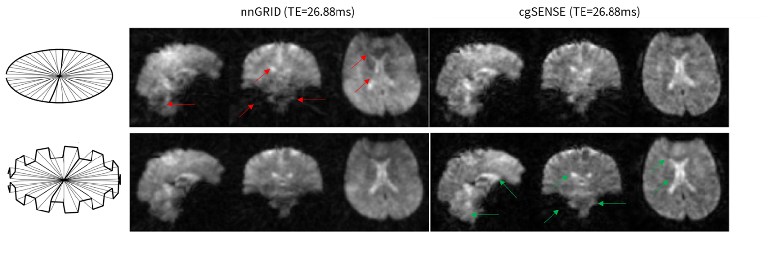
Figure 3 shows dual-echo (FID+2GRE) Looping Star (TE=[0,14.3,28.6]ms) reconstructed with nnGRID and cgSENSE. For cgSENSE, the FID images are illustrated via the first four compressed coil sensitivity maps. Enhanced image quality with the auto-calibrated cgSENSE reconstruction can be appreciated via reduced streaking artifacts, decreased background signal, and increased sharpness. More uniform tSNR maps are obtained for cgSENSE although with reduced maximum magnitude.Figure 4 compares single-echo (FID+GRE) Looping Star (TE=[0,26.88ms]) using the conventional versus the wave trajectory for nnGRID and cgSENSE. Best image quality is obtained by the combination of wave Looping Star encoding and cgSENSE reconstruction (i.e., bottom right subplot) and indicated by arrows in the image.
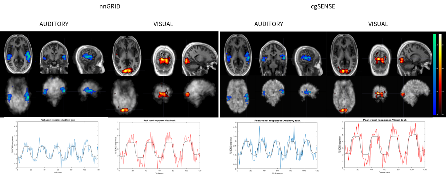
Visual and auditory statistical BOLD activation maps for nnGRID and cgSENSE reconstruction in one representative volunteer are shown in Figure 5 together with the temporal response signal at the peak voxel. Results with both reconstructions were found equivalent in localization but cgSENSE revealed slightly better localized and increased percentage BOLD signal response than nnGRID (~2% for auditory response and ~4% for visual response)
Conclusion
Enhanced image quality in terms of intensity uniformity, sharpness, and reduced streaking artifacts, for silent fMRI using Looping Star has been demonstrated by using auto-calibrated parallel imaging and an enhanced image encoding approach. Future work will focus on further improvement of spatiotemporal encoding efficiency and SNR performance.Acknowledgements
No acknowledgement found.References
1. Wiesinger F, Menini A, Solana AB. Looping star. Magn Reson Med. 2019;81(1):57-68
2. Dionisio-Parra B, Wiesinger F, Sämann PG, Czisch M, Solana AB. Looping star fMRI in cognitive tasks and resting state. J Magn Reson Imaging. 2020;52(3):739-751.
3. Damestani NL, O’Daly O, Solana AB, et al. Revealing the mechanisms behind novel auditory stimuli discrimination: An evaluation of silent functional MRI using looping star. Hum Brain Mapp. 2021 Jun 15;42(9):2833-2850.
4. Ljungberg E, Damestani NL, Wood TC, et al. Silent zero TE MR neuroimaging: current state-of-the-art and future directions. Prog Nucl Magn Reson Spectrosc. 2021;123:73-93.
5. Pruessmann KP, Weiger M, Börnert P, Boesiger P. Advances in sensitivity encoding with arbitrary k-space trajectories. Magn Reson Med. 2001;46(4):638-651.
6. Wood TC, Ljungberg E, Wiesinger F. Radial Interstices Enable Speedy Low-volume Imaging. J. Open Source Softw. 2021; 6:3500.
7. Jenkinson, M., Bannister, P., Brady, J. M. and Smith, S. M. Improved Optimisation for the Robust and Accurate Linear Registration and Motion Correction of Brain Images. NeuroImage, 17(2), 825-841, 2002.
8. M. Jenkinson and S.M. Smith. A global optimisation method for robust affine registration of brain images. Medical Image Analysis, 5(2):143-156, 2001.
9. K.J. Worsley. Statistical analysis of activation images. Ch 14, in Functional MRI: An Introduction to Methods, eds. P. Jezzard, P.M. Matthews and S.M. Smith. OUP, 2001.
Figures