3304
Improved physiology-induced temporal stability achieved with variable-flip-angle FLEET multi-shot Echo Planar Time-resolved Imaging (EPTI)1Athinoula A. Martinos Center for Biomedical Imaging, Massachusetts General Hospital, Boston, MA, United States, 2Department of Radiology, Harvard Medical School, Boston, MA, United States, 3Department of Physics, Carleton University, Ottawa, ON, Canada, 4University of Ottawa Institute of Mental Health Research, Royal Ottawa, Mental Health Centre, Ottawa, ON, Canada, 5Department of Biomedical Engineering, Case School of Engineering, Case Western Reserve University, Cleveland, OH, United States, 6Harvard-MIT Division of Health Sciences and Technology, Massachusetts Institute of Technology, Cambridge, MA, United States
Synopsis
Keywords: fMRI Acquisition, fMRI
Motivation: EPTI is a new highly-efficient imaging approach that addresses limitations of EPI by providing high-resolution distortion- and blurring-free imaging for fMRI. However, shot-to-shot phase-variations induced by physiological processes in conventional multi-shot EPTI can introduce instabilities into the reconstructed time-series data.
Goal(s): Improving physiology-induced temporal stability of multi-shot EPTI.
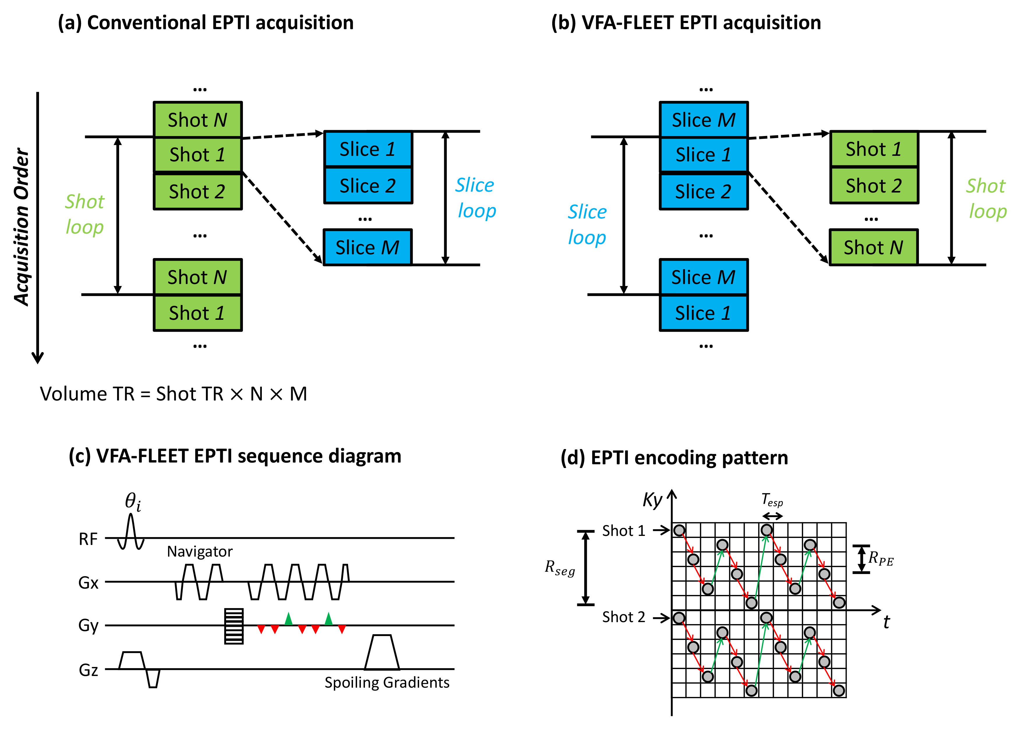
Approach: Combing multi-shot EPTI with the VFA-FLEET method, which minimizes shot-to-shot phase-variations by reordering the multi-shot acquisitions while maximizing the signal level by using a variable-flip-angle train and recursive RF pulse design.
Results: In vivo fMRI data acquired at 7T demonstrate that the physiological instabilities of multi-shot EPTI can be substantially reduced with proposed method.
Impact: Here we test whether temporal instabilities in conventional multi-shot EPTI time-series caused by physiological variability can be reduced by combining EPTI with the variable-flip-angle FLEET method. This combination can improve the usability and robustness of EPTI for high-resolution fMRI studies.
INTRODUCTION
Single-shot echo-planar-imaging (EPI)1 is commonly used for fMRI due to its high-efficiency and stability. However, the intrinsic geometric-distortion and blurring pose challenges for high-resolution studies. Recently introduced Echo Planar Time-resolved Imaging (EPTI)2 provides high-resolution distortion- and blurring-free fMRI with reasonable time efficiency through highly-accelerated multi-shot acquisitions and subspace reconstruction3. Nonetheless, conventional multi-shot EPTI requires some delays between the shots comprising each slice. Phase-variations induced by physiological processes or motion occurring between shots introduce instabilities into the reconstructed time-series data, which cannot be fully-corrected with either navigator-free or navigator-based methods4. It has been shown that the stability of conventional multi-shot EPI (with interleaved segments) can be improved with VFA-FLEET (Variable-Flip-Angle Fast Low-Excitation-angle Echo-planar Technique)5. This method acquires all shots of one slice consecutively before acquiring data of the next slice6, 7, thereby minimizing phase-variations between shots. A VFA-scheme with recursively-designed Shinnar-LeRoux (SLR)8 RF pulses was used to achieve consistent slice profiles across shots, achieving high signal and reduced artifacts. In this study, we integrated VFA-FLEET with gradient-echo EPTI and characterized the influence of shot-to-shot phase-variations on EPTI reconstruction. Experimental results show that the physiological instabilities of EPTI can be substantially reduced with VFA-FLEET.METHODS
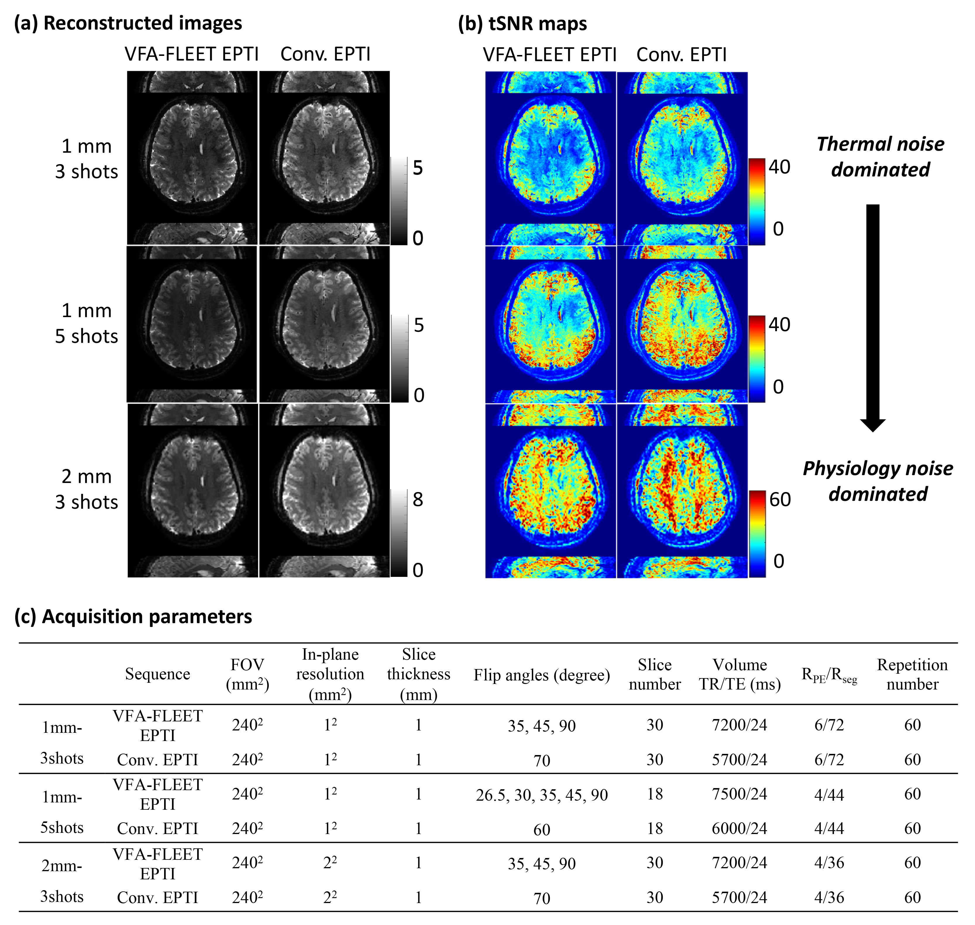
Comparisons of conventional EPTI and VFA-FLEET EPTI are depicted in Figs. 1a and 1b. The flip-angles of different shots in VFA-FLEET were recursively calculated5 as $$$\theta_{i-1}=tan^{-1}(sin(\theta_{i}))$$$(Fig. 1c), with the final flip-angle set to 90°, giving $$$\theta_{i}=$$$ [35°,45°,90°] or [26.5°,30°,35°,45°,90°] for 3- or 5-shot, respectively. For conventional EPTI, SLR pulses with constant Ernst-angles were used.In-vivo experiments were conducted using a whole-body 7T scanner with a home-built 64-channel brain-array-coil9. Two volunteers provided written-informed consent prior to scanning, following all policies of our institution’s Human Subjects Research Committee. Acquisition parameters are shown in Fig. 2c. The acquired data were reconstructed using subspace reconstruction3. Temporal-signal-to-noise-ratio (tSNR) was computed and compared.
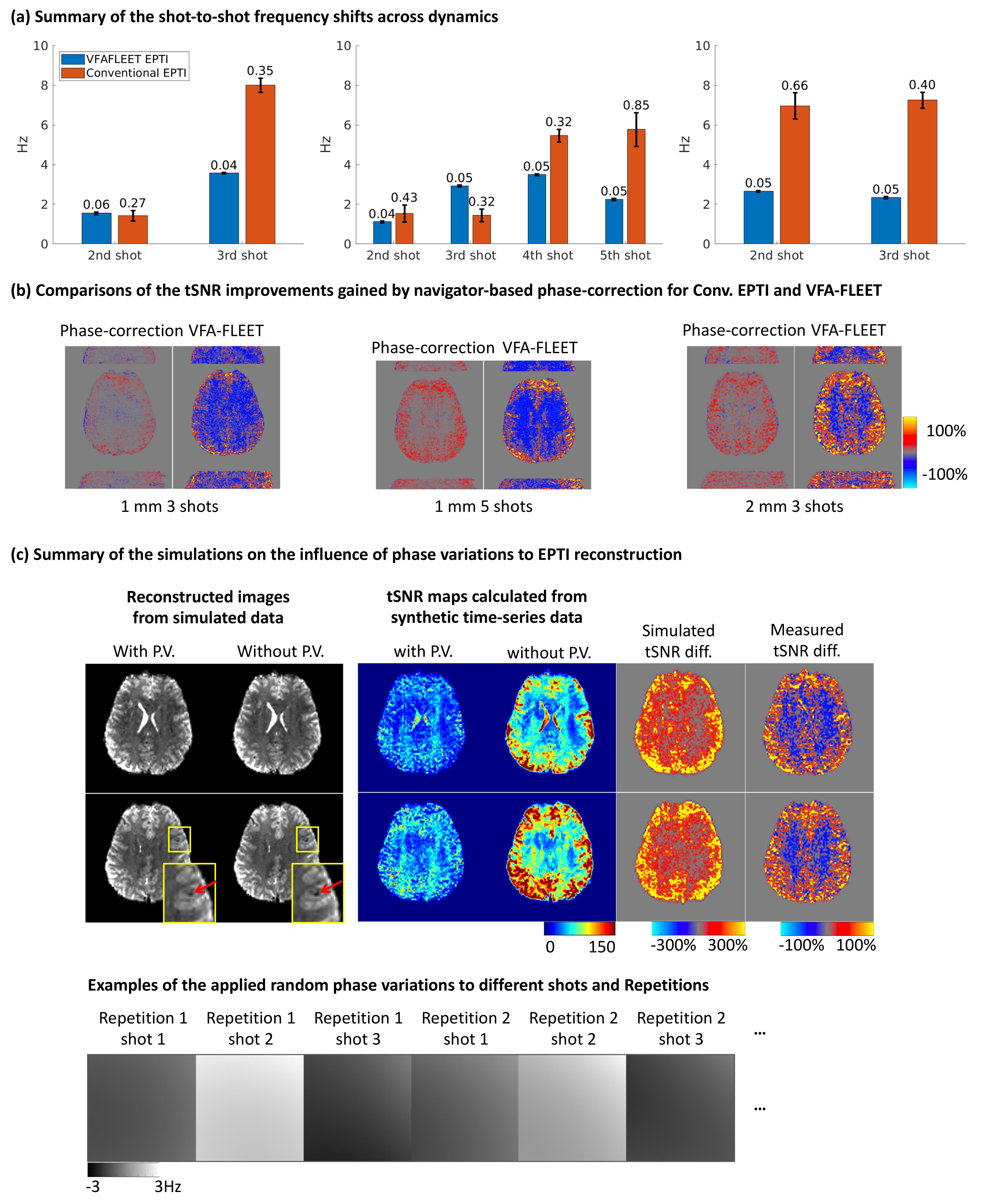
For each dynamic, shot-to-shot frequency-shifts relative to the first shot were estimated from the navigator-echoes. These shifts reflect the levels of phase-variations across shots. Simulations were employed to investigate the influences of phase-variations on EPTI reconstruction. Complex-domain NORDIC (NOise-Reduction-with-Distribution-Corrected)10 was used to remove thermal noise to help evaluate reductions in noise from physiology instabilities. The required thermal-noise level and g-factor maps were estimated with Marchenko-Pastur (MP) PCA method11.
RESULTS and DISCUSSION
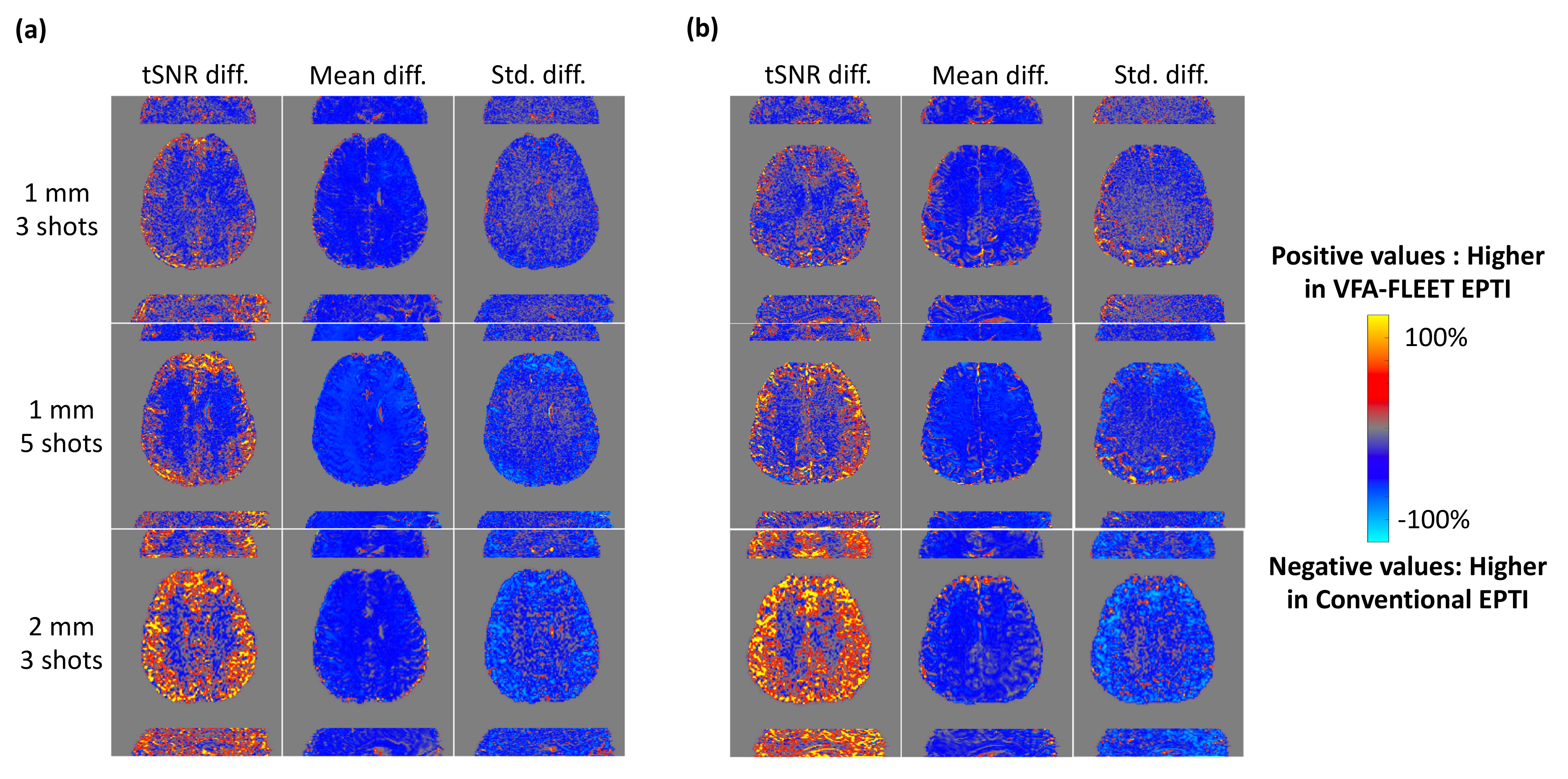
Fig. 2a shows reconstructed images of VFA-FLEET EPTI and conventional EPTI for one representative subject. As voxel sizes increased and acceleration-factors decreased, the tSNR rose for both sequences, and the tSNR maps of conventional EPTI transitioned from thermal-noise-dominated to physiological-noise-dominated (Fig. 2b)12.Fig. 3 displays the relative tSNR differences between VFA-FLEET EPTI and conventional EPTI. As larger voxels and more shots will lead to greater physiological noise vulnerability in conventional EPTI, there was a noticeable enhancement of the increased temporal stability with VFA-FLEET, also evidenced by the relative differences in signal-intensities and standard-deviations. The regions with decreased tSNR aligned with white-matter, and those with increased tSNR predominantly aligned with cortical gray-matter.
The tSNR loss in white-matter is attributed to smaller effective-flip-angles in VFA-FLEET, which can be estimated from the steady-state signal-intensities of conventional and VFA-FLEET EPTI. For example, for the three-shot acquisition, flip-angle-related tSNR decrease was estimated to be 24% for white-matter, matching our measurements. Despite lower signal intensities, VFA-FLEET can still improve the tSNR of gray-matter regions by reducing shot-to-shot phase-variations, as further shown below.
Fig. 4a shows that VFA-FLEET effectively minimizes phase changes between shots and phase variability over time. By employing a navigator-based phase-correction method5, the tSNR of conventional EPTI can be improved, though the improvements are smaller than those achieved with VFA-FLEET (Fig. 4b). Rather than causing temporally-varying ghosting as in interleaved-EPI, phase-variations in EPTI lead to temporally-varying blurring, which manifests as increased temporal variance and tSNR losses. The resulting spatial pattern of improvement is similar to that provided by VFA-FLEET, illustrating that phase-variations can induce tissue-specific temporal stability loss, which is also shown by the simulation results (Fig. 4c). These results suggest that the temporal stability of gray-matter regions was more affected by phase-variations.
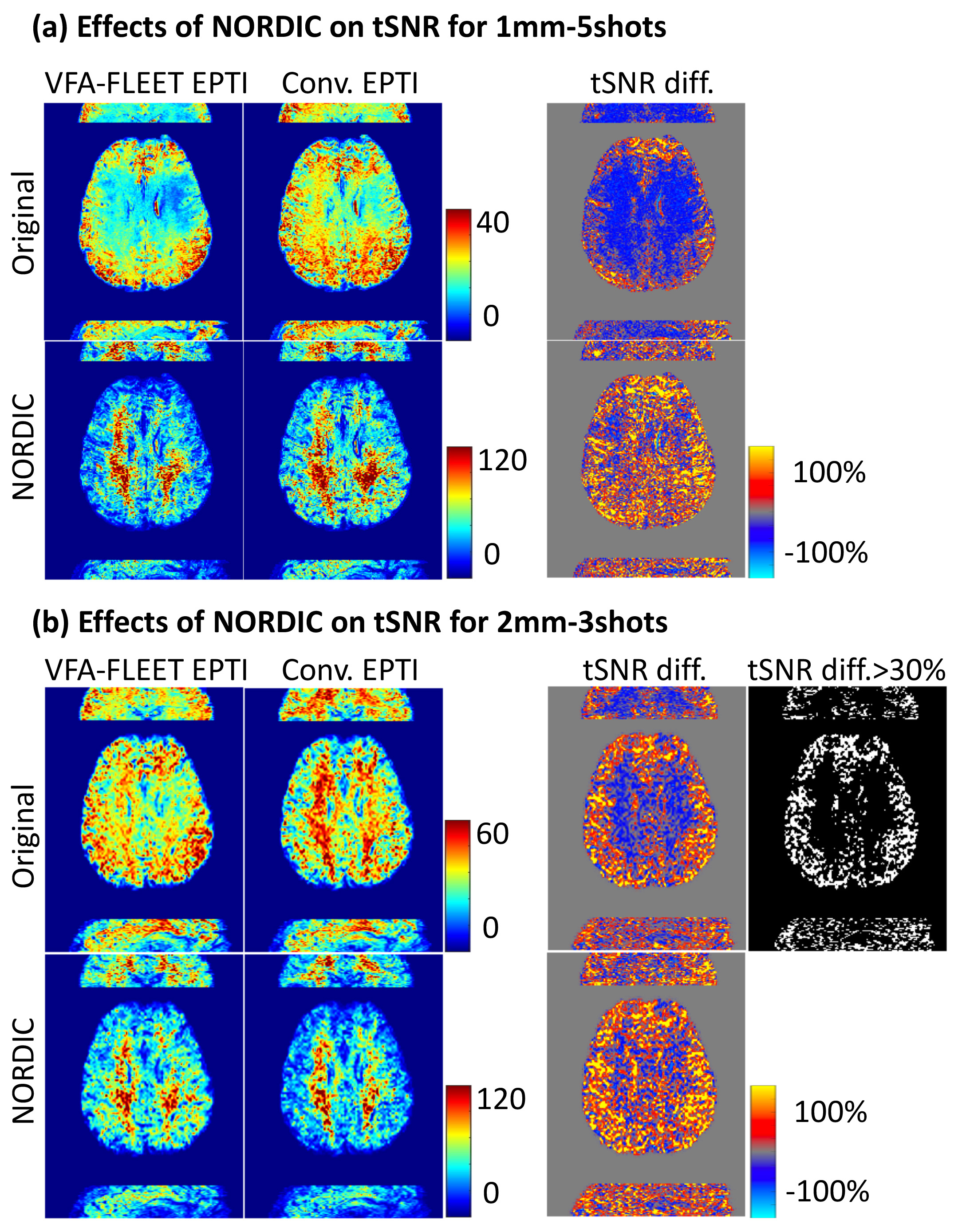
Fig. 5 helps demonstrate that the tSNR improvement from VFA-FLEET resulted from improved physiological stability. After thermal-denoising, tSNR differences between VFA-FLEET and conventional EPTI would be dominated by reduction in physiological instabilities, as shown by the more pronounced tSNR gains from VFA-FLEET after denoising. Given modern thermal-denoising methods, in practice thermal noise may be more straightforward to remove, and thus the improved physiological stability of VFA-FLEET EPTI combined with more effective noise removal in these thermal-noise dominated data can provide far higher sensitivity than conventional EPTI.
Acknowledgements
We would like to thank Estee Perelgut, Sarah Richter and Kyle Droppa for their help with subject recruitment and MRI scanning support, and Drs. Kawin Setsompop, Mukund Balasubramanian and Yulin Chang for his helpful feedback. This work was supported in part by the NIH NIBIB (grants P41-EB030006, R01-EB019437 and R01-EB033206), NCCIH (grant R01-AT011429), by the BRAIN Initiative (NIH NIMH grant R01-MH111419 and NIH NINDS grants U19-NS123717 and U19-NS128613), and by the MGH/HST Athinoula A. Martinos Center for Biomedical Imaging, and by Carleton University; and was made possible by the resources provided by NIH Shared Instrumentation Grant S10-OD023637.References
1. Mansfield P. Multi-planar image formation using NMR spin echoes. Journal of Physics C: Solid State Physics 1977;10:L55.2. Wang F, Dong Z, Reese TG, et al. Echo planar time-resolved imaging (EPTI). Magn Reson Med 2019;81:3599-3615.
3. Dong Z, Wang F, Reese TG, Bilgic B, Setsompop K. Echo planar time-resolved imaging with subspace reconstruction and optimized spatiotemporal encoding. Magn Reson Med 2020;84:2442-2455.
4. Hu Z, Ma X, Truong TK, Song AW, Guo H. Phase-updated regularized SENSE for navigator-free multishot diffusion imaging. Magn Reson Med 2017;78:172-181.
5. Berman AJL, Grissom WA, Witzel T, et al. Ultra-high spatial resolution BOLD fMRI in humans using combined segmented-accelerated VFA-FLEET with a recursive RF pulse design. Magn Reson Med 2021;85:120-139.
6. Chapman B, Turner R, Ordidge RJ, et al. Real‐time movie imaging from a single cardiac cycle by NMR. Magnetic resonance in medicine 1987;5:246-254.
7. Polimeni JR, Bhat H, Witzel T, et al. Reducing sensitivity losses due to respiration and motion in accelerated echo planar imaging by reordering the autocalibration data acquisition. Magnetic resonance in medicine 2016;75:665-679.
8. Pauly J, Le Roux P, Nishimura D, Macovski A. Parameter relations for the Shinnar-Le Roux selective excitation pulse design algorithm (NMR imaging). IEEE transactions on medical imaging 1991;10:53-65.
9. Mareyam A, Kirsch JE, Chang Y, Madan G, Lawrence W. A 64-Channel 7T array coil for accelerated brain MRI. ISMRM; 2020.
10. Vizioli L, Moeller S, Dowdle L, et al. Lowering the thermal noise barrier in functional brain mapping with magnetic resonance imaging. Nature communications 2021;12:5181.
11. Veraart J, Novikov DS, Christiaens D, Ades-Aron B, Sijbers J, Fieremans E. Denoising of diffusion MRI using random matrix theory. NeuroImage 2016;142:394-406.
12. Wald LL, Polimeni JR. Impacting the effect of fMRI noise through hardware and acquisition choices - Implications for controlling false positive rates. NeuroImage 2017;154:15-22.
Figures