3302
T2*-weighted echo-planar imaging of the human brainstem and spinal cord at 7 Tesla with region-wise geometry, timing, and shim parameters1Systems Neuroscience, University Medical Center Hamburg-Eppendorf, Hamburg, Germany, 2Max-Planck-Institute for Human Cognitive and Brain Sciences, Leipzig, Germany
Synopsis
Keywords: fMRI Acquisition, fMRI, z-shim
Motivation: Combined fMRI acquisitions of human brainstem and cervical spinal cord helps to investigate their interaction, e.g., during pain processing.
Goal(s): To test whether combined T2*-weighted EPI of both regions is feasible at 7 Tesla.
Approach: EPI pulse sequence supporting region-wise geometry (FOV, slice thickness, voxel size), timing (echo spacing, echo time), and shim parameters (linear terms and frequency), and flip angles together with a region-wise shim algorithm extended to consider static third-order shim terms.
Results: A reasonable image quality could be obtained in both regions, but strategies to reduce ghosting artifacts in the spinal cord need to be investigated.
Impact: Being able to cover human brainstem and cervical spinal cord regions in a single EPI acquisition at 7 Tesla can help to investigate the interaction of both regions with improved spatial resolution.
Introduction
BOLD-based functional neuroimaging of the human brain is well established and its application to the human spinal cord has become more popular in recent years. To investigate the direct interaction of brain or brainstem and cervical spinal cord regions, both target volumes need to be covered in a single acquisition. This is not only challenging due to the different geometries desired (e.g. FOV, voxel size) and related timing parameters (e.g. bandwidth, TE), but especially because optimum shim settings differ considerably between the two regions. Therefore, corresponding approaches involve slice- or region-wise settings of the linear shim terms to obtain a reasonable image quality in both regions1,2. With their successful application at 3T, the interest to perform such experiments with improved signal-to-noise ratio and spatial resolution at 7T has risen.Here, a T2*-weighted EPI pulse sequence with independent geometry and timing parameters for the two regions and region-wise linear shim and frequency settings1 in combination with a dedicated algorithm for region-wise shim settings3 have been used to acquire brainstem (periaqueductral gray, rostral ventromedial medulla) and cervical spinal cord regions (vertebrae C4-C6) in a single acquisition.
Methods
Measurements were performed on a 7 T whole-body MR system (Terra, Siemens Healthineers) in single-transmit mode equipped with a 24-channel transmit/receive cervical-spine coil (MRI.Tools, Fig. 1a). Healthy volunteers were investigated after their informed consent had been obtained.The EPI pulse sequence (Fig. 1b) with region-wise geometry, timing, and shim settings1 was modified to support region-specific flip angles for the slice-selective RF excitations to consider differences in the B1 transmit field between the two regions. A dedicated algorithm to calculate static second-order shim and region-wise linear shim and frequency settings3 was extended to consider four static third-order shim terms, z3, z2x, z2y, and z(x2-y2), that are available on the MR system used.
Phantom experiments were performed to determine the conversion factors of the values specified on the shim adjustment user interface to the ideal shim terms in physical units considered in the algorithm for all second- and third-order shim terms (Fig. 2), as described previously3.
14/36 slices without gap were used to cover the spinal cord/brainstem region (Fig. 3) with voxel sizes of 1.0×1.0×3.0/1.5×1.5×1.5 mm3, FOVs of 128×150/225x225 mm2, receiver bandwidths of 1450/1956 Hz per pixel, and TEs of 25/25 ms together with a partial Fourier factor of 7/8 and PAT (GRAPPA) acceleration factor of 3 (54 reference lines, interleaved acquisition in three shots) for both volumes yielding a TR of 2606 ms. Slice-specific z-shim settings were used for the spinal cord slices that were determined from a reference acquisition with 31 equidistant z-shim settings compensating gradients between +0.3/–0.3 mT m-1 as described previously1. 20 measurements were performed and averaged.
Results and Discussion
Figure 4 shows 28 slices in the brainstem of a healthy volunteer. Significant intensity variations within and between slices are observed. To some extent, they could be assigned to the single-side coil geometry and related variations in the transmit and receive efficiency; but most likely they also reflect typical and unavoidable B1 inhomogeneities in this part of the brain due to the short RF wave length at 7T. Nevertheless, and despite being worse compared to a brainstem-only acquisition, the overall image quality in the brainstem target region seems to be sufficient for fMRI applications.12 spinal cord slices from the same acquisition are presented in Fig. 5. The most prominent problems are ghosting artifacts that are more pronounced than in typical spinal-cord-only acquisitions at this field strength. They could be related to frequency offsets due to increased field inhomogeneities in the spinal cord region because second- and third-order shim terms are not optimized for it but chosen to provide a reasonable field homogeneity also in the brainstem which could decrease the field homogeneity in the spinal cord region. Probably, slice-specific frequency settings as in a previous 3-T work2 could reduce such offsets and help to ameliorate these problems. Another source for the ghosting could be the acquisition scheme for the PAT reference data (segmented EPI) that has been shown to have a significant impact on the image quality of spinal cord EPI4. Thus, it would be worth to test FLASH acquisitions4 for the reference data.
Conclusion
With region-wise linear shim, frequency, and flip angle settings, combined fMRI of the human brainstem and cervical spinal cord may be feasible at 7T. However, it may benefit from further extensions like slice-specific frequency settings or alternative PAT reference data acquisition schemes that are currently being implemented.Acknowledgements
FE is supported by the Max Planck Society and the European Research Council (under the European Union’s Horizon 2020 research and innovation programme; grant agreement No 758974).References
1. Finsterbusch J, Sprenger C, Büchel C. Combined T2*-weighted measurements of the human brain and cervical spinal cord with a dynamic shim update. NeuroImage 2013; 79: 153-161.
2. Islam H, Law CSW, Weber KA, Mackey SC, Glover GH. Dynamic per slice shimming for simultaneous brain and spinal cord fMRI. Magn. Reson. Med. 2019; 81, 825-838.
3. Chu Y, Fricke B, Finsterbusch. Improving T2*-weighted human cortico-spinal acquisitions with a dedicated algorithm for region-wise shimming. Neuroimage 2023; 268: 119868.
4. Seifert AC, Xu J. Impact of autocalibration method on accelerated EPI of the cervical spinal cord at 7 T. Magn Reson Med 2022; 88: 2583-2591.
Figures
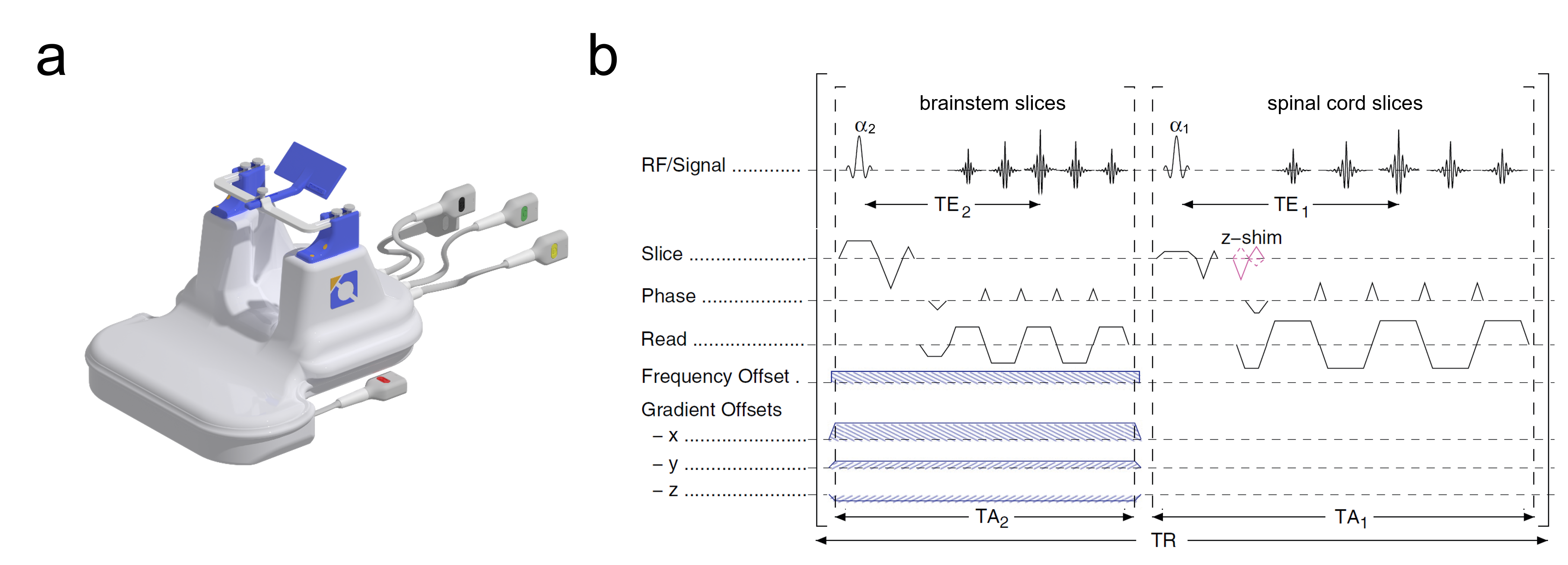
Fig. 1: (a) Picture of the 24-channel transmit/receive cervical-spine coil and (b) basic EPI pulse sequence used in the present study. The sequence supports region-specific geometry (FOV, voxel size and slice thickness), timing (TE, echo spacing), and shim (linear terms and frequency) parameters and flip angles.
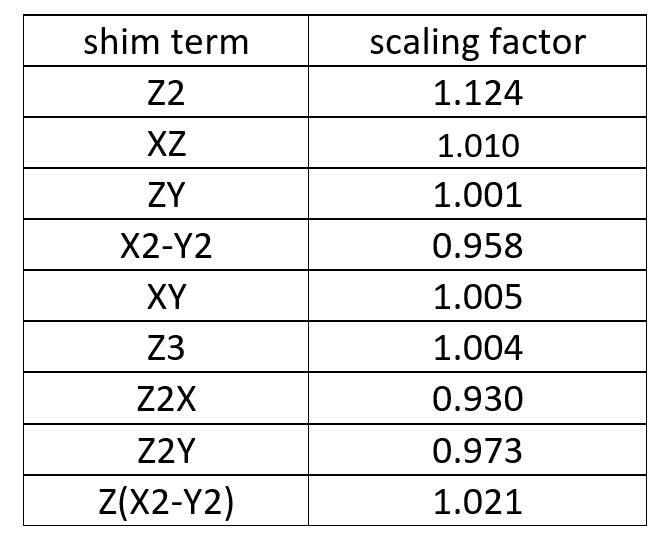
Fig. 2: Scaling factors to convert UI shim parameters into the physical units used in the shim algorithm3 for the 7T system used.
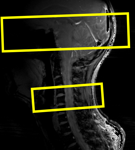
Fig. 3: Sketch of the position and orientation of the slice groups on a localizer image.
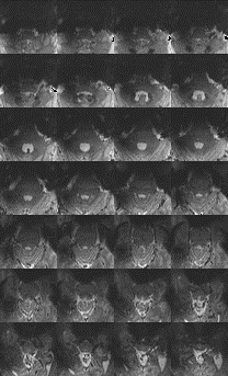
Fig. 4: 28 of 36 slices of the brain stem region acquired with a spatial resolution of 1.5×1.5×1.5 mm3.
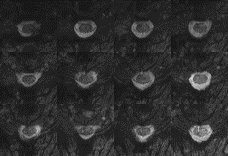
Fig. 5: 12 of 14 slices of the cervical spinal cord region acquired with a spatial resolution of 1.0×1.0×3.0 mm3.