3301
Zoomed imaging with calibration-free destructive B1+ shimming for rapid fMRI1Spinoza Centre for Neuroimaging, Royal Netherlands Academy of Arts and Sciences, Amsterdam, Netherlands, 2Computational Cognitive Neuroscience and Neuroimaging, Netherlands Institute for Neuroscience, Amsterdam, Netherlands, 3Biomedical Engineering and Physics, MS Center Amsterdam UMC location VUmc, Amsterdam, Netherlands
Synopsis
Keywords: fMRI Acquisition, fMRI, B1-shim
Motivation: High-resolution MRI enables precise studies of specific brain regions but this comes at a cost of increased acquisition time.
Goal(s): We examined if calibration-free destructive interferences can be introduced with simple static B1-shims for zoomed fMRI acquisitions.
Approach: We optimised destructive B1 phase offsets and implemented these whilst assessing image quality in MPRAGE, tSNR in an EPI timeseries, flip angle distributions and |B1|.
Results: Calibration-free destructive shims allowed reducing the field-of-view (50%) and acquisition-time (35%), while retaining good signal (higher B1 and similar tSNR) in the target (visual cortex).
Impact: The calibration-free destructive B1 shims can be applied across brain regions with standard head coils and therefore translate across neuroscientific studies, while reducing acquisition-time and signal contributions from noise-prone areas. This may flexibly improve the spatial resolution and signal quality.
Introduction
High-field MRI enables increased spatial resolution, used to image previously-uncharted brain regions, e.g. cortical columns and layers1–3. High-resolution comes at a cost of increased acquisition times. To keep the acquisition time within the constraints of clinically- and neuroscience-relevant human imaging, acceleration techniques such as parallel imaging or partial-Fourier are commonly used4–6.Alternatively and since the scientific target when employing high-spatial resolution is typically a specific region of interest (ROI), the size of the excitation slab can be reduced through slab-specific-excitation/zoomed acquisitions7,8. These minimise fold-over artefacts utilising saturation slabs, local B0 shims or dynamic pTX excitation. Nevertheless, these approaches come at the cost of increased SAR or individual optimisation.
Dynamic or static parallel-excitation approaches are frequently used at high-field MRI to reduce destructive interference of the B1-field, but are typically individually-optimised and thus time-inefficient9,10. Recently, group-optimised kt-point pulses11,12 and group-optimized static B1-shims, were suggested, taking advantage of the largely-invariant human head geometry.
Here we examined if destructive interferences could be introduced with simple static B1-shims to reduce the necessary excitation field-of-view. We further examined if variability in head geometry allows generalisation across individuals. Such shims may provide a novel way to easily reduce the acquisition time or reduce unwanted signal contributions.
Methods
15 participants were scanned on a Philips Achieva (8Tx/32Rx whole-head coil). Participants were divided into three groups (Fig.1A). Group-1 (n=7, design) was used to design the destructive shim for different ROIs. Group-2 (n=5, simulate) was used to simulate the resulting B1+ field, testing consistency across participants and ROIs. For Group-3 (n=4, acquire) new EPI and MPRAGE data was acquired using the destructive shim for one of the ROIs. Acquisitions are summarised in Fig.1B.Shim design
Destructive shims were optimised using the quadrature B1+ maps of the participants in group 1. The optimised B1 field is given by:
$$\mathrm{B1}_{\text{optimised shim}} = \sum_{t=1}^{t=8} \mathrm{B1}_{\text{quadrature shim}} \cdot e^{ix_t}$$ Where xt (-π < xt < π) is the phase offset for a given transmit channel.
For each participant three ROIs were defined (Fig.2A). For each ROI, the shim vector was calculated with a minmax approach across participants in group 1: $$\min\left(\max\left(\sum_{\text{Participant}=1}^{\text{Participant}=7} \left(\bar{B1}_{\text{ROI}}^2 \cdot \sigma_{\text{B1 ROI}}\right)\right)\right)$$
The mean |B1+| in the target ROI was calculated using the resulting shim vector (Fig.2A).
Shim simulation
To assess whether the shim was universal, the obtained shim vectors were used to simulate B1+ patterns using B1+ maps from group 2 (Fig.2B).
Shim acquisition
To test the shim in practice, new data was acquired for group 3, all using the destructive and the quadrature B1-shims. For both shims, images with a standard FOV and a zoomed FOV (reducing scan time) were acquired. We assessed image quality, tSNR and flip angle distributions in the visual cortex.
Results
The destructive shim (optimised over group 1 data) reduced the mean |B1| for all participants in group 2 consistently across ROIs (Fig.2).Zoomed acquisitions with the quadrature shim showed fold-over in the back of the brain (Fig.3B). The destructive shim successfully destroyed signal in the frontal lobe in both anatomical and functional acquisitions (Fig.3A), thereby removing the fold-over in the back of the brain in the zoomed acquisitions (Fig.3B).
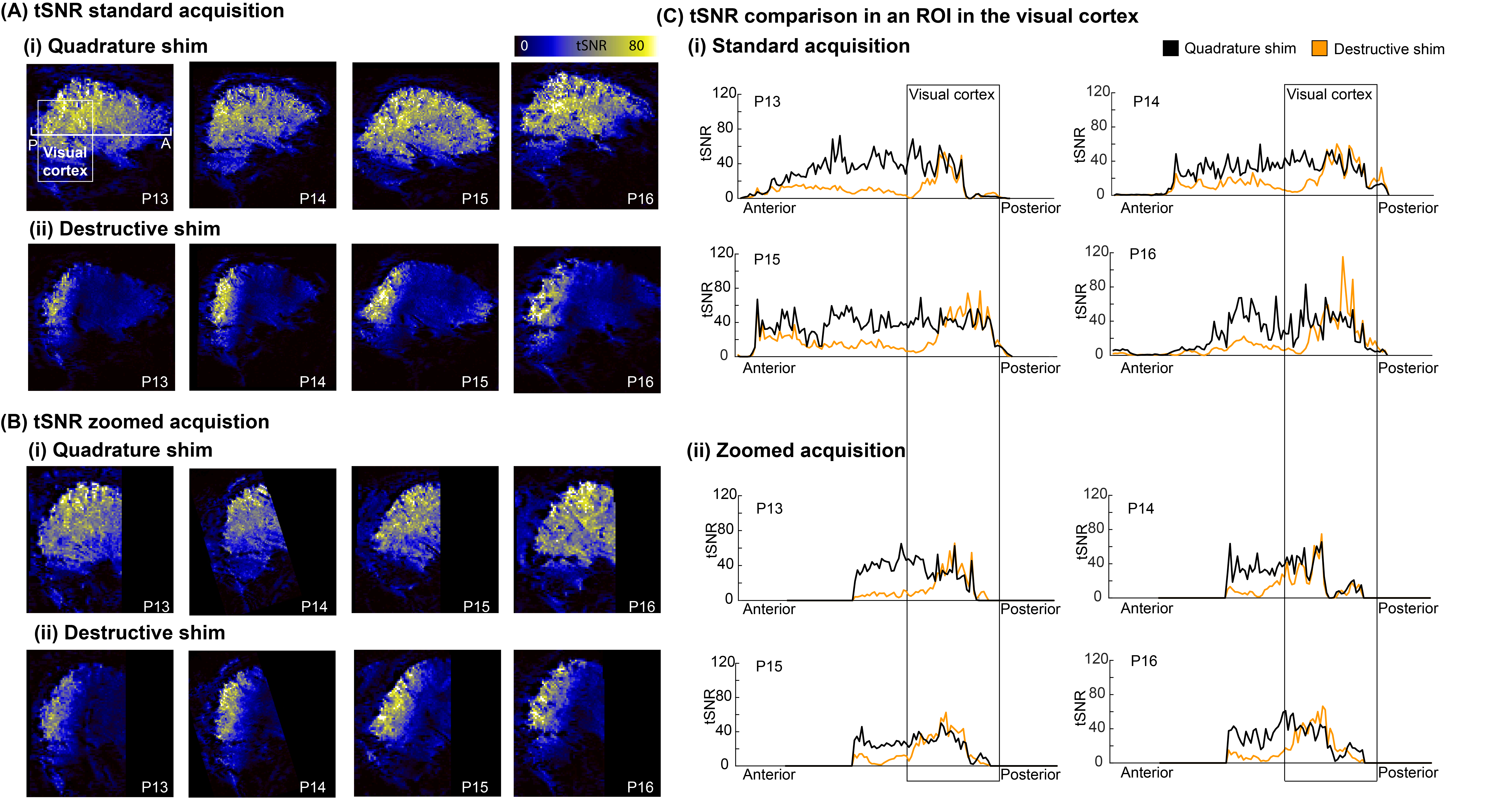
Flip-angle distributions were consistent across participants (Fig.4). The destructive shim could successfully reduce scan time without compromising image quality, this is illustrated by the high flip-angles in visual cortex and similar tSNR compared to the quadrature shim in this area (Fig.5).
Discussion and Conclusion
Reducing acquisition times in a user-friendly way remains a key-target for high-field MRI. We introduced a universal phase-shim with destructive interferences that allow reducing the FOV while minimising fold-over artefacts. In our example, we reduced FOV by 50% and acquisition time by 35% while retaining good signal in our target region (higher B1+ and similar tSNR). The destructive shim reduced signal contributions from areas with large vessels and the eyes.Importantly, we show that such approaches can be applied with a simple phase-shim and without further optimisation across individuals. This minimises patient burden and is directly translatable across sequences (i.e. 2D/slab-non-selective-3D/slab-selective-3D). Moreover, it has the potential to increase SAR flexibility through electric-field modeling of the given shims by the coil vendor. Note that our approach can gain further flexibility through combined phase-and-magnitude shimming and explicit optimisation of both combined and destructive interferences in distinct ROIs. We expect that this allows our method to translate across brain regions and therefore research interests.
Acknowledgements
This work is supported by a Dutch Research Council (NWO) grant (OCENW.XS22.4.007), an Amsterdam Brain and Cognition grant (T0922) and a Dutch Research Council (NWO)TTW VIDI grant (VI.Vidi.198.016).References
1. Dumoulin SO. Layers of Neuroscience. Neuron. 2017;96(6):1205-1206. doi:10.1016/J.NEURON.2017.12.0042. Dumoulin SO, Wandell BA. Population receptive field estimates in human visual cortex. Neuroimage. 2008;39(2):647-660. doi:10.1016/j.neuroimage.2007.09.034
3. Olman CA, Harel N, Feinberg DA, He S, Zhang P. Layer-Specific fMRI Reflects Different Neuronal Computations at Different Depths in Human V1. PLoS ONE. 2012;7(3):32536. doi:10.1371/journal.pone.0032536
4. Klaas P. Pruessmann, Markus Weiger, Markus B. Scheidegger, Boesiger P. SENSE: Sensitivity encoding for fast MRI. Published online 1999. Accessed August 16, 2022. https://onlinelibrary.wiley.com/doi/epdf/10.1002/%28SICI%291522-2594%28199911%2942%3A5%3C952%3A%3AAID-MRM16%3E3.0.CO%3B2-S
5. Sodickson DK, Manning WJ. Simultaneous acquisition of spatial harmonics (SMASH): Fast imaging with radiofrequency coil arrays. Magnetic Resonance in Medicine. 1997;38(4):591-603. doi:10.1002/mrm.1910380414
6. Griswold MA, Jakob PM, Heidemann RM, et al. Generalized autocalibrating partially parallel acquisitions (GRAPPA). Magnetic Resonance in Medicine. 2002;47(6):1202-1210. doi:10.1002/MRM.10171
7. van der Zwaag W, Reynaud O, Narsude M, Gallichan D, Marques JP. High spatio-temporal resolution in functional MRI with 3D echo planar imaging using cylindrical excitation and a CAIPIRINHA undersampling pattern. Magnetic Resonance in Medicine. 2018;79(5):2589-2596. doi:10.1002/mrm.26906
8. Pfeuffer J, van de Moortele PF, Yacoub E, et al. Zoomed functional imaging in the human brain at 7 Tesla with simultaneous high spatial and high temporal resolution. Neuroimage. 2002;17(1):272-286. doi:10.1006/nimg.2002.1103
9. Pfaffenrot V, Brunheim S, Rietsch SHG, et al. An 8/15-channel Tx/Rx head neck RF coil combination with region-specific B1+ shimming for whole-brain MRI focused on the cerebellum at 7T. Magnetic Resonance in Medicine. 2018;80(3):1252-1265. doi:10.1002/MRM.27125
10. Beqiri A, Price AN, Padormo F, Hajnal JV, Malik SJ. Extended RF shimming: Sequence-level parallel transmission optimization applied to steady-state free precession MRI of the heart. NMR in Biomedicine. 2017;30(6). doi:10.1002/NBM.3701
11. Gras V, Vignaud A, Amadon A, Le Bihan D, Boulant N. Universal pulses: A new concept for calibration-free parallel transmission. Magnetic Resonance in Medicine. 2017;77(2):635-643. doi:10.1002/MRM.26148
12. Aigner CS, Dietrich S, Schaeffter T, Schmitter S. Calibration-free pTx of the human heart at 7T via 3D universal pulses. Magnetic Resonance in Medicine. 2022;87(1):70-84. doi:10.1002/MRM.28952
13. Heidemann RM, Anwander A, Feiweier T, Knösche TR, Turner R. k-space and q-space: combining ultra-high spatial and angular resolution in diffusion imaging using ZOOPPA at 7 T. Neuroimage. 2012;60(2):967-978. doi:10.1016/j.neuroimage.2011.12.081
14. Mooiweer R, Sbrizzi A, Raaijmakers AJE, van den Berg CAT, Luijten PR, Hoogduin H. Combining a reduced field of excitation with SENSE-based parallel imaging for maximum imaging efficiency. Magn Reson Med. 2017;78(1):88-96. doi:10.1002/mrm.26346
15. Winkelmann R, Börnert P, Nehrke K, Dössel O. Efficient foldover suppression using SENSE. MAGMA. 2005;18(2):63-68. doi:10.1007/s10334-004-0081-5
16. High spatio‐temporal resolution in functional MRI with 3D echo planar imaging using cylindrical excitation and a CAIPIRINHA undersampling pattern - van der Zwaag - 2018 - Magnetic Resonance in Medicine - Wiley Online Library. Accessed November 7, 2023. https://onlinelibrary.wiley.com/doi/10.1002/mrm.26906
17. Berrington A, Považan M, Mirfin C, et al. Calibration-free regional RF shims for MRS. Magnetic Resonance in Medicine. 2021;86(2):611-624. doi:10.1002/MRM.28749
Figures
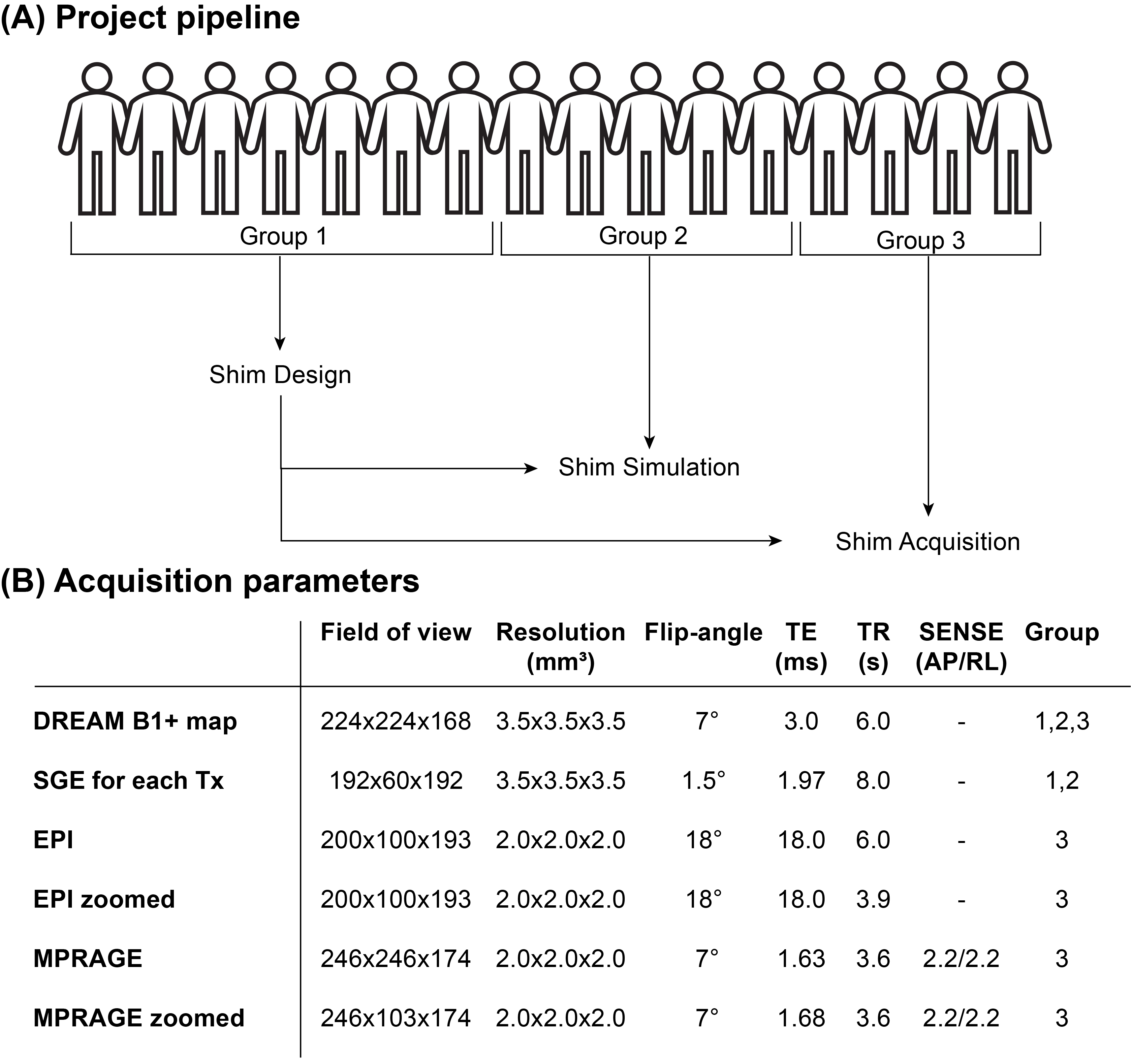
Figure 1: (A) The pipeline of the project. Group 1 (N=7) was used to optimise shimming offsets. Group 2 (N=5) was used to simulate the universal implementation of the offsets. Group 3 (N=4) was used to acquire new data showing the implementation of the shim. (B) The parameters of the obtained acquisitions and for which group they were obtained. All acquisitions for group 3 were obtained with the quadrature and the destructive B1 shim. *SGE=Spoiled Gradient Echo.
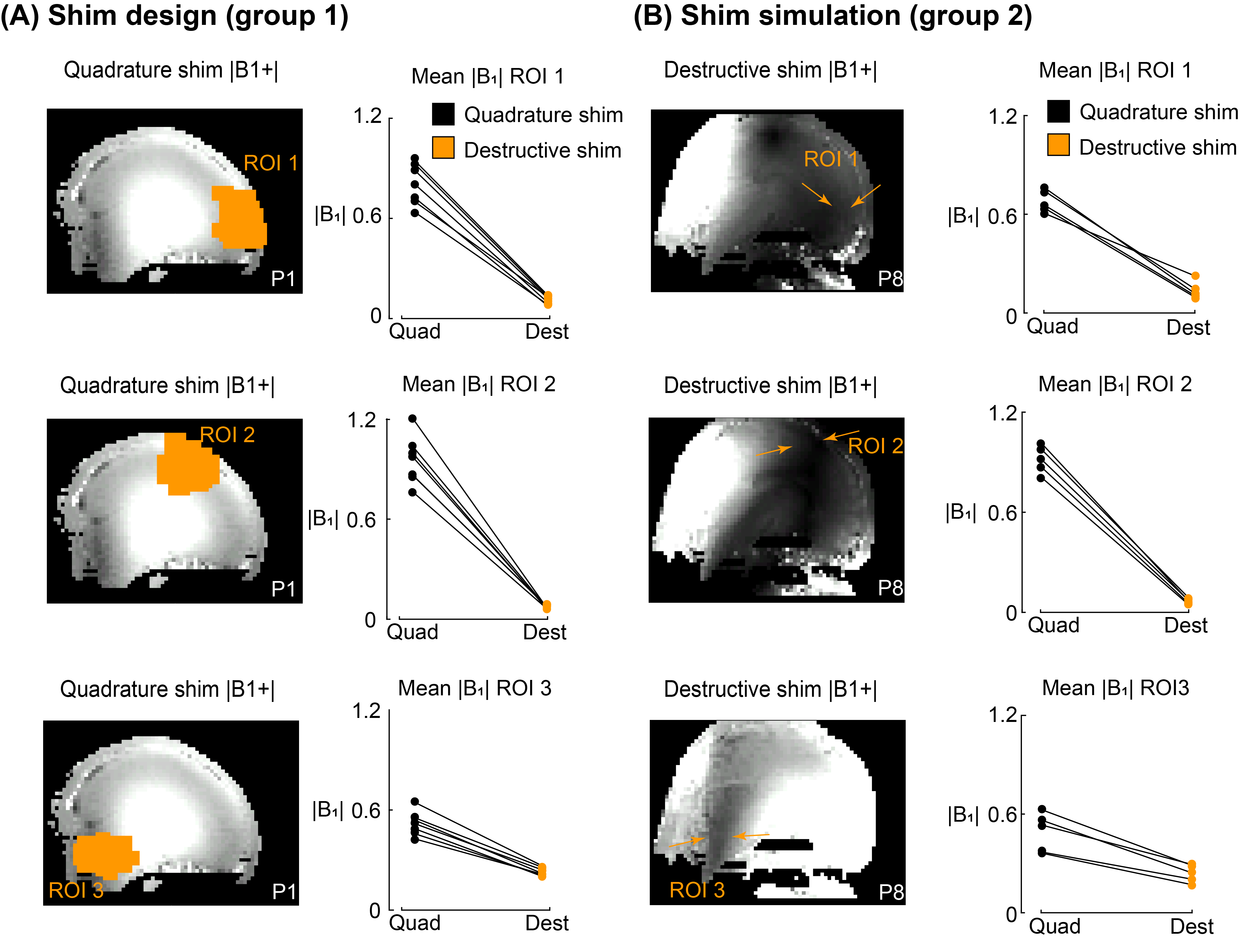
Figure 2: (A) The B1+ map with a quadrature B1 shim of a participant from group 1. The ROI used for the destructive shim is overlaid in orange. Each row shows the implementation of the shim on a different ROI. The mean B1+ for all participants in group 1 is plotted before and after the destructive shim. (B) The resulting B1+ map with the destructive shim of a participant from group 2. The arrows indicate the decreased signal in the ROI. The mean B1+ before and after the implementation of the destructive shim for all participants in group 2 is plotted.
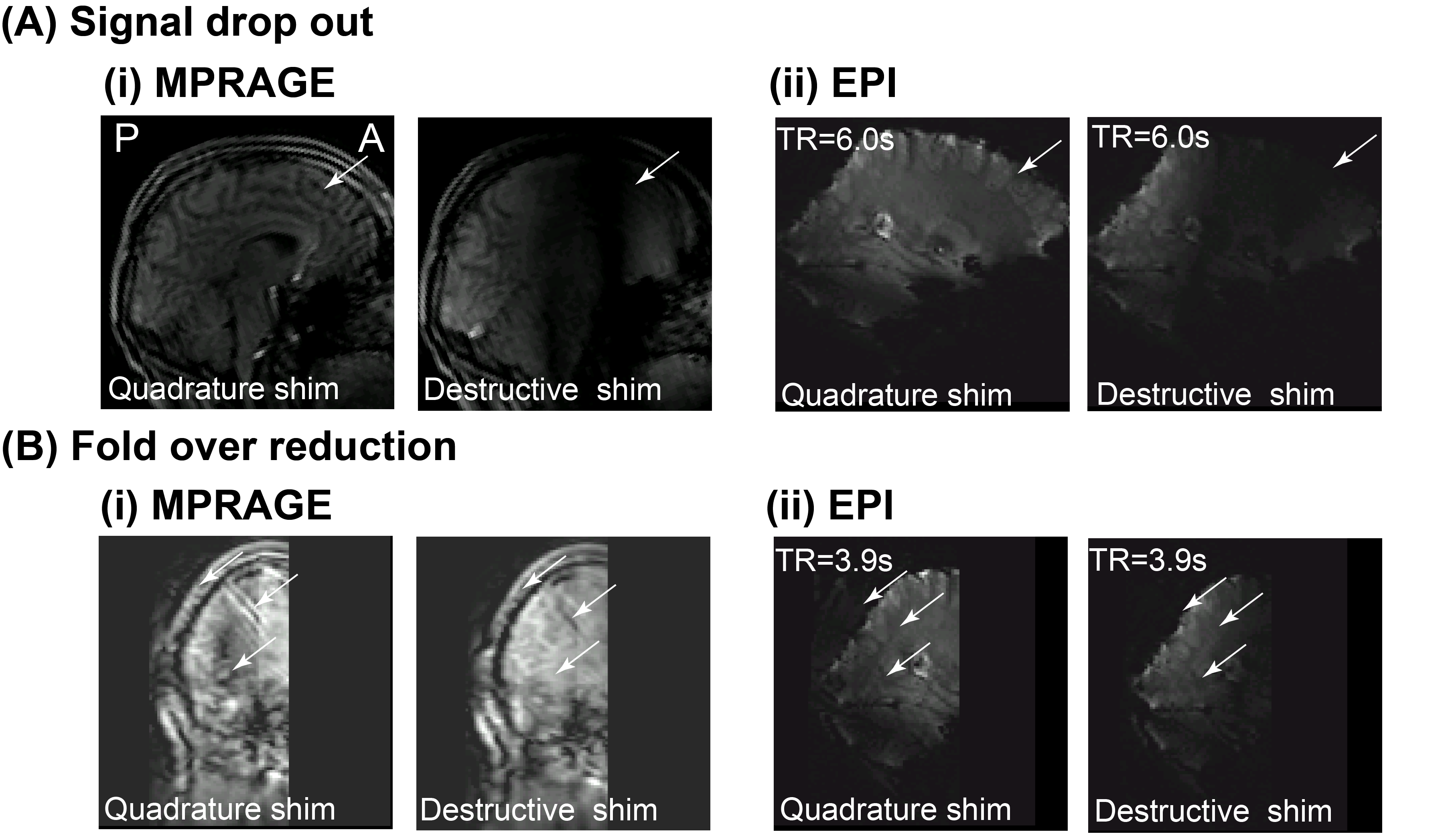
Figure 3: (A) An example of the signal drop out when the destructive shim is used compared to a quadrature shim for an MPRAGE (i) and EPI (ii) acquisition. (B) A comparison of the fold over artefact present in the quadrature shim but not using the destructive shim for an MPRAGE (i) and EPI (ii) acquisition. Arrows highlight the location of the fold over or the lack thereof.
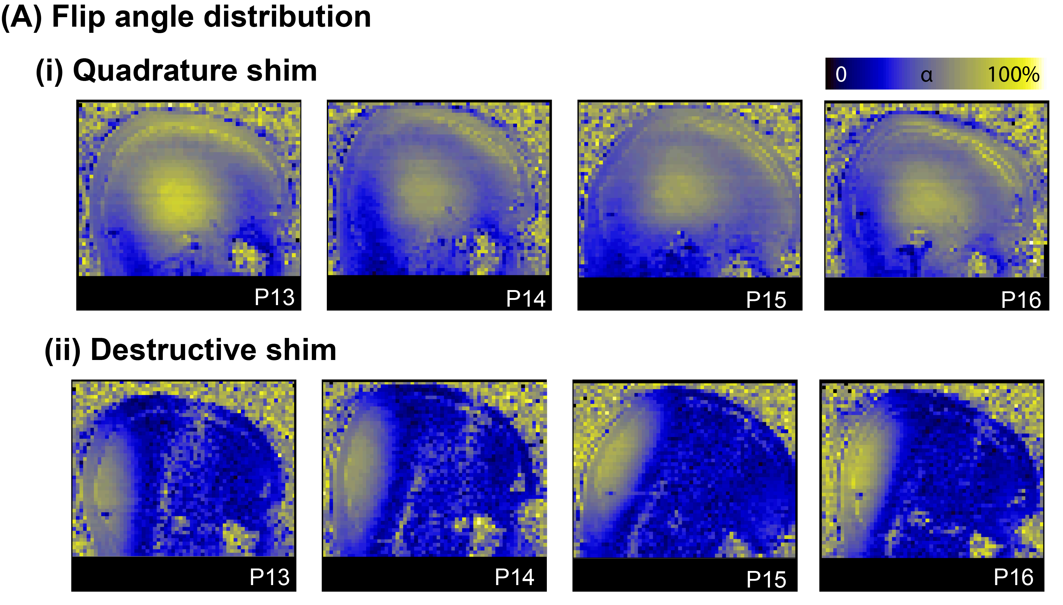
Figure 4: (A)The flip angle distribution as a result of the quadrature shim for all participants in group 3. (B)The flip angle distribution as a result of the destructive shim for all participants in group 3.