3297
MR Imaging of tumor vascular normalization: DCE and IVIM1Department of Radiology, Affiliated Hospital of Guizhou Medical University, Guiyang, China, 2Philips Healthcare, Shanghai, China
Synopsis
Keywords: Parallel Imaging, Cancer
Motivation: The prognosis for patients with advanced nasopharyngeal carcinoma is often poor. Although anti-angiogenic therapy can enhance the effectiveness of chemo-radiotherapy, drug resistance remains a challenging issue. Accurately monitoring the time window for vascular normalization is crucial.
Goal(s): The objective of this study was to assess the value of DCE-MRI and IVIM-MRI in dynamically monitoring tumor vascular normalization.
Approach: By applying different antiangiogenic therapies to preclinical models, this study evaluated the effectiveness of DCE and IVIM in monitoring tumor vascular normalization (TVN).
Results: Preliminary results suggest that IVIM-MRI can potentially replace DCE-MRI in monitoring tumor vascular normalization.
Impact: This study holds profound implications for the clinical application of anti-angiogenesis therapy. It provides valuable insights into the effectiveness of combination therapies and offers a deeper understanding of vascular normalization indicators. These findings could potentially improve treatment strategies.
Introduction
Antiangiogenic therapy shows promise as a treatment for locally advanced nasopharyngeal carcinoma. Studies indicate that Galunisertib can enhance the antitumor effect of BEV. This therapy promotes tumor vascular normalization (TVN), which reduces tumor hypoxia and increases tumor sensitivity to radiotherapy and chemotherapy [1]. Administering radiotherapy and chemotherapy during the TVN time window effectively improves the antitumor effect [2]. Therefore, accurate monitoring of the TVN time window is crucial for the clinical application of combination therapy. Dynamic contrast-enhanced (DCE)-MRI is a functional imaging technique used in clinical trials to evaluate the effects of anti-angiogenic drugs [3]. In patients with primary rectal and breast cancer, changes in vascular heterogeneity were quantified using Ktrans distribution and correlated with prognosis [4]. However, the use of contrast media increases the risk of adverse events [5], which may limit the frequent use of DCE-MRI technology. In recent years, the rapid development of diffusion-weighted imaging technology has enabled Intravoxel incoherent motion (IVIM)-MRI to demonstrate excellent ability in evaluating tissue microvascular perfusion and tissue space diffusion characteristics [6]. This study discusses the application value of DCE-MRI and IVIM-MRI multimodal MRI imaging techniques for dynamically monitoring TVN efficacy and tumor response to treatment. Our findings may contribute to the clinical application of anti-angiogenic therapies.Materials and Methods
100 subcutaneous xenograft tumor models of nasopharyngeal carcinoma were randomly divided into 4 groups (25 in each group): the control group, galunisertib group, BEV group, and galunisertib + BEVY group. MRI and histopathological analysis were performed at different time points (day 0, 3, 7, 14, and 25). The MRI experiment was conducted using a 3.0T scanner (Elition X, Philips Healthcare, Best, the Netherlands) equipped with an eight-channel mouse coil (HC607P, Hezi Medical Technology Co., Ltd., Wuxi, China). The scanning sequence included T1-weighted imaging (T1WI), T2-weighted imaging (T2WI), IVIM, and DCE. The IVIM parameters were evaluated using the bi-exponential model, generating corresponding parameter maps. The IVIM parameters included the slow apparent diffusion coefficient (ADC) (D), fast ADC (D*), and fast ADC fraction (f). The DCE-MRI images were processed using the IntelliSpace Portal 9.0 software (ISP, Philips). The quantitative parameter Ktrans was calculated using the Extended Tofts-Ketty pharmacokinetic model, generating a map. The correlation between MRI parameters and pathological parameters was determined using Pearson’s correlation analysis.Results
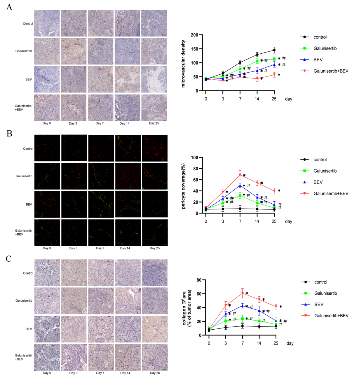
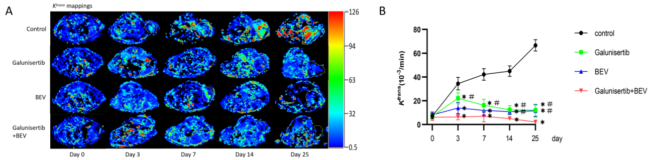
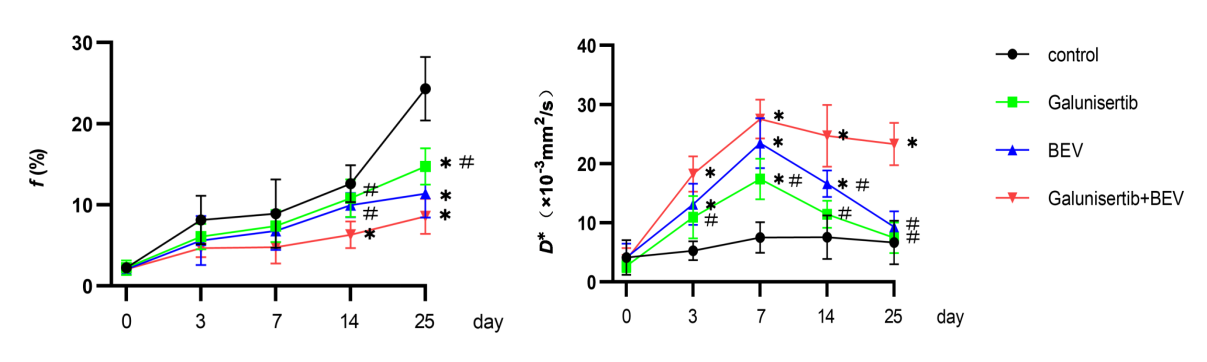
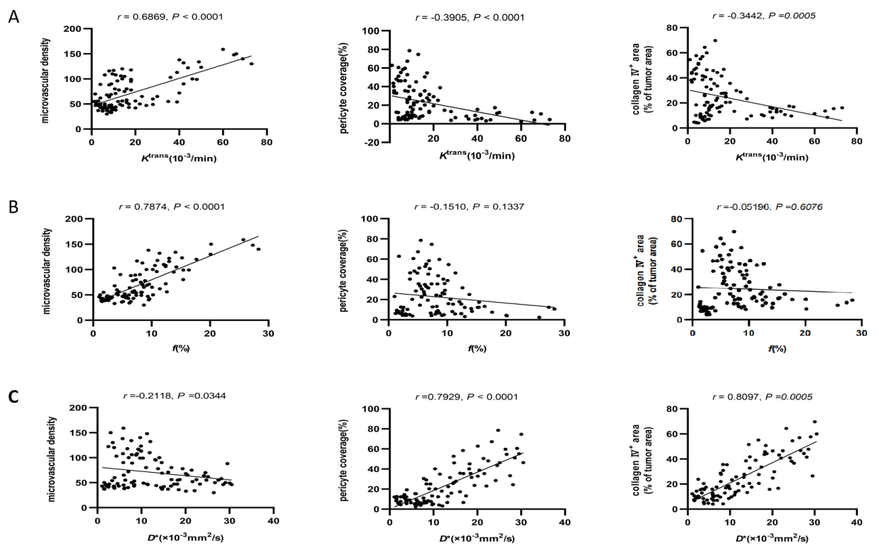
After 14 and 25 days of treatment, the combined therapy of Galunisertib + BEV significantly reduced tumor microvascular density (MVD) compared to either treatment alone. (Fig. 1A). Vascular normalization indicators, such as the pericellular coverage index (PCI) and collagen IV, increased after the combination therapy, peaking on day 7 and remaining high until day 25. (P<0.05). The latter group's indicators decreased to near the baseline level by day 25 (Fig. 1B, C). The DCE-MRI parameter Ktrans value (Fig. 2) and the IVIM-MRI parameter f value (Fig. 3) showed a significant positive correlation with MVD (Fig. 4). Ktrans was weakly correlated with the PCI and collagen IV markers of vascular normalization (Fig. 4). The IVIM-MRI parameter D* was strongly associated with the PCI and collagen IV markers, but only weakly associated with MVD (Fig. 4).Discussion
In this study, we demonstrated that Galunisertib can enhance the effect of TVN induced by BEV, prolong the time window, continuously alleviate tumor hypoxia, and ultimately delay tumor growth. Dynamic monitoring of TVN progression can help determine the window period of TVN and provide effective guidance for the implementation of other combination therapy programs. The MRI parameter Ktrans is a crucial factor that reflects the volume transfer coefficient between plasma and contrast agent in the extravascular space [7]. A high Ktrans value indicates active vascular growth, high vascular permeability, and high malignancy of the tumor [8]. The f value represents the perfusion fraction, which indicates the percentage of rapid diffusion within a voxel in the overall diffusion effect. As in previous studies [9], we observed a significant positive correlation between the f value and MVD, which, along with f, reflects tumor angiogenesis. Another study found a significant association between D* and PCI or collagen IV, suggesting that elevated D* can reflect improved tumor vascular function [10].Conclusion
Our study demonstrates that the combination of Galunisertib and BEV is more effective than any individual therapy in promoting tumor vascular normalization. Multiparameter MRI has the potential to replace pathological indicators in monitoring the occurrence of the time window for tumor vascular normalization. IVIM-MRI can be used instead of DCE-MRI for non-invasive and real-time monitoring of tumor vascular normalization, and it shows a strong correlation with pathological indicators.Acknowledgements
No acknowledgement found.References
[1] Gong X, et al. The Treatment Combining Antiangiogenesis with Chemoradiotherapy Impinges on the Peripheral Circulation Vascular Endothelial Cells and Therapeutic Effect in the Patients with Locally Advanced Nasopharyngeal Carcinoma. Biomed Res Int. 2022 Jul;2022:1787854.
[2] Zheng R, et al. Targeting tumor vascularization: promising strategies for vascular normalization. J Cancer Res Clin Oncol. 2021 Sep;147(9):2489-2505.
[3] Xue W, et al. Aberrant glioblastoma neovascularization patterns and their correlation with DCE-MRI-derived parameters following temozolomide and bevacizumab treatment. Sci Rep. 2017 Oct;7(1):13894.
[4] Liu L, et al. Correlation of DCE-MRI Perfusion Parameters and Molecular Biology of Breast Infiltrating Ductal Carcinoma. Front Oncol. 2021 Oct;11:561735.
[5] Rose TA Jr, Choi JW. Intravenous Imaging Contrast Media Complications: The Basics That Every Clinician Needs to Know. Am J Med. 2015 Sep;128(9):943-9.
[6] Le Bihan D. What can we see with IVIM MRI? Neuroimage. 2019 Feb;187:56-67.
[7] Xue W, et al. Aberrant glioblastoma neovascularization patterns and their correlation with DCE-MRI-derived parameters following temozolomide and bevacizumab treatment. Sci Rep. 2017 Oct;7(1):13894.
[8] Meng F, et al. The diagnostic efficiency of the perfusion-related parameters in assessing the vascular disrupting agent (CA4P) response in a rabbit VX2 liver tumor model. Acta Radiol. 2022 Sep;63(9):1147-1156.
[9] Li B, Xu D, Zhou J, Wang SC, Cai YX, Li H, Xu HB. Monitoring Bevacizumab-Induced Tumor Vascular Normalization by Intravoxel Incoherent Motion Diffusion-Weighted MRI. J Magn Reson Imaging. 2022 Aug;56(2):427-439.
[10] Zhang J, et al. Dual inhibition of PFKFB3 and VEGF normalizes tumor vasculature, reduces lactate production, and improves chemotherapy in glioblastoma: insights from protein expression profiling and MRI. Theranostics. 2020 Jun;10(16):7245-7259.
Figures