3294
Rapid gradient-recalled echo line scanning with crisp line profiles for human MRI at 7 Tesla1Australian Institute for Bioengineering and Nanotechnology, University of Queensland, St Lucia, Australia, 2Department of Radiology, Harvard Medical School, Boston, MA, United States, 33Athinoula A. Martinos Center for Biomedical Imaging, Massachusetts General Hospital, Charlestown, MA, United States, 4Harvard-MIT Division of Health Sciences and Technology, Massachusetts Institute of Technology, Cambridge, MA, United States
Synopsis
Keywords: New Signal Preparation Schemes, Pulse Sequence Design, Line Scanning
Motivation: Line-scanning trades coverage for speed and resolution in a single direction, suitable for the study of mesoscale structure and functional dynamics across cortical depth. However, at ultra high-field strength, the sampling rate is constrained by the specific absorption rate.
Goal(s): Facilitate Rapid gradient-recalled echo line scanning with crisp line profiles for human MRI at 7 Tesla.
Approach: A new line-scan Tip-Down Tip-Up based preparation module.
Results: Using the proposed preparation module, it is possible to perform gradient-recalled echo line scanning experiments with short TR and crisp line profiles.
Impact: Using the proposed preparation module, it is possible to perform gradient-recalled echo line scanning experiments with short TR and crisp line profiles. Such advances can be used to study functional dynamics across cortical depth with high sampling rates.
Introduction
When pushed to the extreme, reduced field-of-view techniques converge to a single line in k-space. In this limit, the excited magnetization resembles a ‘stack of pancakes’, where the diameter of the pancakes corresponds to the line width and their thickness corresponds to the resolution along the line. Effectively, line-scanning trades coverage for speed and resolution in a single direction, suitable for the study of mesoscale structure and functional dynamics across cortical depth1-6.In gradient-recalled echo line scanning, the line is carved out of a slice using saturation pulses. This process, called ‘Outer Volume Suppression (OVS)’ requires careful planning. Saturation bands must be placed strategically to prevent unwanted signals from leaking into the line, and the saturation pulses must be tuned to balance line profile and Specific Absorption Rate (SAR). Saturation pulses with a large Time BandWidth (TBW) product can produce crisp line profiles, but their high SAR contribution limits the minimal achievable TR. Yet, to unravel the intricate functional dynamics across cortical depth, high sampling rates are desired.
In this work, we demonstrate a new approach to gradient-recalled echo line scanning, offering crisp line profiles, shorter repetition times, and lower SAR.
Methods
We propose to use a preparation-module consisting of a tip-down and tip-up pulse pair (Fig.1). First a rectangular pulse (0.5ms) tips the magnetization everywhere onto the transverse plane (90°). Subsequently a slice-selective pulse (SLR, 3ms, TBW=8), is used to tip the magnetization back up along a single plane (−90°). After gradient spoiling, the desired line is revealed by exciting a slice orthogonal to the tip-up pulse.All experiments were performed at 7 Tesla (Siemens Magnetom 7 Tesla Plus, Germany), using a 32-channel head coil (Nova Medical, USA). The fidelity of the line profiles was evaluated in an 18-cm spherical water phantom, comparing two configurations of the product sequence (saturation pulses: 4ms, TBW=4.2), the double saturation method6, and the proposed method. All experiments excited a 3mm diameter line, either through the top of the phantom (8cm diameter, Fig.2a-d) or the center for the phantom (18cm diameter, Fig.2e-h). To resolve the line profile a 3D cartesian readout was used (0.5x0.5x5mm3, TR=450ms, FA=20°).
Lines through the hand area in the motor cortex (M1) of a human subject were collected using configurations f-h (Fig. 2) to assess in-vivo performance. To visualize the potential contamination from residual out-of-line signal, 2D cartesian sampling was used (0.5x1.0mm3,TR=450ms or minimum, TE=20ms). All methods used saturation flip-angles of nominally 90° and an excitation flip-angle close to the Ernst angle. The study was approved by the local human research ethics committee in accordance with national guidelines.
Results & Discussion
Fig.3 shows lines profiles excited using the different methods. Although two 4-cm wide saturation bands should be enough to carve out a clean line through the top of the phantom, the dull profiles of the saturation bands in the product sequence allow signal to leak in. Consequently, the effective line width exceeded 10mm and the edges of the phantom remained visible (Fig.3a). Knowing the shape of these saturation bands, their separation was optimized to best approximate the 3mm beam and avoid residual signal from the phantom edges (Fig.3b). The double-saturation method was designed for an 8cm FOV allowing the high-bandwidth saturation pulses to carve out a well-defined beam through the top of the phantom (Fig.3c). However, compared to the product implementation, the SAR was 3.3x higher. Although slightly less rectangular than the double saturation prepared line, the proposed method produced a good line profile using 3x less SAR (Fig.3d).The bottom row in Fig.3 shows beams excited through the center of the phantom. When using OVS based methods, the larger sample cross-sections necessitate wider (Fig.2a vs. 2d) or additional saturation bands (Fig. 2a vs 2e-f). Wider saturation-bands have stretched out profiles, making the line broader and less crisp (Fig.3e), whereas additional saturation bands require more SAR(Fig.3f-g). The proposed method, on the other hand, requires no additional planning or parameter modifications and maintains its line shape and SAR characteristics (Fig.3h).
Fig.4 shows exemplary lines measured in-vivo. Using the proposed method, 3x shorter TR were possible without line profile degradation. Reduced SAR allows faster sampling of functional dynamics and increased temporal degrees of freedom, without compromising the saturation angle. Moreover, shorter TR also enhance robustness to B1+ nonuniformity as any residual magnetization outside of the slice is driven into a steady state with a smaller longitudinal component.
Conclusion
Using the proposed Tip-Down-Tip-Up preparation module, it is possible to perform gradient-recalled echo line scanning experiments with short TR and crisp line profiles.Acknowledgements
This work was supported by the Australian government through the Australian Research Council (ARC) Future fellowship grant FT200100329, by the NIH NIBIB (grants P41-EB030006 and R01-EB019437), and by the BRAIN Initiative (NIH NINDS grant U19-NS123717).References
- Balasubramanian M, Mulkern R V, Polimeni JR. In vivo irreversible and reversible transverse relaxation rates in human cerebral cortex via line scans at 7 T with 250 micron resolution perpendicular to the cortical surface. Magn Reson Imaging. 2022;90:44-52.
- Balasubramanian M, Mulkern R V, Neil JJ, Maier SE, Polimeni JR. Probing in vivo cortical myeloarchitecture in humans via line-scan diffusion acquisitions at 7 T with 250-500 micron radial resolution. Magn Reson Med. 2021;85(1):390-403. doi:10.1002/mrm.28419
- Raimondo L, Knapen T, Oliveira ĺcaro AF, et al. A line through the brain: implementation of human line-scanning at 7T for ultra-high spatiotemporal resolution fMRI. Journal of Cerebral Blood Flow and Metabolism. 2021;41(11):2831-2843. doi:10.1177/0271678X211037266
- Raimondo L, Priovoulos N, Passarinho C, et al. Robust high spatio-temporal line-scanning fMRI in humans at 7T using multi-echo readouts, denoising and prospective motion correction. J Neurosci Methods. 2023;384:109746. doi:10.1016/j.jneumeth.2022.109746
- Yu X, Qian C, Chen D yow, Dodd SJ, Koretsky AP. Deciphering laminar-specific neural inputs with line-scanning fMRI. Nat Methods. 2014;11(1):55-58. doi:10.1038/nmeth.2730
- Morgan, A.T., Nothnagel, N., Petro, L.S., Goense, J. and Muckli, L., 2020. High-resolution line-scanning reveals distinct visual response properties across human cortical layers. BioRxiv, pp.2020-06. doi: 10.1101/2020.06.30.179762
Figures
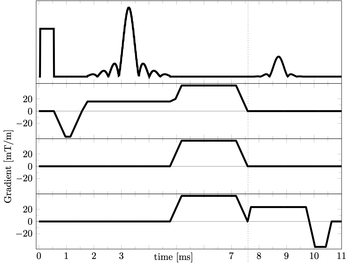
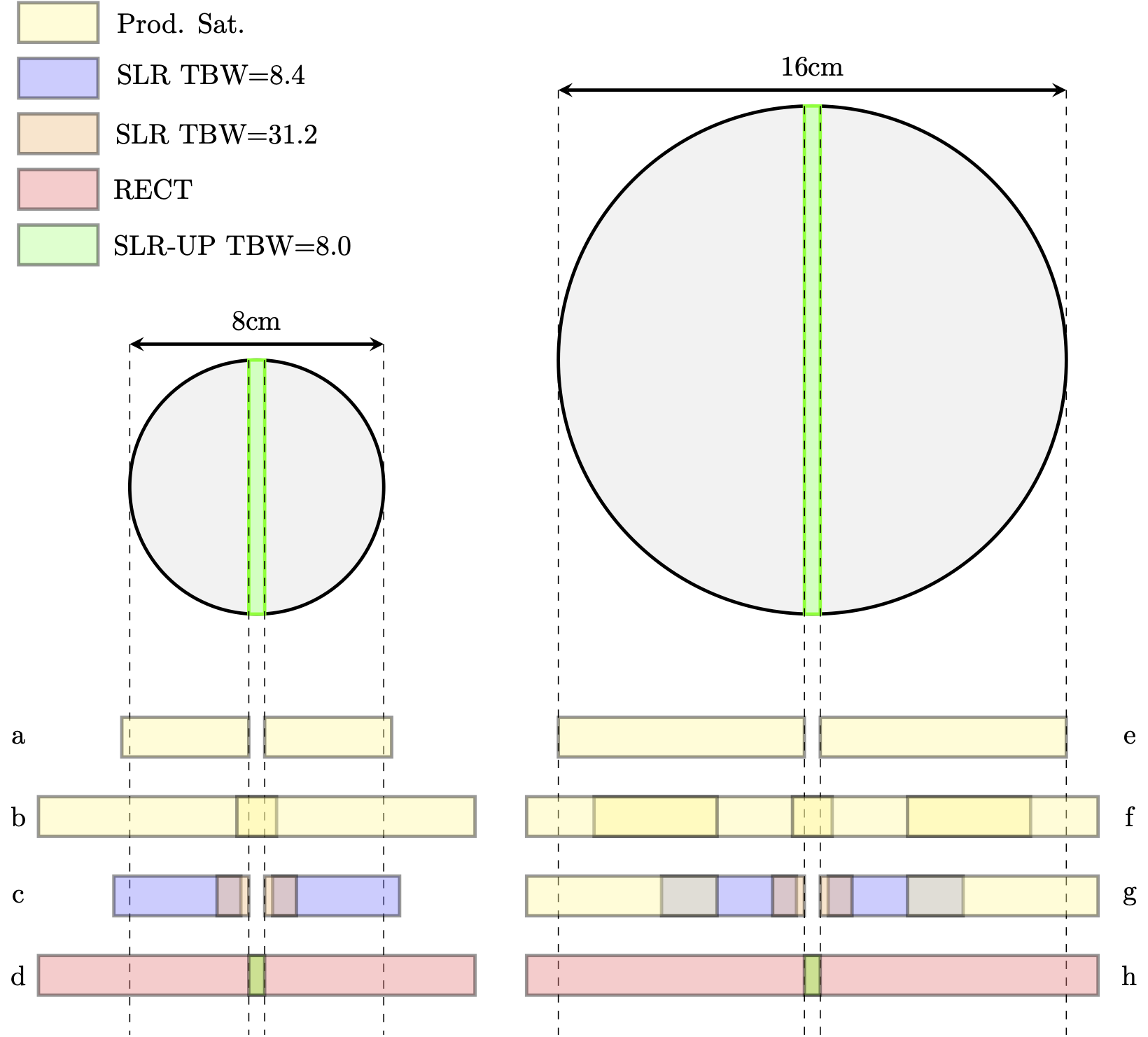
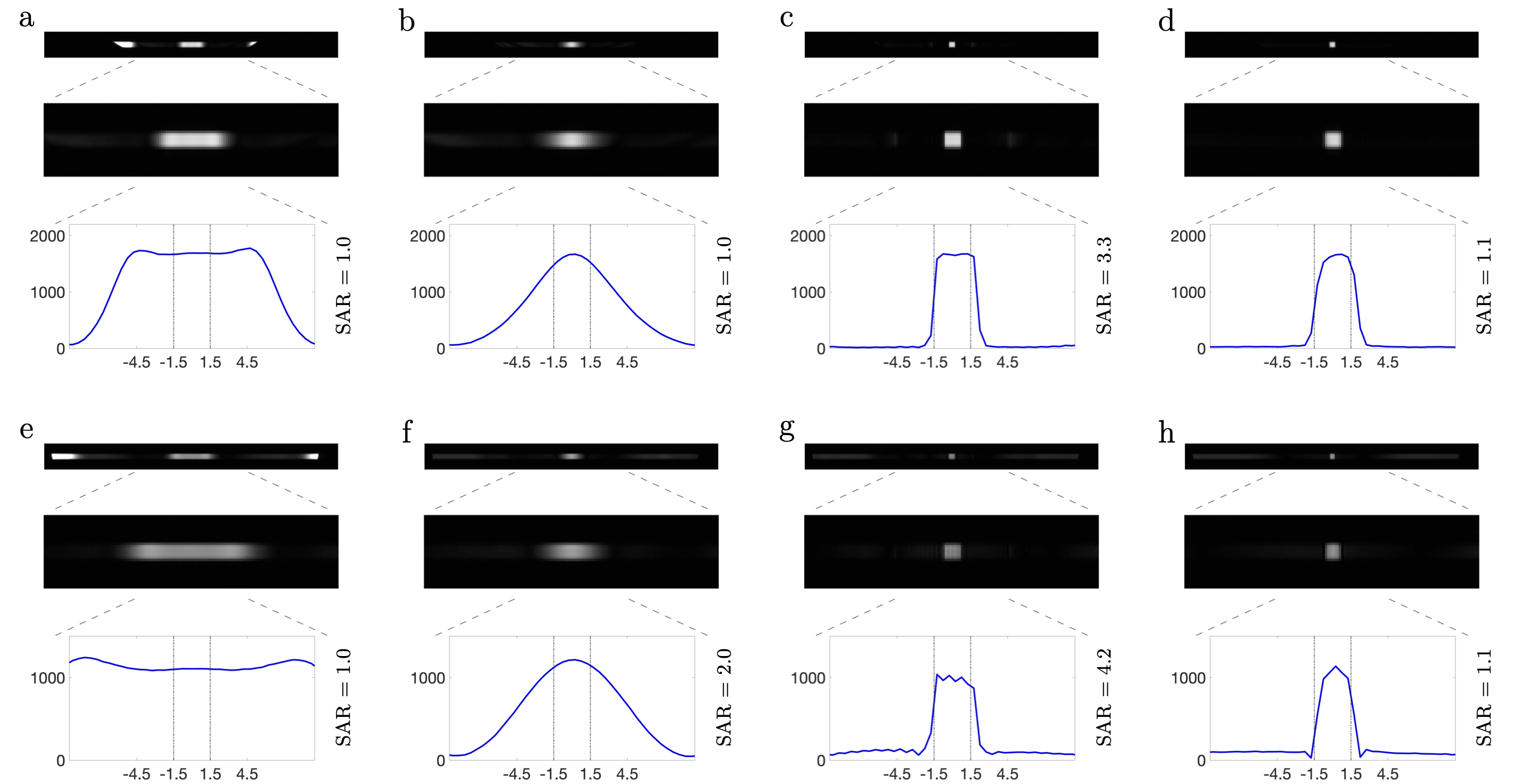
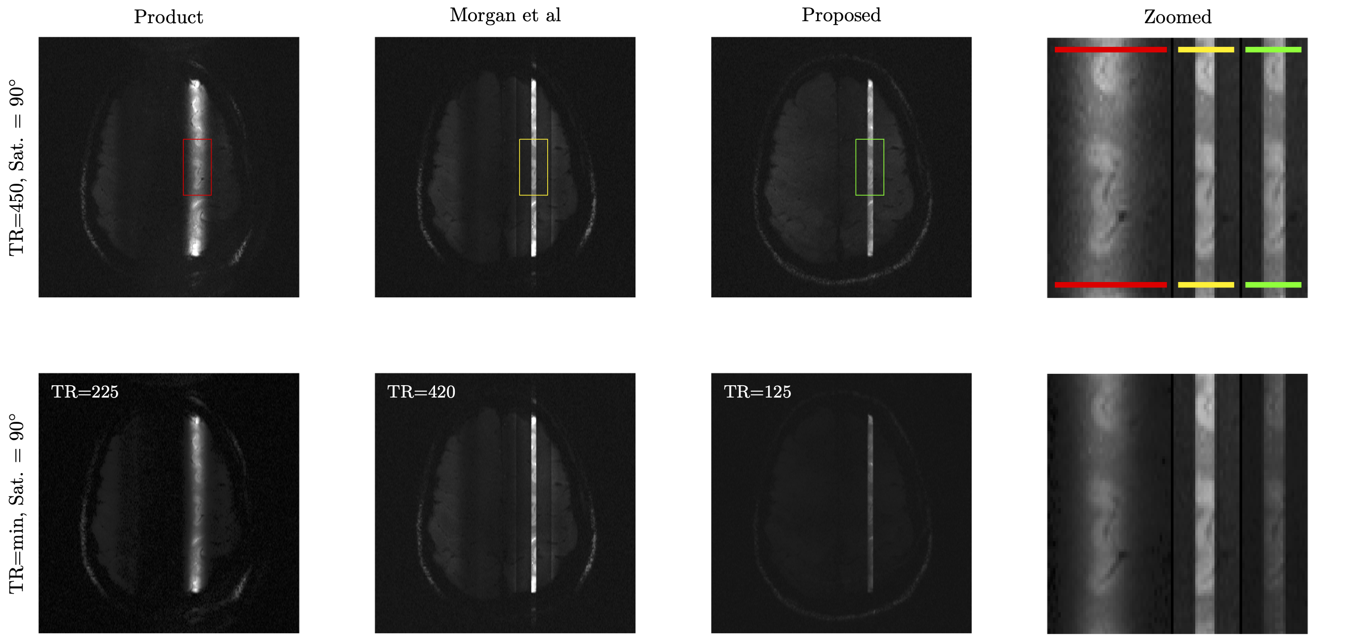
Figure 4: In-vivo evaluation of background suppression using different line scanning methods. The first column shows results obtained using the product sequence (Fig. 2f). The second column shows the double saturation method. The third column shows the proposed method. The fourth column shows zoomed section of the line side by side. The top row shows results obtained with a TR of 450ms. The middle row shows results obtained with minimum TR allowed. Note that the signal amplitude decreases with shorter TR due to the reduced longitudinal magnetisation in the steady state.