3290
Compartmentalised reconstruction of cardiac 31P concentric ring MRSI at 7T1OCMR, RDM Cardiovascular Medicine, University of Oxford, Oxford, United Kingdom, 2School of Biomedical Engineering and Imaging Sciences, Faculty of Life Sciences and Medicine, King's College London, London, United Kingdom, 3The MR Research Centre and the PET Centre, Aarhus University, Aarhus, Denmark, 4Wellcome Centre for Integrative Neuroimaging, FMRIB, Nuffield Department of Clinical Neurosciences, University of Oxford, Oxford, United Kingdom, 5Department of Imaging Methods, Institute of Measurement Science, Slovak Academy of Sciences, Bratislava, Slovakia
Synopsis
Keywords: New Trajectories & Spatial Encoding Methods, Spectroscopy, Ultra high field
Motivation: Cardiac 31P MRS allows the probing of metabolism in various heart diseases, however, commonly employed 3D MRSI techniques are slow even at 7 T.
Goal(s): Our work aims to evaluate the use of compartment-based localised spectroscopy using a linear algebraic model (SLAM).
Approach: 31P MRSI data was collected using concentric ring trajectory acquisitions and SLAM was used to reconstruct 31P signal from the myocardium.
Results: We show increased SNR and reduced uncertainty in determining the cardiac PCr signal, in addition to also reducing the repeatability of PCr/ATP ratio measurements when compared to a 2.5 minute, NUFFT-reconstructed CRT protocol.
Impact: Repeatable and high-SNR 31P acquisitions will allow the probing of cardiac energetics in patient populations that were previously unable to attend prolonged scan sessions at ultra-high field strengths.
Introduction
Phosphocreatine to adenosine triphosphate concentration ratio (PCr/ATP) measurements using phosphorus magnetic resonance spectroscopy (31P-MRS) offer a unique insight into the energy metabolism of the failing human heart in vivo (1,2). 31P-MRS can also be used to describe the potentially more sensitive chemical kinetics of the oxidative phosphorylation system. Shorter scan times are desirable for both applications, to fit 31P-MRS into a clinical protocol. Whilst moving from 3 T to 7 T carries the advantage of a potential 2.8-fold (3) increase in SNR, this can only be achieved with the most commonly used, time-consuming point-by-point Cartesian sampling (Fourier-transform (FT) based MRSI) scheme. A new method employing a fast concentric ring readout trajectory (CRT) has recently been described (4), and its repeatability has been established (5). While a CRT-based acquisition can measure PCr/ATP maps as FT-based MRSI sampling in a fraction of the time (with matched resolution) or with higher spatial resolution (in a matched time), data interpretation may still be subject to the way relevant septal cardiac voxels are selected (6). Localised spectroscopy using a linear algebraic model (SLAM) allows the selection of anatomical compartments that contribute to the 31P signal (7), thus minimising any variation in the measured PCr/ATP due to suboptimal voxel selection, effectively permitting the acquisition of spectra from arbitrarily shaped compartments. The aim of this study was thus to compare voxel-based and compartment-based PCr/ATP values for three different CRT protocols.Methods
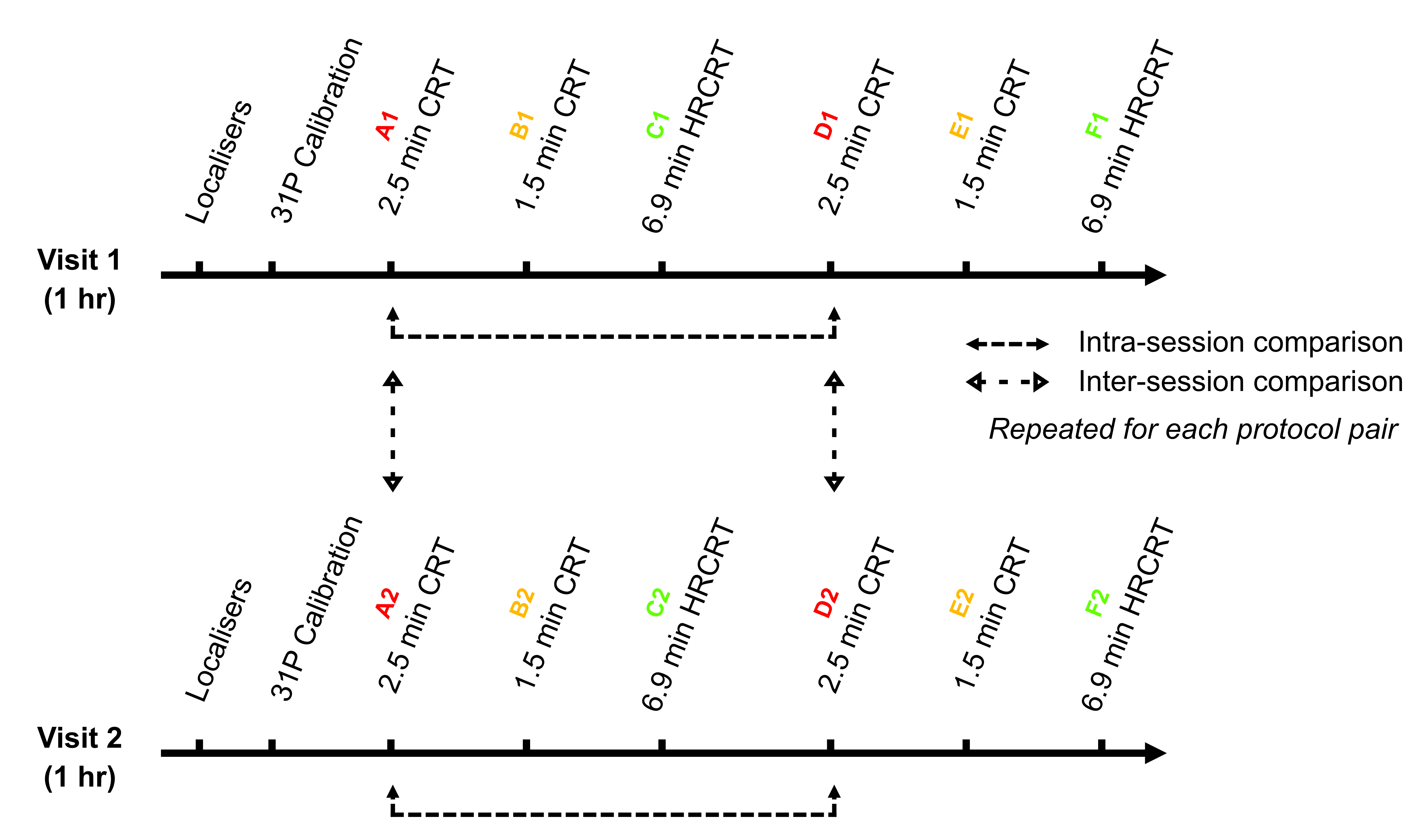
Three CRT sequences were evaluated: two with a 10×10×10 matrix size employing 12 (2:31 min) and 10 rings (1:37 min) respectively, and one with a matrix size of 12×12×12 using 19 rings (6:55 min, labeled as high-res CRT [HRCRT]). These three sequences were run twice per session in two sessions over two different days, 72 hours apart, to evaluate intra- and inter-session repeatability (Figure 1). Five healthy participants (2 females, 61±9 kg, 27±6 years) were scanned supine in a Siemens Magnetom 7 T scanner (Siemens Healthineers, Germany) equipped with a square surface transmit and 16-channel receive array coil (Rapid Biomedical, Germany) positioned over the heart (6). CRT was run with 240×240×200 mm3 FOV, 1 s TR, 2778 Hz bandwidth, and 720 time samples. CONCEPT data were reconstructed offline using the non‐uniform FFT (NUFFT) toolbox with min‐max Kaiser‐Bessel kernel interpolation and twofold oversampling in MATLAB (MathWorks, Natick, USA). Individual coil data was combined using the WSVD algorithm (8). Regridded k-space data and a compartment mask of the myocardium drawn on a central slice were fed into the SLAM algorithm to produce a single, SNR-maximised 31P myocardial spectrum. CSI/MRSI data fitting was performed using the OXSA toolbox (9). PCr/γ‐ATP ratios were corrected for partial saturation and blood contamination, in mid-septal voxels of a mid-slice of the heart for NUFFT-reconstructed spectra and in the myocardial compartment-specific spectrum reconstructed by SLAM. All corrections were calculated per subject and per session. Single voxel analysis of the NUFFT-reconstructed 2.5-minute CRT acquisition was used as a reference measurement since the reproducibility of this method has been previously established (5). Agreement with the NUFFT-reconstructed 2.5-minute CRT acquisition was assessed using paired t-tests with Bonferroni-Holm correction for multiple comparisons, and Bland-Altman plots between SLAM-reconstructed acquisitions and the 2.5-minute CRT with NUFFT acquisition. The intra- and inter-session coefficients of repeatability (CoR) were calculated from SD of the signed differences in PCr/ATP between two scans for each subject according to CoR = SDintrasubject × $$$\sqrt{2}$$$ × 1.96. A lower CoR reflects higher repeatability.Results and Discussion
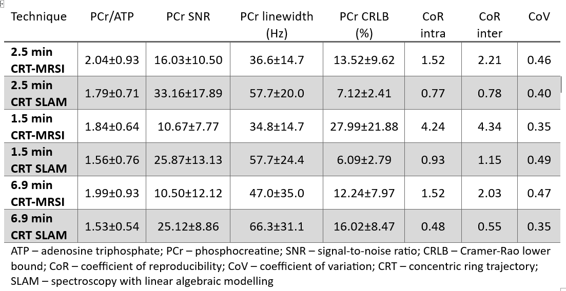
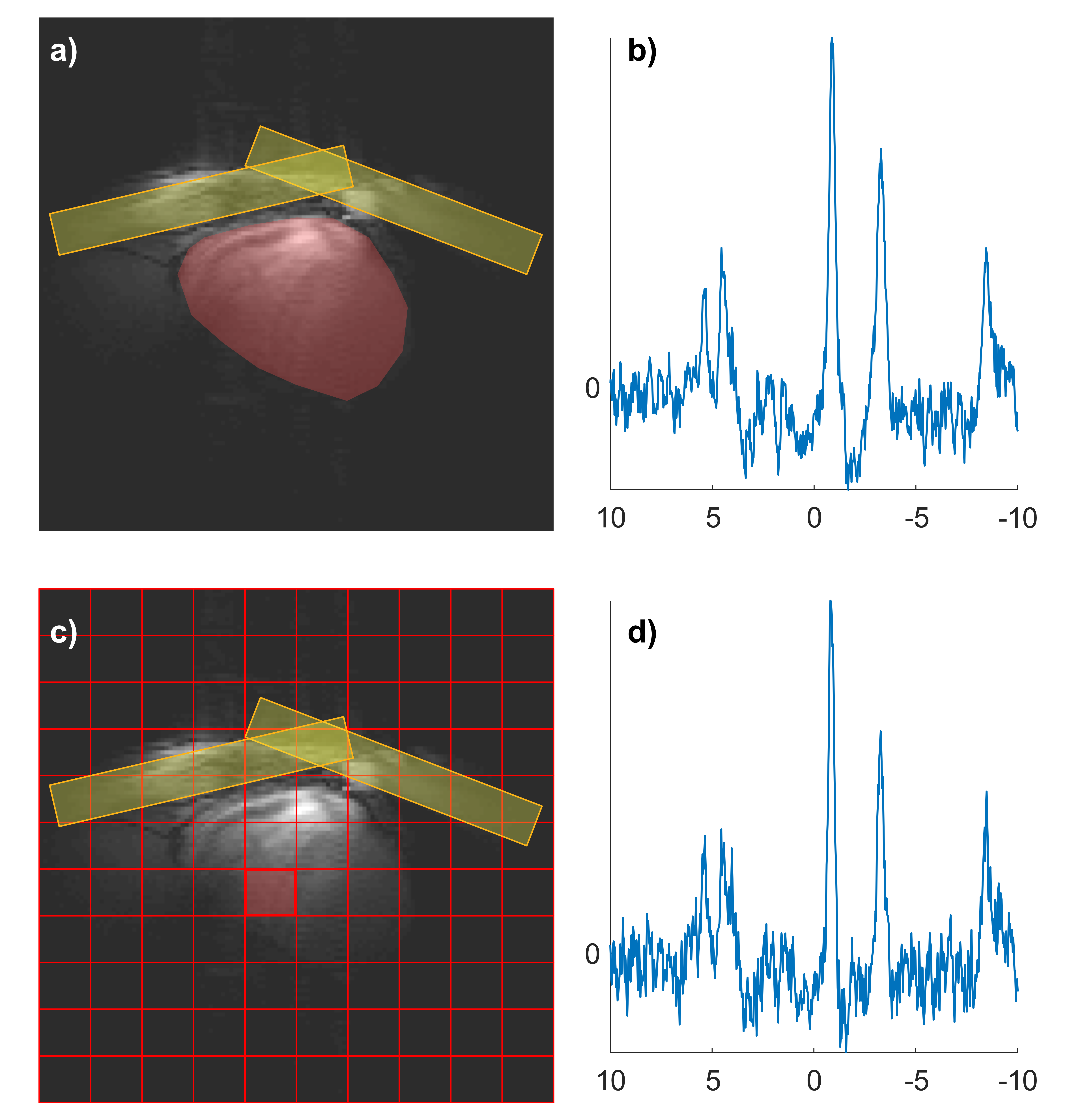
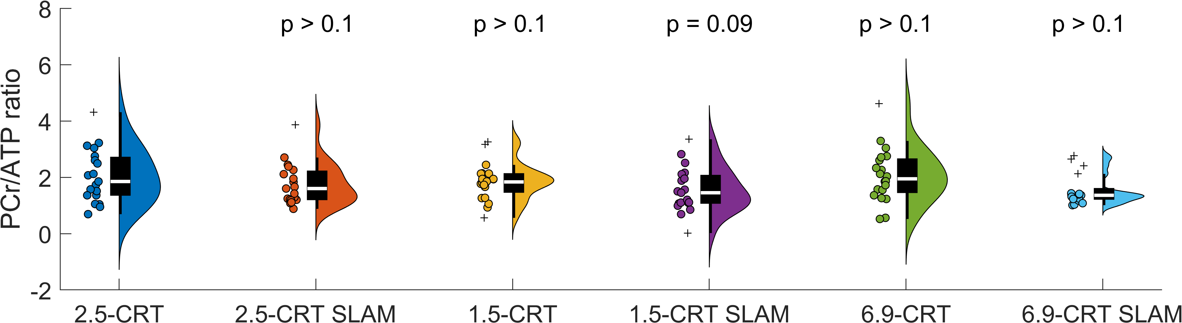
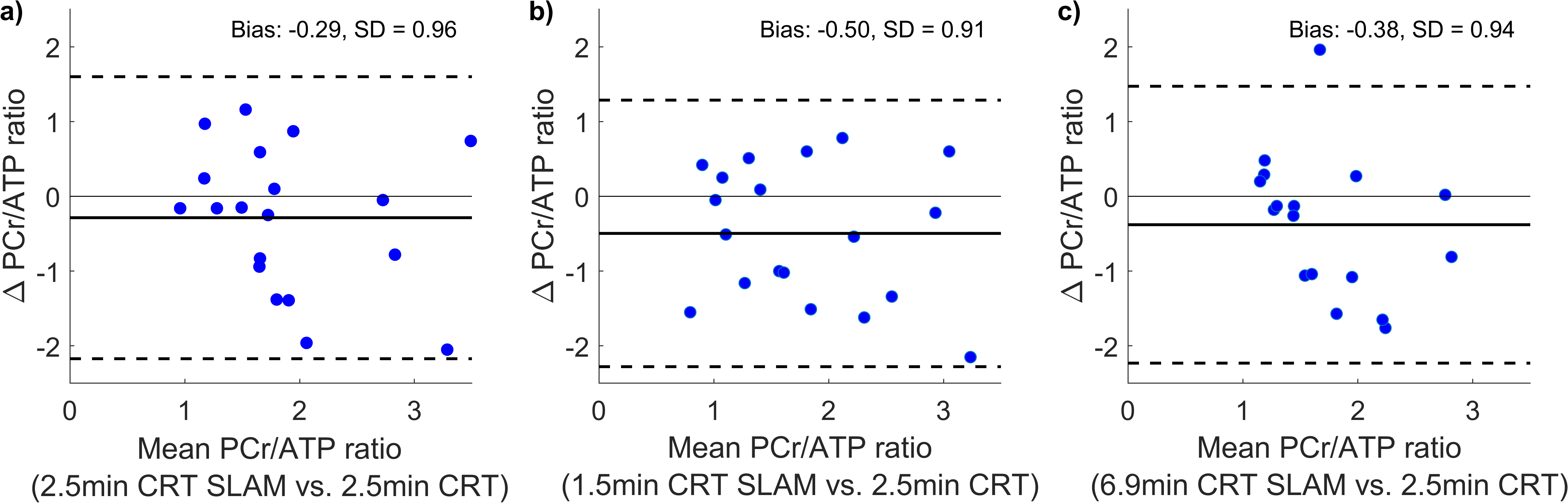
Example spectra from both NUFFT- and SLAM-reconstructed 2.5-minute CRT acquisition are shown on Figure 2. Neither PCr/ATP ratio determined from SLAM-reconstructed spectra, nor the NUFFT-reconstructed spectra were significantly different from the 2.5-minute NUFFT-reconstructed CRT acquisition (Table 1 and Figure 3). There was good agreement between the NUFFT-reconstructed 2.5-minute CRT acquisition and all the SLAM-reconstructed PCr/ATP ratios, with SLAM showing negative bias compared to NUFTT-based reconstruction (Figure 4). All SLAM-reconstructed PCr/ATP ratios had lower coefficients of repeatability than NUFFT-reconstructed PCr/ATP ratios. While we noticed line broadening in SLAM-reconstructed spectra, these can be ascribed to the increased size of the volume of interest as well as the B0 field inhomogeneity due to the lack of shimming.Conclusions
In agreement with the literature (10), the SLAM reconstruction provided lower PCr/ATP ratios than the NUFFT-based reconstruction with lower coefficients of reproducibility than NUFFT-reconstructed spectra, but also higher SNR of the phosphocreatine resonance. This implies that segmented, SLAM CRT 31P MRS may be a more reproducible, less operator-dependent method to quantify cardiac energetics at 7T.Acknowledgements
LV and FEM are supported by a Sir Henry Dale Fellowship of the Wellcome Trust and the Royal Society [221805/Z/20/Z]. LV would also like to acknowledge the support of the Slovak Grant Agencies VEGA [2/0004/23] and APVV [21–0299]. JJM would like to acknowledge the Novo Fonden, Ref. NNF21OC0068683. WTC is supported by the Wellcome Trust [225924/Z/22/Z].References
1. Watson WD, Miller JJJ, Lewis A, et al. Use of cardiac magnetic resonance to detect changes in metabolism in heart failure. Cardiovasc Diagnosis Ther Vol 10, No 3 (June 27, 2020) Cardiovasc Diagnosis Ther 2020. https://cdt.amegroups.org/article/view/37723.
2. Monga S, Valkovič L, Tyler D, et al. Insights Into the Metabolic Aspects of Aortic Stenosis With the Use of Magnetic Resonance Imaging. JACC Cardiovasc Imaging 2022; 15: 2112–26.
3. Rodgers CT, Clarke WT, Snyder C, Vaughan JT, Neubauer S, Robson MD. Human cardiac 31P magnetic resonance spectroscopy at 7 tesla. Magn Reson Med 2014; 72: 304–15.
4. Clarke WT, Hingerl L, Strasser B, Bogner W, Valkovič L, Rodgers CT. Three-dimensional, 2.5-minute, 7T phosphorus magnetic resonance spectroscopic imaging of the human heart using concentric rings. NMR Biomed 2022; : e4813.
5. Mózes FE, Clarke WT, Valkovic L. Reproducibility of 7T human cardiac 3D 31P-MRSI using concentric ring k-space trajectories (CRT). In: Proc. Intl. Soc. Mag. Reson. Med. 2023: 4730.
6. Ellis J, Valkovič L, Purvis LAB, Clarke WT, Rodgers CT. Reproducibility of human cardiac phosphorus MRS (31P-MRS) at 7 T. NMR Biomed 2019; 32: e4095.
7. Zhang Y, Gabr RE, Schär M, Weiss RG, Bottomley PA. Magnetic resonance Spectroscopy with Linear Algebraic Modeling (SLAM) for higher speed and sensitivity. J Magn Reson 2012; 218: 66–76.
8. Rodgers CT, Robson MD. Receive array magnetic resonance spectroscopy: Whitened singular value decomposition (WSVD) gives optimal Bayesian solution. Magn Reson Med 2010; 63: 881–91.
9. Purvis LAB, Clarke WT, Biasiolli L, Valkovič L, Robson MD, Rodgers CT. OXSA: An open-source magnetic resonance spectroscopy analysis toolbox in MATLAB. PLoS One 2017; 12: e0185356.
10. Tyler A, Ellis J, Lau JYC, et al. Compartment-based reconstruction of 3D acquisition-weighted 31P cardiac magnetic resonance spectroscopic imaging at 7 T: A reproducibility study. NMR Biomed 2023; 36: e4950.
Figures