3289
An Advanced (k,T)-acquisition scheme to largely improve sensitivity and resolution for dynamic deuterium MRSI application in human brain at 7T1CMRR, Department of Radiology, University of Minnesota, Minneapolis, MN, United States, 2Beckman Institute, University of Urbana Champaign, Urbana, IL, United States
Synopsis
Keywords: New Trajectories & Spatial Encoding Methods, Deuterium
Motivation: Further improving the spatial resolution and sensitivity of human brain deuterium MRSI (DMRSI).
Goal(s): To develop an advanced (k,T)-acquisition scheme and processing pipeline to improve the SNR and resolution of dynamic DMRSI at 7T.
Approach: We designed and implemented the new DMRSI pulse sequence on an FDA-approved 7T clinical scanner equipped with a 4-channel 2H-1H dual-frequency transreceiver head array coil; evaluated and compared the new (k,T)-CSI sequence and the FSW-CSI sequence performing high-resolution dynamic DMRSI.
Results: The new DMRSI method significantly improves the SNR and spatial resolution and achieved better contrast between human GM, WM and CSF.
Impact: We developed an advanced DMRSI method for high-resolution and high-fidelity whole-brain DMRSI in humans at 7T. It enhances our ability to study neuroenergetics and metabolic reprograming associated with brain tumors and other neurological disorders, and provides clinical value for diagnosis.
INTRODUCTION
Deuterium MRS Imaging (DMRSI) has shown great promise in assessing glucose metabolism and the measurement of metabolic rates in the human brain under normal and diseased states.1,2 It allows dynamic imaging of deuterated glucose, glutamate and glutamine (Glx), lactate and HDO after an oral administration of [6,6’-2H2]-glucose.1 However, the low gyromagnetic ratio of deuterium and low concentration of deuterated metabolites result in a poor signal-to-noise ratio (SNR). Fourier-series window-based CSI (FSW-CSI) sequences have been commonly used for X-nuclear MRSI application with improved SNR, but at the cost of significant image degradation and low spatial resolution.3–5 Here, we have developed a novel DMRSI pulse sequence to simultaneously achieve high spatial resolution and high signal sensitivity. The sequence is rigorously evaluated in both phantom and human experiments at 7T. We demonstrate a largely improved spatial resolution and sensitivity as compared to the traditional FSW-CSI method.METHODS
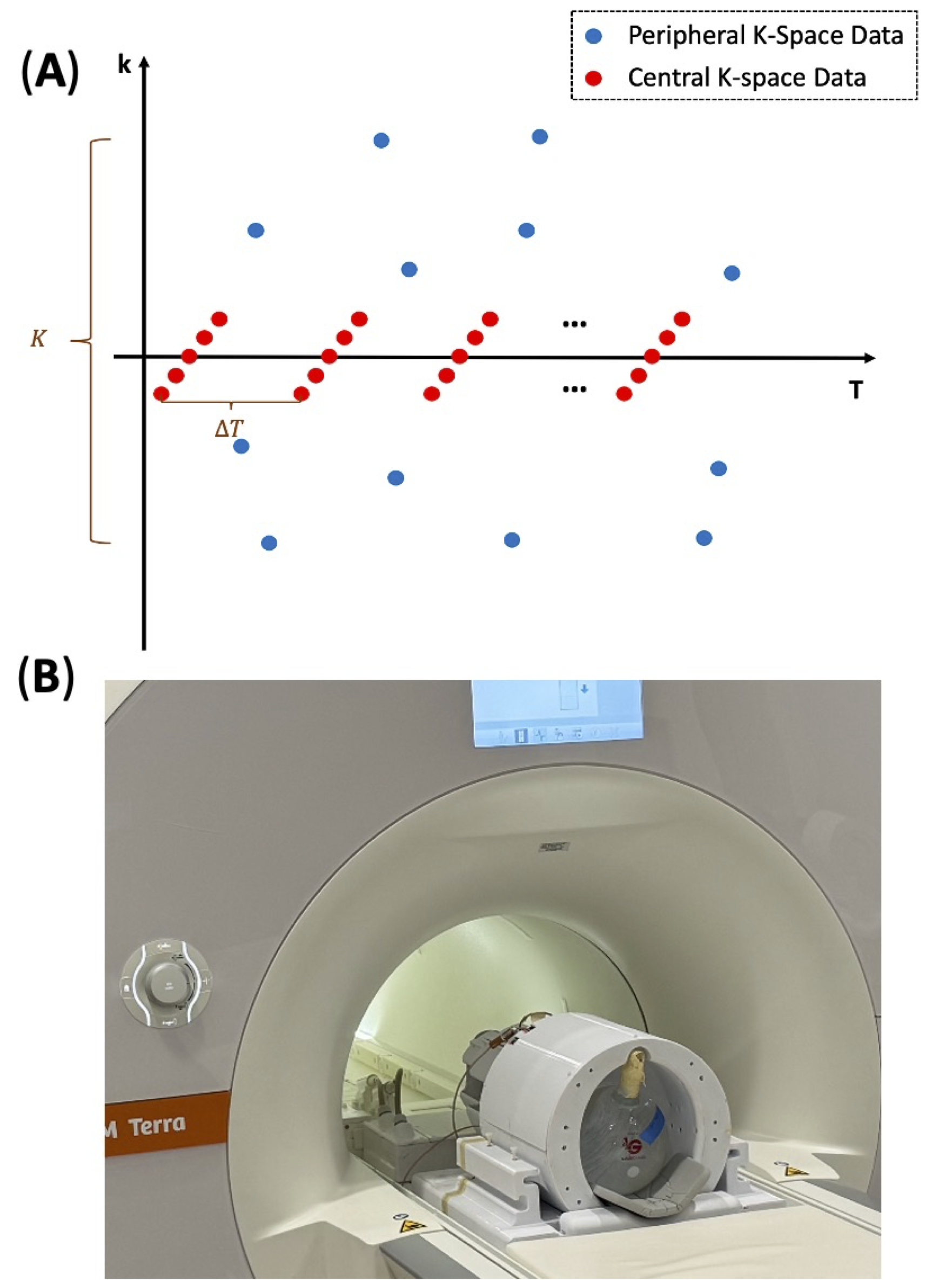
The (k,T)-acquisition scheme as shown in Fig. 1A, acquires two complementary data sets: a central k-space dataset D1 and a peripheral k-space dataset D2. For the dataset D1, it covers only a limited region of central k-space but fully samples the (t,T)-space; and contributes to image formation in two aspects: (1) it will be used to determine the spectral and temporal subspace structures that satisfy the Nyquist and resolution requirements; and (2) it will be used to determine the spatial coefficients to enhance SNR. For the dataset D2, it under-samples the peripheral k-space region in a pseudo random fashion, such that the sampled k-space locations for a specific T are randomly distributed but “complementary” for different Ts. These sparsely sampled data are used to determine the spatial coefficients to achieve the desired spatial resolution. To reconstruct the desired spatial-spectral-temporal image function, the SPICE data processing scheme is adopted which uses a subspace model to effectively leverage both D1 and D2 data for image formation. This new method can achieve 3D whole-brain DMRSI with nominal spatial resolution of 0.9 mL (with minimum spatial blurring) and temporal resolution of 2.5 min.2H/1H images using a 4-channel 2H-1H dual-frequency transreceiver head array coil6 were acquired on an FDA approved 7T-Terra clinical scanner (Siemens, Germany). The (k,T)-method sampled the central k-space in a 5×5×3 matrix regularly and outer k-Space randomly in ratio of 40(center):6(periphery). 20 volumes were acquired for FSW-CSI and kT-CSI, respectively, using the same repetition time (TR) of 173 ms/TE 0.6ms, Ernst-angle of 56° and 19×19×15 matrix. Natural abundance acquisitions of HDO in a head-sized phantom and a human brain (female, 22 y) were performed. A 1H TurboFLASH T1 weighted anatomical scan was acquired with isotropic 1 mm spatial resolution. The study was approved by the University of Minnesota IRB.
RESULTS AND DISCUSSION
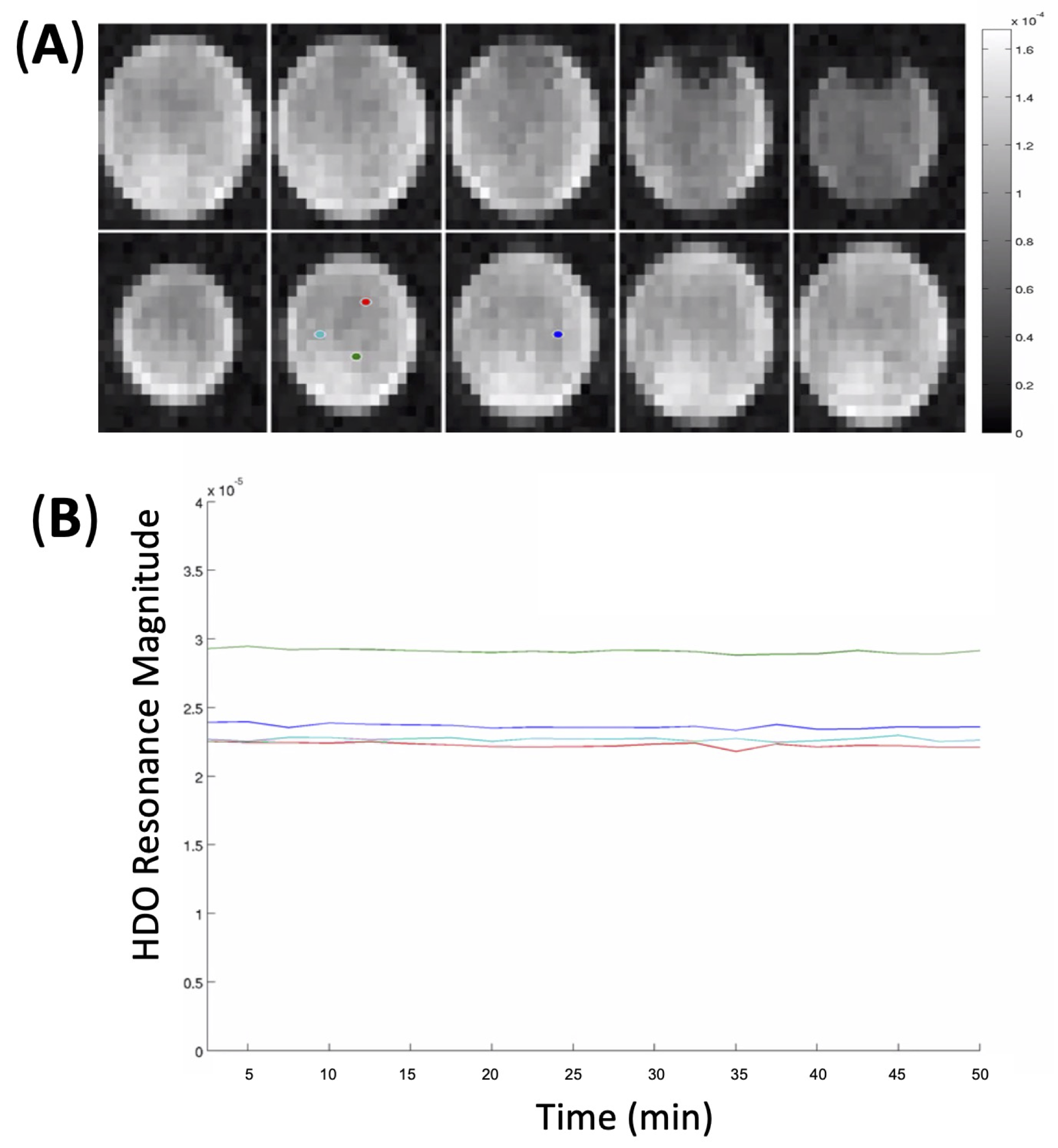
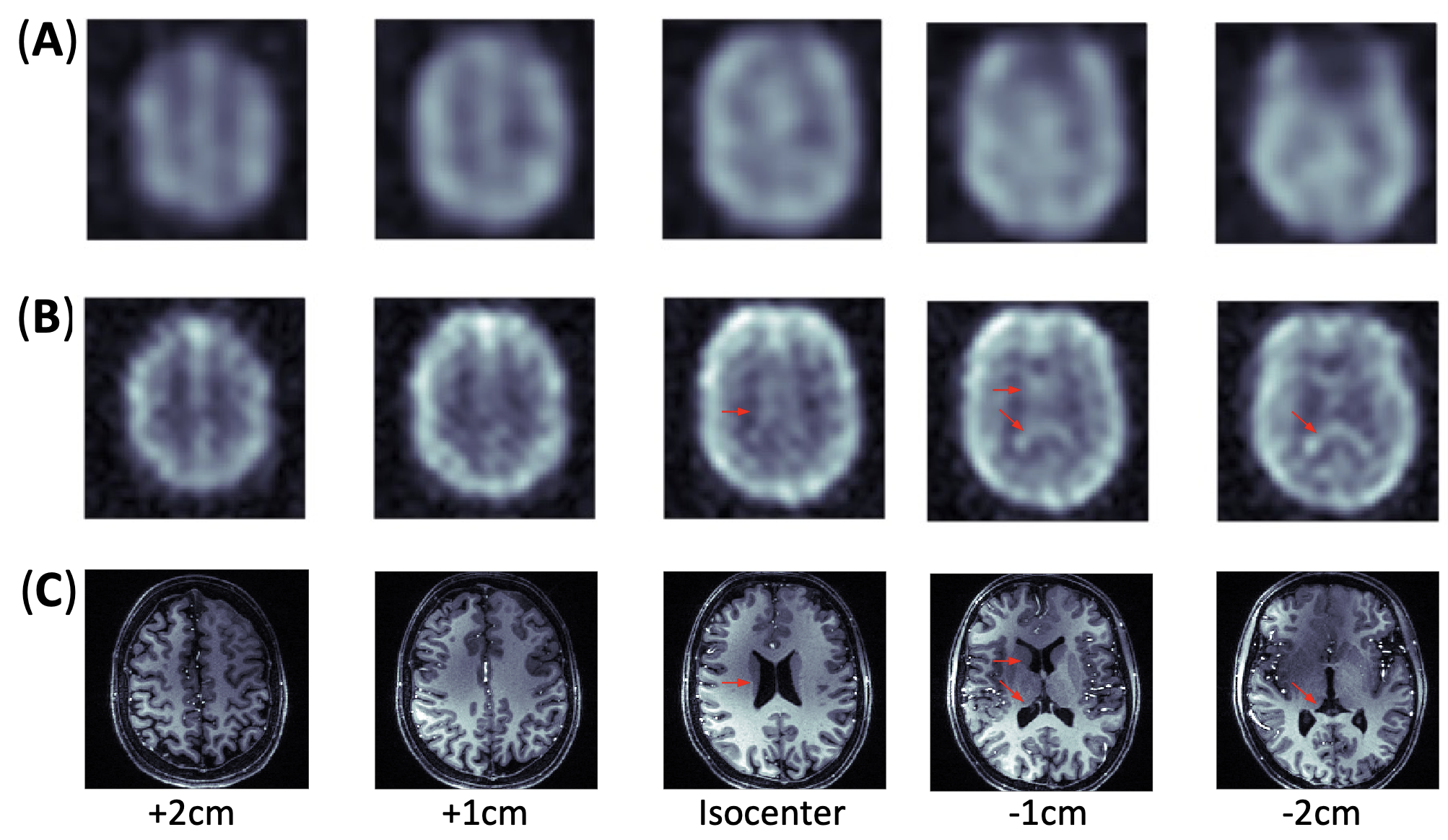
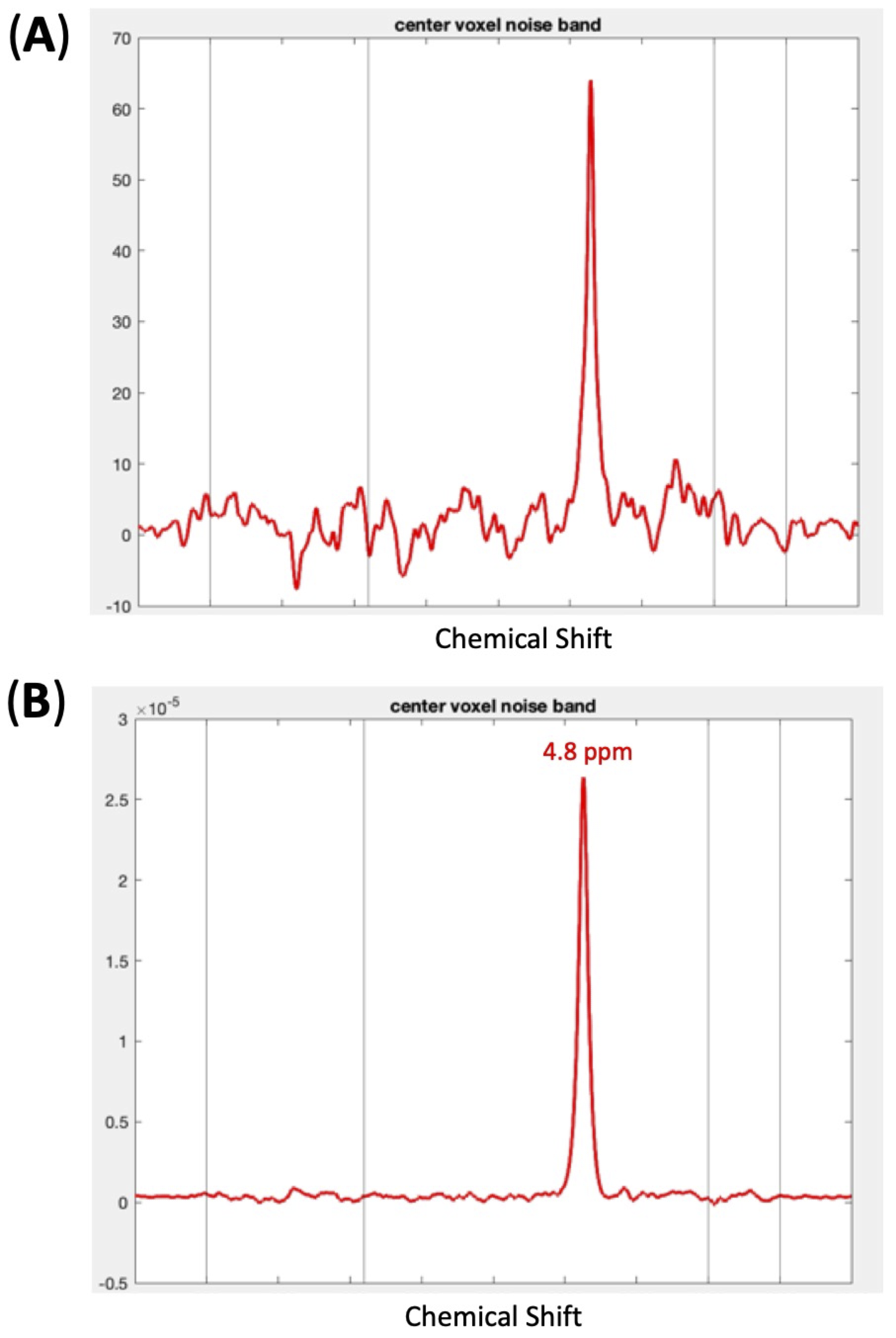
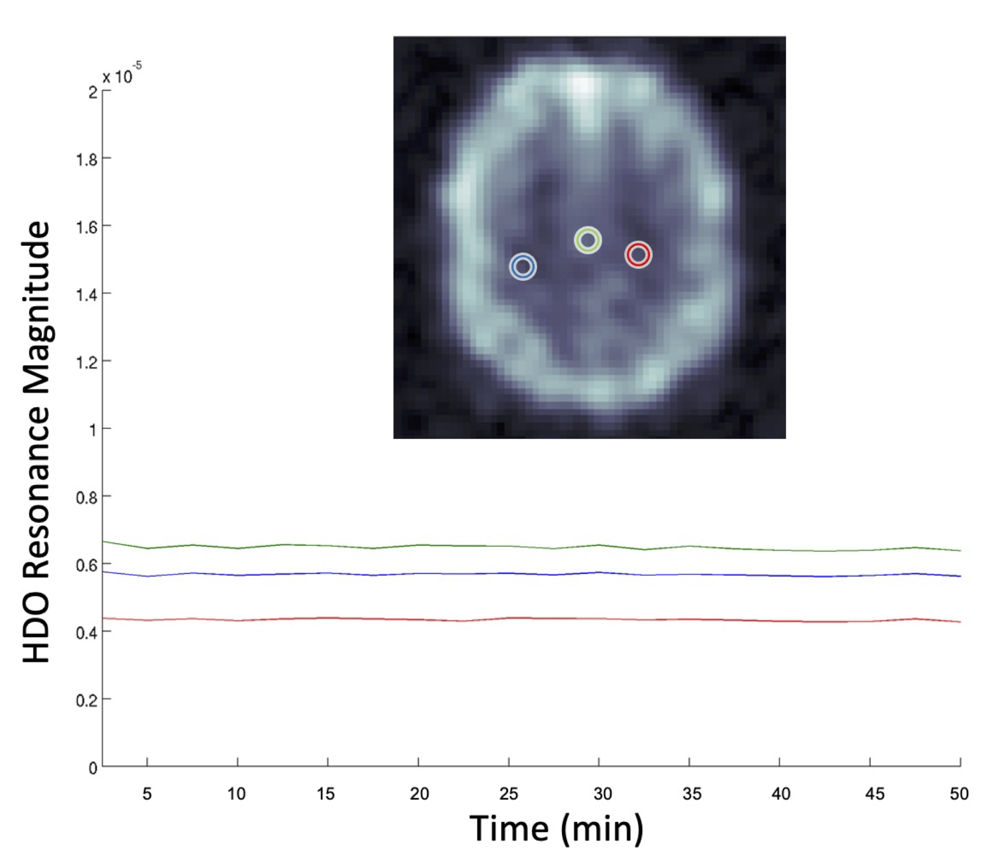
Figure 1 shows a schematic representation of k-Space sampling (A) and experimental setup (B). Figure 2 exhibits the distribution of HDO water signal measured using the kT-CSI method in the phantom and representative HDO time courses of sample voxels. Excellent SNR and uniform images with sharp edges of the phantom (indicating a better spatial resolution) were observed. Figure 3 shows co-registrated FSW-CSI, kT-CSI and corresponding T1-weighted anatomical MRI acquired from one subject. The kT-CSI provides a much better contrast for identifying human gray and white matters as well as CSF in ventricles. Figure 4 shows the CSI-FSW spectrum in (A) and kT-CSI spectrum in (B) from a representative voxel, clearly kT-CSI has better SNR. HDO time courses of brain voxels were generated (see Fig. 5), and a small temporal fluctuation was detected, indicating high temporal stability. The nominal voxel size was similar between the kT-CSI (0.9 cc) and FSW-CSI (0.7 cc) as applied in this study. However, the true voxel size is usually > 3 times larger than the nominal voxel size for FSW-CSI owing the truncation of peripheral k-space acquisitions.7 We anticipate that the true voxel size should be close to the nominal voxel size for kT-CSI since full k-space lines were acquired. This notion is supported by the observation of sharper images of phantom (Fig. 2) and human brain (Fig. 3) acquired with kT-CSI sequence. We will conduct a simulation study to determine the true voxel size for the kT-CSI.CONCLUSION
In this study, we developed and validated a new DMRSI method with several-fold improvements in sensitivity (roughly 3-4 fold) and much higher spatial resolution and imaging specificity. This technological advance will provide a sensitive metabolic imaging tool for studying metabolic reprograming under normal and diseased states, and will be particularly useful for mapping the Warburg effects and intra-tumor heterogeneity in brain tumors.Acknowledgements
This work was supported in part by NIH grants of R01 CA240953, R01 NS133006, U01 EB026978, S10 OD025256, P41 EB027061.References
1. Lu M, Zhu XH, Zhang Y, Low W, Chen W. Simultaneous Assessment of Abnormal Glycolysis and Oxidative Metabolisms in Brain Tumor using In Vivo Deuterium 2H MRS Imaging. In: Proc Intl Soc Mag Reson Med. ; 2016:3962.
2. Lu M, Zhu XH, Zhang Y, Mateescu G, Chen W. Quantitative assessment of brain glucose metabolic rates using in vivo deuterium magnetic resonance spectroscopy. J Cereb Blood Flow Metab. 2017;37(11):3518-3530. doi:10.1177/0271678X17706444
3. Hendrich K, Hu XP, Menon RS, et al. Spectroscopic Imaging of Circular Voxels with a Two-Dimensional Fourier-Series Window Technique. J Magn Reson B. 1994;105(3):225-232. doi:10.1006/jmrb.1994.1128
4. Koch T, Brix G, Lorenz WJ. Theoretical Description, Measurement, and Correction of Localization Errors in 31P Chemical-Shift Imaging. J Magn Reson B. 1994;104(3):199-211. doi:10.1006/jmrb.1994.1077
5. Vikhoff-Baaz B, Starck G, Ljungberg M, Lagerstrand K, Forssell-Aronsson E, Ekholm S. Effects of k-space filtering and image interpolation on image fidelity in 1H MRSI. Magn Reson Imaging. 2001;19(9):1227-1234. doi:10.1016/S0730-725X(01)00456-8
6. Li X, Soon SH, Waks M, Wiesner H, Zhu XH, Chen W. Comparing 8-channel and 4-channel head array coils for 7T human brain Deuterium MRS imaging applications. In: Proc Intl Soc Mag Reson Med. ISMRM; 2023:3727.
7. Zhu XH, Qiao H, Du F, et al. Quantitative imaging of energy expenditure in human brain. NeuroImage. 2012;60(4):2107-2117. doi:10.1016/j.neuroimage.2012.02.013
Figures