3287
Open SPEN using Pulseq1Electrical Engineering and Information Technology, South Westphalia University of Applied Sciences, Luedenscheid, Germany, 2Division of Medical Physics, Department of Diagnostic and Interventional Radiology, University Medical Center Freiburg, Faculty of Medicine, University of Freiburg, Freiburg, Germany, 3Department of Radiology and Nuclear Medicine, St. Olav's University Hospital, Trondheim, Norway
Synopsis
Keywords: New Trajectories & Spatial Encoding Methods, Pulse Sequence Design, SPEN, open-source
Motivation: SPEN is an alternative encoding method with various advantages. However, SPEN sequences are not easily accessible.
Goal(s): The aim of this project is to make SPEN in the open-source framework Pulseq openly available. This enables reproducibility and availability of SPEN.
Approach: The SPEN sequence was developed in MATLAB using the Pulseq framework and is openly accessible via a GitHub repository. In addition, a sequence example is developed as a guide for the use of SPEN.
Results: A SPEN sequence is openly available for the vendor-independent execution on different platforms.
Impact: The implementation in Pulseq significantly increases the accessibility of SPEN. This hopefully leads to more SPEN-related studies and to a more widespread use in clinical applications.
Introduction
SPatio-Temporal-ENcoding (SPEN) is an alternative encoding method relying on a quadratic phase profile1-3. This enables to establish a correlation between the signal intensity during the acquisition and the spin density at a single position 1-3. Due to the experimental state of SPEN, it is not easily accessible and the implementation on different MRI systems from different vendors and different software versions is associated with complications. Therefore, the aim of this project is to make the advantages of SPEN openly available in Pulseq 4 and providing all necessary scripts on a GitHub repository 5. This enables to implement SPEN without additional effort on various MRI platforms. For this purpose, an exemplary SPEN-Spin-Echo sequence is developed and made available as instruction for using SPEN 1,3,6.Materials and Methods
A 3T Siemens Trio (Siemens Healthineers, Erlangen, Germany) with the latest version of the Pulseq interpreter (v1.4.1) and MATLAB version R2023b were used 4,7. The mapVBVD-toolbox was used to read in raw-data 9. For the implementation of SPEN, a 90°-chirped-RF-pulse with a linear frequency sweep was designed by modifying the existing "makeAdiabaticPulse.m" Pulseq-function so that a flip angle can be defined. In addition, the source code of the sigpy-package "sigpy.mri.rf.adiabatic.wurst" 10 was adapted that it can be interpreted by Pulseq. This helps avoiding possible compatibility problems between the MATLAB and Python versions. A trapezoidal gradient was used to generate the required quadratic phase profile in combination with the chirped-RF pulse. The desired SPEN direction is defined by the direction of the SPEN gradient. A conventional Spin-Echo sequence with SPEN in the former frequency encoding direction and phase encoding was implemented as an example sequence. The created sequence file was installed and executed on the scanner. The raw data was reconstructed offline using a one-dimensional Fourier-transform along the phase encoding direction. A Fresnel-convolution method was applied along the SPEN direction to correctly reconstruct the image and suppress wave patterns due to the cyclic definition of the fast-Fourier-transform 6. Imaging data was acquired on a phantom with a 32-channel head coil. The sequence parameters are: TE=9ms, TR=600ms, TA=120s, FOV=256mm, slice thickness=5mm, resolution=200×200, sweep bandwidth=100kHz. All necessary scripts are provided on GitHub 5 and only need to be in the MATLAB path together with the current Pulseq-Toolbox 4 and mapVBVD-Toolbox 9.Results and Discussion
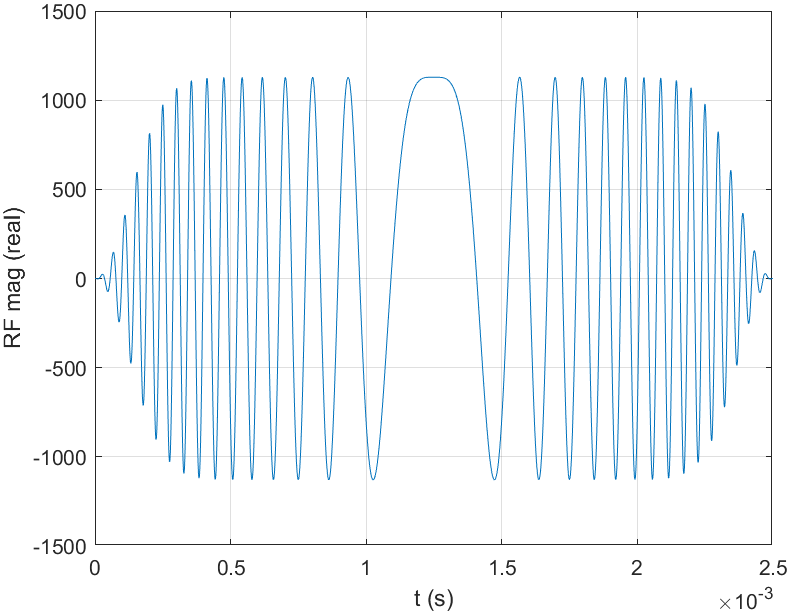
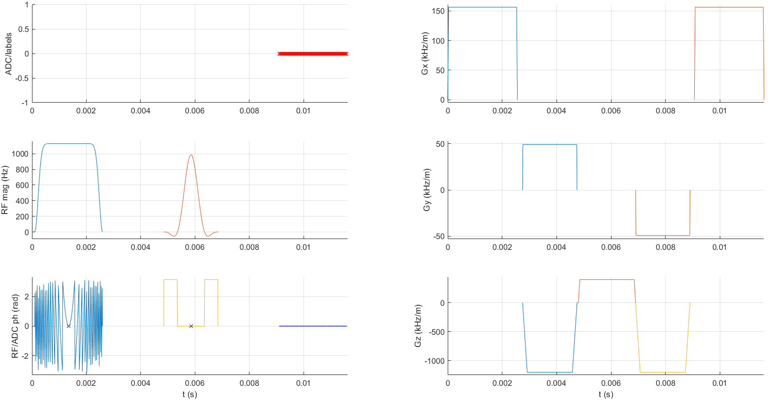
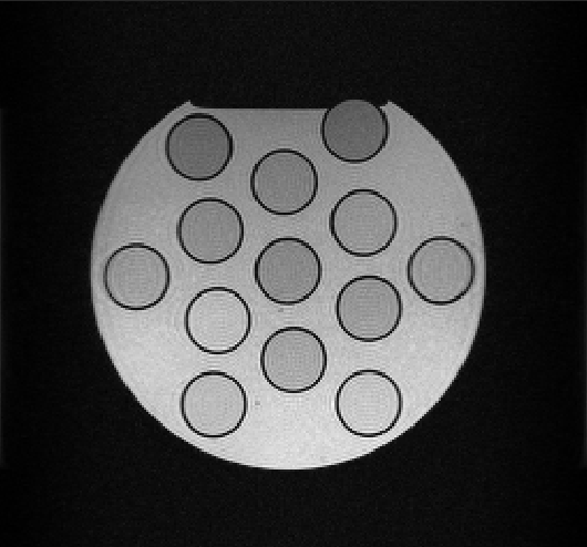
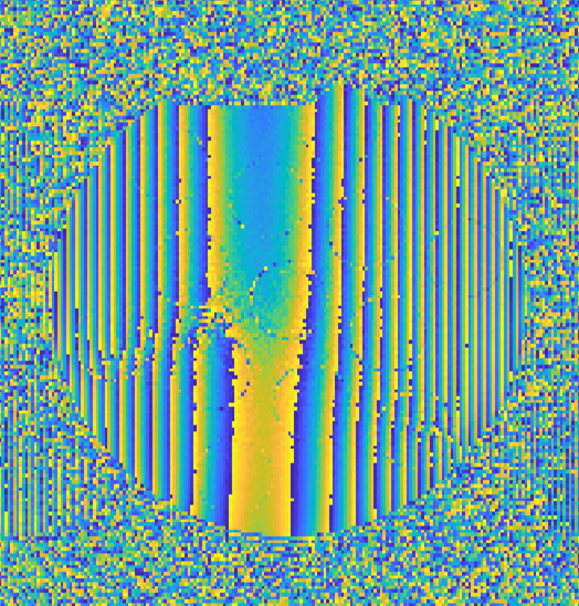
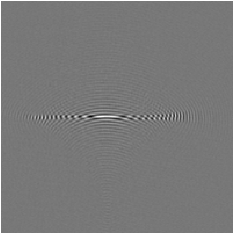
The generated linear chirped-RF-pulse is depicted in Fig.1. One echo of the developed SPEN-Spin-Echo sequence is shown in Fig.2. Due to the linear frequency sweep in both directions, as shown in Fig.1, the quadratic phase profile can be induced with the simultaneous SPEN-gradient, as shown in Fig.4 and Fig. 5. The crusher gradients before and after the refocusing pulse in Fig.2 are necessary because the SPEN excitation is not slice-selective and therefore all other spins outside the target slice should be dephased. The quadratic phase profile which is characteristic for SPEN and the fully reconstructed image can be seen in Fig.3 and Fig.4. The reconstructed image is characterized by a high contrast and high SNR. In addition, no apparent artifacts are visible, except for the ringing artifacts at sharp contrast transitions. However, these are not caused by SPEN and can be suppressed by appropriate filtering.Outlook
The full potential of SPEN becomes available in single-shot MRI. Therefore we plan to implement single-shot SPEN MRI as a next step for comparison to single-shot EPI. In addition we plan to induce the quadratic phase necessary for SPEN by using a quadratic gradient profile using additional gradient hardware 11. By using a pulsed z2 gradient instead of chirped-RF-pulses, the resolution and robustness to field inhomogeneities can be improved without increasing the specific absorption rate. Moreover, introducing a quadratic phase with an additional nonlinear gradient does not require additional time compared to the specialized RF pulse. Furthermore, a specific slice can be excited with a selective 90°-sinc-pulse rather than the entire volume as with the non-selective chirped-RF-pulse, which also leads to an improved signal 2. In addition, a SPEN-EPI-SE with a chirped-RF-pulse and a SPEN-EPI-SE with a quadratic gradient will be added to allow a direct comparison between a Fourier-encoded and spatio-temporally-encoded EPI sequence.Conclusion
A SPatio-Temporally-ENcoded (SPEN) sequence was implemented with Pulseq in MATLAB and is openly available. Image data was acquired and images were successfully reconstructed to demonstrate the method. This provides a good basis for further studies, as accessibility to SPEN is now much easier through the provided scripts on GitHub.Acknowledgements
No acknowledgement found.References
1. Pipe, J. G. (1995), 'Spatial encoding and reconstruction in MRI with quadratic phase profiles.', Magnetic resonance in medicine 33, 24-33.
2. Marhabaie, S., Bodenhausen, G. and Pelupessy, P. (2020), 'Spatio-temporal encoding by quadratic gradients in magnetic resonance imaging', Journal of Magnetic Resonance Open 4-5, 100008.
3. Ben-Eliezer, N., Solomon, E., Harel, E., Nevo, N. and Frydman, L. (2012), 'Fully refocused multi-shot spatiotemporally encoded MRI: robust imaging in the presence of metallic implants', Magnetic Resonance Materials in Physics, Biology and Medicine 25(6), 433--442.
4. Pulseq: Open-source pulse sequences. Available at: https://pulseq.github.io/ (Accessed 8 November 2023).
5. Open-SPEN. Available at: https://github.com/andih98/Open-SPEN (Accessed 8 November 2023).
6. Zaitsev, M., Schultz, G., Hennig, J., Gruetter, R. and Gallichan, D. (2014), 'Parallel imaging with phase scrambling', Magnetic Resonance in Medicine 73(4), 1407--1419.
7. MATLAB R2023b. Available at: https://de.mathworks.com/products/new_products/latest_features.html (Accessed 8 November 2023).
8. MATLAB Documentation. Available at: https://de.mathworks.com/help/matlab/ (Accessed 8 November 2023).
9. mapvBVD. Available at: https://github.com/pehses/mapVBVD(Accessed 8 November 2023).
10. sigpy Documentation. Available at: https://sigpy.readthedocs.io/en/latest/(Accessed 8 November 2023).
11. Littin, S., Jia, F., Layton, K. J., Kroboth, S., Yu, H., Hennig, J. and Zaitsev, M. (2017), 'Development and implementation of an 84-channel matrix gradient coil', Magnetic Resonance in Medicine 79(2), 1181--1191.
Figures