3284
Super Resolution Using Sparse Sampling at Portable Ultra-Low Field MR1NINDS, NIH/Oxford, Bethesda, MD, United States, 2Nuffield Department of Clinical Neurosciences, Oxford University, Oxford, United Kingdom, 3NINDS, NIH, Bethesda, MD, United States, 4Hyperfine Inc, Guilford, CT, United States, 5NIH, Bethesda, MD, United States
Synopsis
Keywords: New Trajectories & Spatial Encoding Methods, Low-Field MRI, Super Resolution
Motivation: Ultra-low field (ULF) MRI is cost-effective and portable but has limited signal-to-noise ratio (SNR) and lower resolution.
Goal(s): To develop a Fourier-based Super Resolution (FouSR) approach to enhance the resolution of ULF MRI images by combining spatial frequencies from two orthogonal ULF images.
Approach: In a standard phantom and a cohort of 10 participants with multiple sclerosis (MS) we compared FouSR to ULF coronal and axial images and their average.
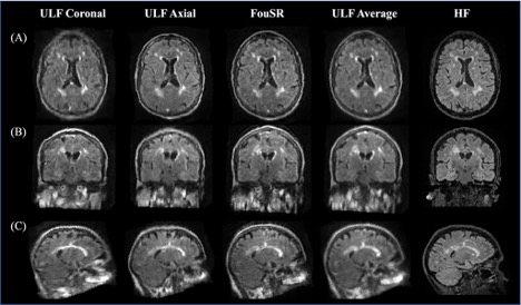
Results: FouSR demonstrated improved image sharpness and lesion delineation without sacrificing SNR. Visual assessments of in-vivo data by an experienced MS neurologist supported the superior image quality of FouSR.
Impact: The presented Fourier-based Super Resolution (FouSR) approach improves image resolution of ultra-low field MRI, providing sharper images for radiological assessment.
Introduction
Ultra-low field (ULF) magnetic resonance imaging (MRI) is more accessible than conventional MRI (1.5T, 3T) due to its cost-effectiveness, reduced power requirements, and portability1,2. However, ULF has reduced image quality and contrast- and signal-to-noise ratios (CNR, SNR). Often, reduced SNR is compensated by averaging multiple scans or lowering the overall resolution of the scan. Alternatively, super-resolution (SR) techniques can address the ULF MRI resolution constraints3,4, thereby affording more detailed and sharper visualization of anatomical structures. Interpolation-based SR, an approach that estimates sub voxel positions through voxel value interpolation, has shown promise in improving the quality of ULF MRI4 but can be time-consuming. Frequency domain SR approaches transform low-resolution images to the discrete Fourier transform (DFT) domain, allowing precise motion parameter estimation planar rotation and shifts through phase correlation analyses5,6. Transforming MRI data into the frequency space is valuable as it separates low-frequency components, carrying contrast information, from high-frequency components containing high-resolution details.In this study, we present a novel Fourier-based Super Resolution (FouSR) approach designed to enhance the resolution of ULF MRI images with minimal increase in total scan time. We hypothesized that FouSR effectively recovers information from under-sampled slice directions, thereby improving the delineation of multiple sclerosis (MS) lesions and other significant anatomical features.
Methods
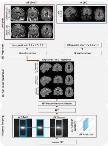
Paired ULF (Hyperfine SWOOP, 0.064 tesla, 3D FLAIR, TR=4000ms, TE=166.72ms, TI=1426ms, scan time=8.38 min) and high field (Siemens, Skyra, 3 tesla) FLAIR scans were collected on the same day from a phantom and in a cohort of 10 participants with MS or suspected MS (6 female; mean ± SD age: 44 ± 4). ULF scans were acquired along both coronal and axial planes, featuring an in-plane resolution of 1.7mm x 1.7mm with a slice thickness of 5mm.After data acquisition, ULF and HF were resampled to 1.7mm using nearest neighbor interpolation, skull-stripped7,8 nonlinearly registered to the 3T image and normalized to the 98th percentile (Fig. 1). These pre-processed images were fast Fourier transformed, and the missing high-frequency components in the under-sampled direction of the ULF Coronal were replaced with those from the ULF Axial (Fig. 1D). The new frequencies were then inversely fast Fourier transformed to create the image. FouSR was evaluated against the resampled and registered ULF coronal and axial scans and their average (ULF Average).
Results
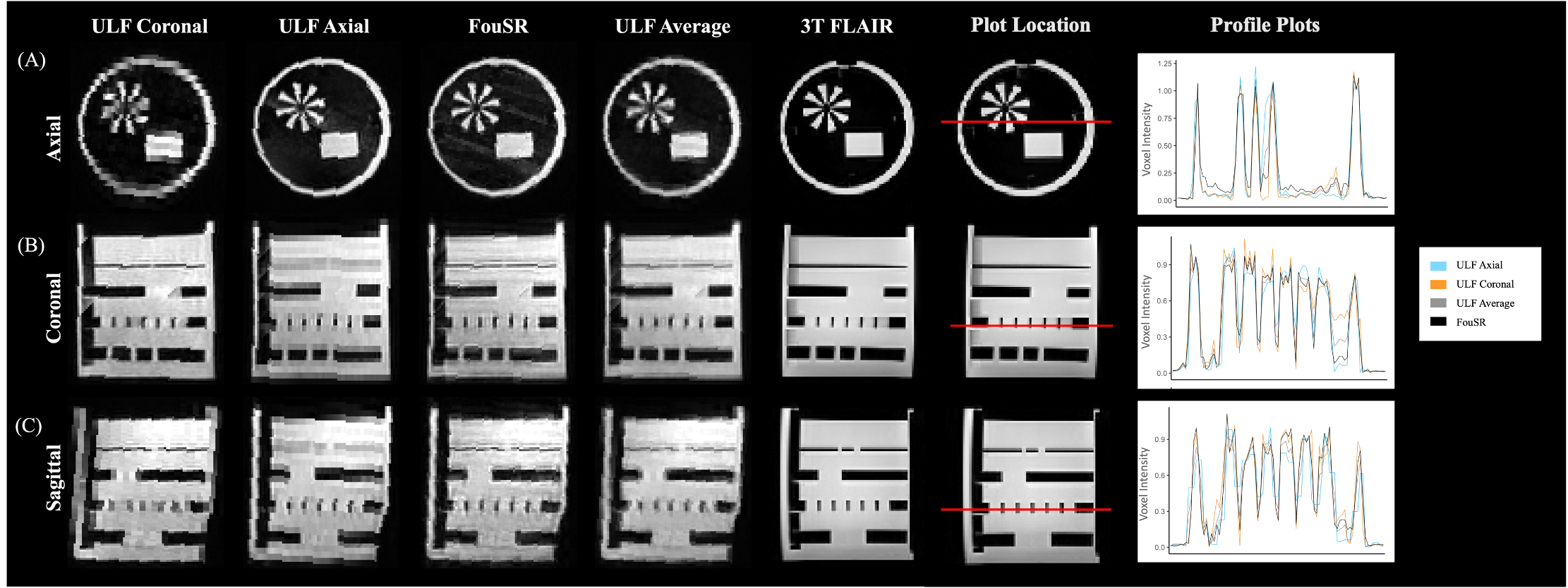
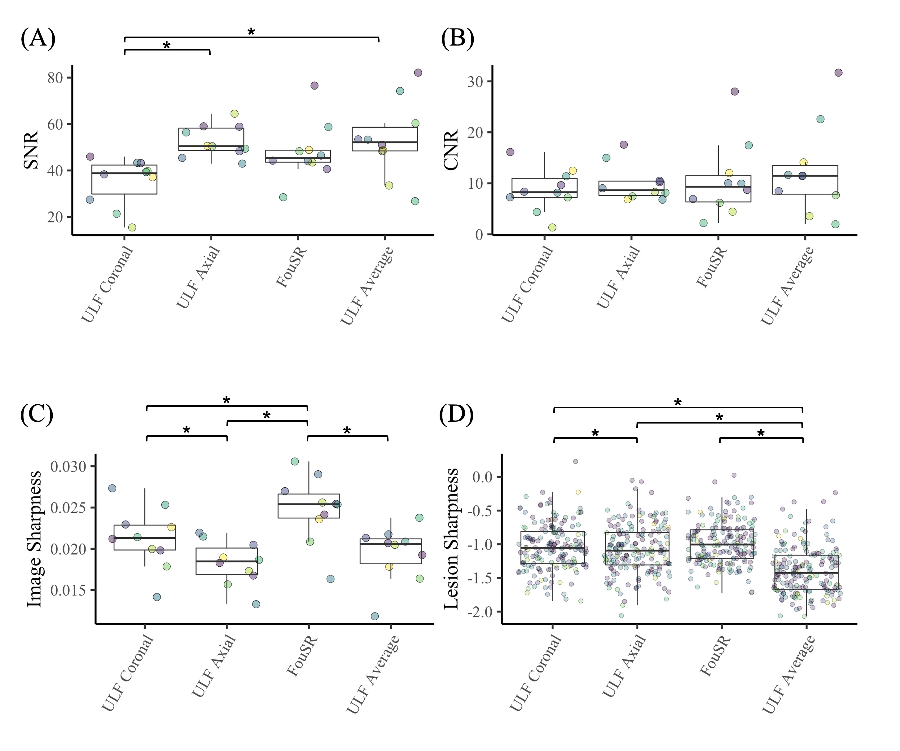
Intensity profiles of ULF Coronal, ULF Axial, ULF Average, and FouSR images of phantom were compared (Fig. 2A). FouSR enhanced shape boundaries with sharper slopes (Fig. 2A) while still preserving ULF Coronal signal intensity (Fig. 2B). Both ULF Average and FouSR matched high signal areas of the in-plane ULF scan.In participant scans, FouSR did not exhibit significant differences in SNR or contrast-to-noise ratio compared to other methods (Fig. 4A/B). However, FouSR demonstrated superior image sharpness (0.025±0.0040) compared to all other techniques (ULF Coronal 0.021±0.0037, q=5.9, p-adj=0.011; ULF Axial 0.018 0.0026, q=11.1, p-adj=0.0001; ULF Average 0.019 0.0034, q=24.2, p-adj.<0.000) and yielded higher lesion sharpness (-0.97±0.31) when compared to the ULF Average (-1.02±0.37, t(543)=-10.174, p=<0.0001) (Fig. 4C/D).
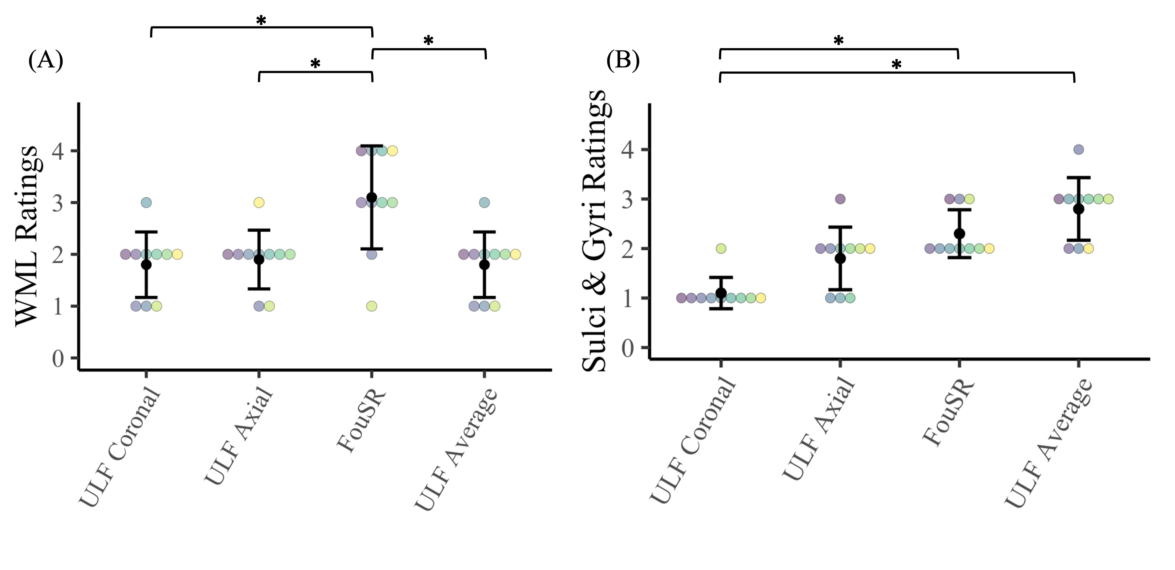
An experienced MS neurologist visually rated WML and Sulci & Gyri (1-Poor, 2-Acceptable, 3-Good, 5-Superior) using a pre-defined scale with detailed guidelines. In these ratings, FouSR had increased scores for white matter lesions (3.1±0.9) compared to all other methods (ULF Coronal 1.8±0.6, Z=3.2 p-adj.=0.008; ULF Axial 1.9±0.57, Z=2.9, p-adj=0.019; ULF Average 1.8 0.63, Z=3.2, p-adj=0.008) and better discrimination of sulcal and gyral structures (2.3±0.48) compared to ULF coronal images (1.1±0.32, Z=3.1, p-adj=0.011) (Fig. 5).
Discussion
FouSR demonstrates the potential for clinically useful FLAIR images in MS without relying on deep learning or synthetic data generation. It derives information directly from existing ULF acquisitions, enhancing generalizability across ULF MRI contrasts and diseases. The FouSR algorithm can also be implemented on the scanner with changes to the k-space trajectory, offering a substantial increase in slice resolution with a proposed ~20% decrease in acquisition timeAcknowledgements
No acknowledgement found.References
1. Liu Y, Leong ATL, Zhao Y, et al. A low-cost and shielding-free ultra-low-field brain MRI scanner. Nat Commun. Dec 14 2021;12(1):7238. doi:10.1038/s41467-021-27317-1
2. Arnold TC, Freeman CW, Litt B, Stein JM. Low-field MRI: Clinical promise and challenges. J Magn Reson Imaging. Jan 2023;57(1):25-44. doi:10.1002/jmri.28408
3. Arnold TC, Tu D, Okar SV, et al. Sensitivity of portable low-field magnetic resonance imaging for multiple sclerosis lesions. Neuroimage Clin. 2022;35:103101. doi:10.1016/j.nicl.2022.103101
4. Deoni SCL, O'Muircheartaigh J, Ljungberg E, Huentelman M, Williams SCR. Simultaneous high-resolution T(2) -weighted imaging and quantitative T(2) mapping at low magnetic field strengths using a multiple TE and multi-orientation acquisition approach. Magn Reson Med. Sep 2022;88(3):1273-1281. doi:10.1002/mrm.29273
5. Thapa D, Raahemifar K, Bobier WR, Lakshminarayanan V. Comparison of super-resolution algorithms applied to retinal images. J Biomed Opt. May 2014;19(5):056002. doi:10.1117/1.JBO.19.5.056002
6. Vandewalle P, Susstrunk S, Vetterli M. A frequency domain approach to registration of aliased images with application to super-resolution. Eurasip J Appl Sig P. 2006;doi:Artn 7145910.1155/Asp/2006/71459
7. Hoopes A, Mora JS, Dalca AV, Fischl B, Hoffmann M. SynthStrip: skull-stripping for any brain image. Neuroimage. Oct 15 2022;260:119474. doi:10.1016/j.neuroimage.2022.119474
8. Jenkinson M, Beckmann CF, Behrens TE, Woolrich MW, Smith SM. Fsl. Neuroimage. Aug 15 2012;62(2):782-790. doi:10.1016/j.neuroimage.2011.09.015
Figures