3282
M(RF)2 – Improving MRF encoding speed with tailored spatiotemporal excitation patterns1Electrical Engineering, Stanford University, Stanford, CA, United States, 2Radiology, Stanford University, Stanford, CA, United States
Synopsis
Keywords: New Trajectories & Spatial Encoding Methods, Image Reconstruction, Pulse sequence design
Motivation: Current Magnetic Resonance Fingerprinting techniques have shown promising results but have room for improvement in terms of acquisition speed and resolution.
Goal(s): Explore the potential of a new spatiotemporal encoding dimension unique to MRF, leveraging tailored spatiotemporal excitation patterns.
Approach: Employ spatially and temporally varying excitation patterns to create distinct signal evolution dictionaries at different spatial locations and reconstruct using multiple subspaces. To address computational challenges, we introduce a clustering method that reduces the number of subspaces, making the problem tractable.
Results: 2D reconstruction experiments demonstrate the approach's potential and show enhanced accuracy, particularly in T2 reconstruction.
Impact: We propose tailored spatiotemporal excitation encoding for Magnetic Resonance Fingerprinting. By leveraging controlled excitation patterns, we create spatially varying dictionaries that enhance the encoding capabilities, allowing for faster and higher-resolution imaging.
Introduction
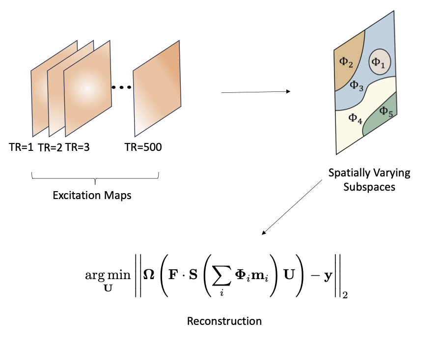
Magnetic resonance fingerprinting (MRF) has rapidly evolved since it was first introduced, enabling fast and accurate quantitative reconstruction2,3 of T1, T2, and proton-density maps by employing a quasi-random sequence of excitations, resulting in distinct signal evolutions for different tissues (fingerprints). The prevailing reconstruction method3,6 leverages a subspace generated from a simulated dictionary and follows a two-stage process. First, the subspace coefficients maps are recovered by solving,$$\boldsymbol{\hat{U}}\;=\;\operatorname*{argmin}_ {\boldsymbol{U}}\left\|\boldsymbol{\Omega\left(FS\Phi\boldsymbol{U}\right)}-\boldsymbol{y}\right\|_2+\lambda R(\boldsymbol{U}),$$where $$$\boldsymbol{\Phi}$$$, $$$\boldsymbol{S}$$$, $$$\boldsymbol{F}$$$, and $$$\boldsymbol{\Omega}$$$ represent the subspace, sensitivity, Fourier, and subsampling operators, respectively, $$$\boldsymbol{y}$$$ are k-space samples and $$$R(\boldsymbol{U})$$$ is a regularization term. The subsequent step involves dictionary matching between the reconstructed coefficients and a pre-computed dictionary to get the quantitative maps.While MRF has already demonstrated impressive results, the unique acquisition method opens up another potential dimension for spatial encoding, which remains largely unexplored but can lead to further enhancements in acquisition time and resolution. In conventional MRI, spatial data is typically encoded using gradients and coils. This work proposes employing spatiotemporally varying excitation patterns to create distinct signal evolution dictionaries at different spatial locations (Figure 1). This additional dimension enables further unaliasing and enhances system conditioning, resulting in a more accurate reconstruction. It is worth noting that using different dictionaries has been explored in the context of simultaneous multi-slice as a means of slice separation4,5. Also, modeling B1+ variations in the dictionary and designing excitations to avoid B1 nulls in the image were previously explored8. However, our approach extends these ideas to encompass more flexible spatiotemporal excitation patterns within a slice or a volume, purposely creating spatially varying dictionaries for improved encoding.
Methods
We call our method MRFRF as we use varying RF excitation patterns with MRF. Numerous methods enable the creation of controlled excitation inhomogeneity (selective excitation, parallel transmission, and dynamic shimming, to name a few). We assume the availability of one of these methods and focus on the reconstruction part, demonstrating its feasibility and potential benefits. The corresponding subspace reconstruction problem is,$$\operatorname*{argmin}_{\{\boldsymbol{U_i}\}_{i=1}^{M}}\left\|\boldsymbol{\Omega\left(FS\sum_{i=1}^{M}\left(\Phi_{i}U_i\right)\right)}-\boldsymbol{y}\right\|_2=\operatorname*{argmin}_{\boldsymbol{U}} \left\|\boldsymbol{\Omega\left(FS\sum_{i=1}^{M}\left(\Phi_{i}m_i\right)U\right)}-\boldsymbol{y}\right\|_2,$$where we have divided the spatial locations into $$$M$$$ groups, each associated with a different subspace $$$\boldsymbol{\Phi}_i$$$, and $$$\boldsymbol{m}_i$$$ denotes a mask keeping the i’th spatial zone.Using a different subspace per spatial zone enables the reconstruction algorithm to reject undersampling artifacts from other regions with different subspaces, thereby improving the conditioning of the problem. The subsequent dictionary matching step utilizes a spatially varying dictionary to reject aliasing artifacts further.
A key challenge arises from the need to solve $$$M$$$ intertwined subspace reconstruction problems, potentially becoming computationally burdensome for large $$$M$$$. To address this, we propose clustering the voxels into a smaller set of $$$N\ll M$$$ subspaces that closely resemble the true subspace at each voxel location. Specifically, we cluster the different flip angle excitation trains using K-means and calculate the corresponding subspaces. Subsequently, we assign each voxel to the nearest subspace based on its excitation flip angle train. We then solve the optimization problem using the conjugate gradient algorithm.
Results
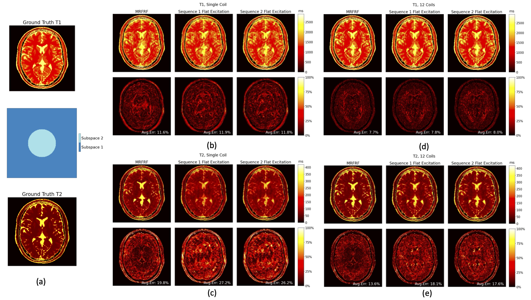
We used a 500-timepoint IR-FISP sequence with 16x undersampled variable-density spiral readouts, where a different interleave is utilized per TR. To examine the capability of MRFRF in improving spatial encoding, we select only the odd eight spiral interleaves out of the 16 to create a challenging encoding case. We add Gaussian noise and report the reconstructed T1/T2 normalized accuracies with and without coils for the following 2D experiments.- Excitation with two sequences – To provide a clear demonstration of the proposed method, we divided a slice into two regions and excited each with a different sequence. Figure 2 compares the results with the case where the whole slice is excited by a single sequence and shows the improved reconstruction.
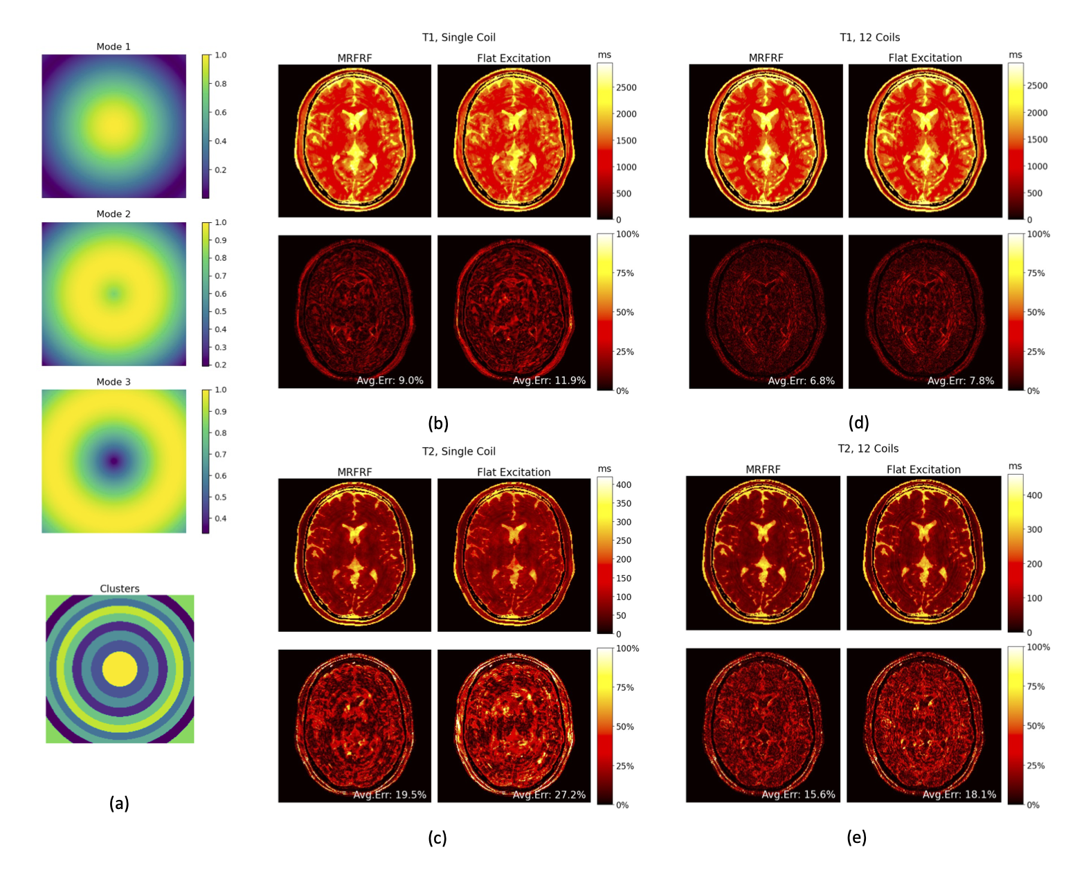
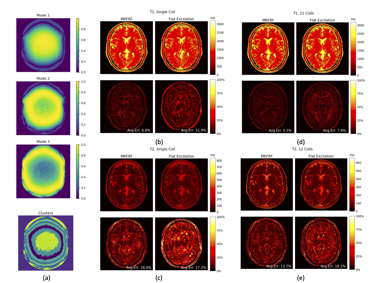
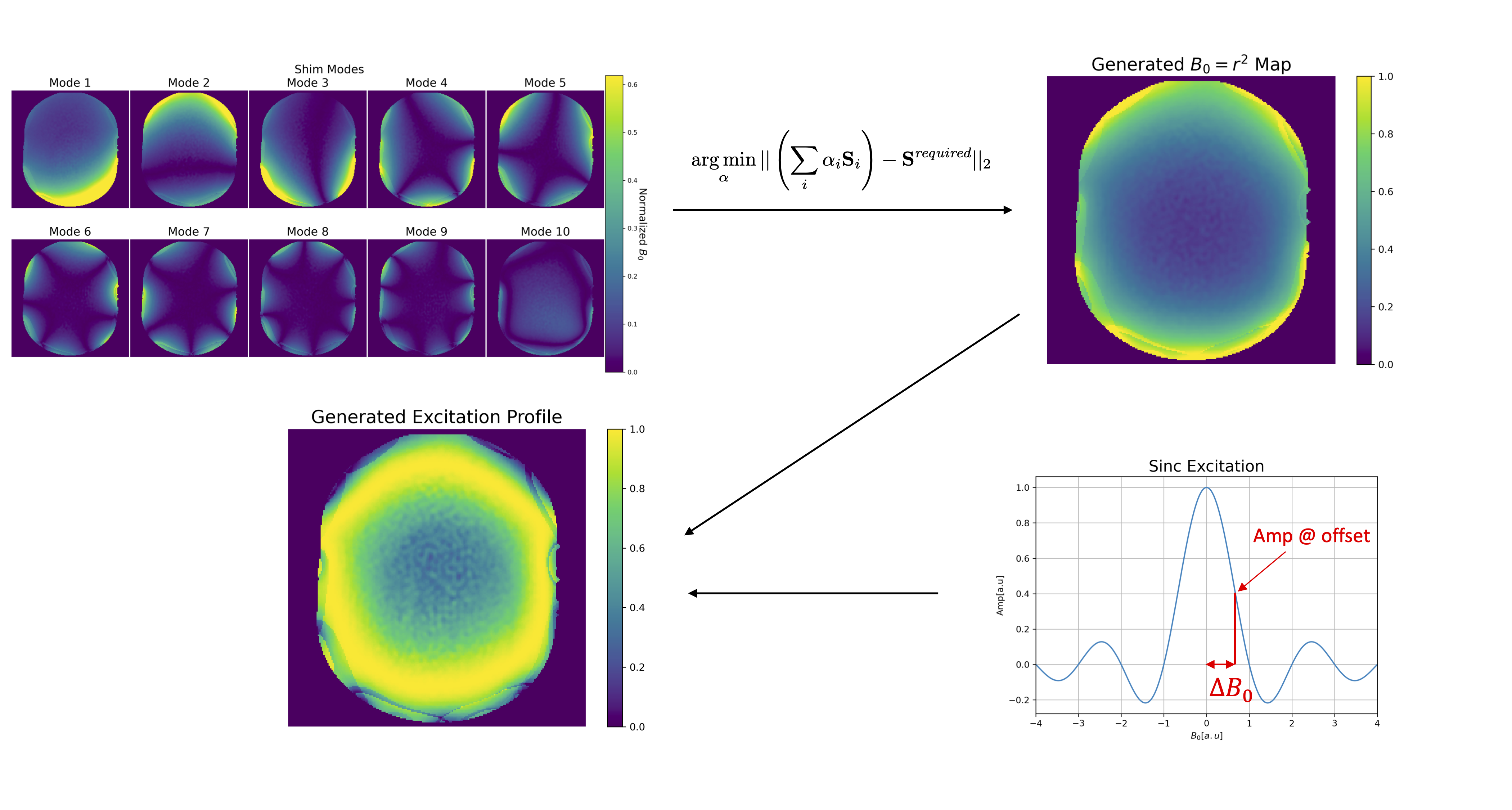
- Spatiotemporal varying excitation pattern – In Figure 3, we alternate three ideal ring-like excitation patterns, while in Figure 4, we used realistic ring excitations. Figure 5 demonstrates one way of achieving the excitation patterns from Figure 4 using a dynamic shim array7 (other methods, such as PTX, are also applicable).
Discussion
This work presents an exciting new approach to further improve spatial encoding in MRF, enabling faster acquisition and higher resolution. The simulation results demonstrate our method's feasibility and potential to enhance reconstruction accuracy. As part of ongoing research, we are exploring more complex excitation patterns and different hardware configurations enabling them. We are also exploring ways of optimizing the patterns and sequence jointly. Lastly, we are working on extensions to 3D-MRF and developing efficient reconstruction techniques that will enable smooth interpolation of different subspaces, facilitating accurate continuous subspace reconstruction and enabling utilization of general complex excitation patterns.Acknowledgements
This work was supported by the National Institutes of Health under grants R01MH116173, R01EB019437, U01EB025162, P41EB030006, R01EB033206, and U24NS129893.References
Ma D, Gulani V, Seiberlich N, Liu K, Sunshine JL, Duerk JL, Griswold MA. Magnetic resonance fingerprinting. Nature. 2013 Mar 14;495(7440):187-92. doi: 10.1038/nature11971. PMID: 23486058; PMCID: PMC3602925.
Cao X, Liao C, Iyer SS, Wang Z, Zhou Z, Dai E, Liberman G, Dong Z, Gong T, He H, Zhong J, Bilgic B, Setsompop K. Optimized multi-axis spiral projection MR fingerprinting with subspace reconstruction for rapid whole-brain high-isotropic-resolution quantitative imaging. Magn Reson Med. 2022 Jul;88(1):133-150. doi: 10.1002/mrm.29194. Epub 2022 Feb 24. PMID: 35199877.
Zhao B, Setsompop K, Adalsteinsson E, Gagoski B, Ye H, Ma D, Jiang Y, Ellen Grant P, Griswold MA, Wald LL. Improved magnetic resonance fingerprinting reconstruction with low-rank and subspace modeling. Magn Reson Med. 2018 Feb;79(2):933-942. doi: 10.1002/mrm.26701. Epub 2017 Apr 15. PMID: 28411394; PMCID: PMC5641478.
Bo Zhao, Bilgic B, Adalsteinsson E, Griswold MA, Wald LL, Setsompop K. Simultaneous multislice magnetic resonance fingerprinting with low-rank and subspace modeling. Annu Int Conf IEEE Eng Med Biol Soc. 2017 Jul;2017:3264-3268. doi: 10.1109/EMBC.2017.8037553. PMID: 29060594; PMCID: PMC5895455.
Jiang Y, Ma D, Bhat H, Ye H, Cauley SF, Wald LL, Setsompop K, Griswold MA. Use of pattern recognition for unaliasing simultaneously acquired slices in simultaneous multislice MR fingerprinting. Magn Reson Med. 2017 Nov;78(5):1870-1876. doi: 10.1002/mrm.26572. Epub 2016 Dec 26. PMID: 28019022; PMCID: PMC5484752.
Assländer J, Cloos MA, Knoll F, Sodickson DK, Hennig J, Lattanzi R. Low rank alternating direction method of multipliers reconstruction for MR fingerprinting. Magn Reson Med. 2018 Jan;79(1):83-96. doi: 10.1002/mrm.26639. Epub 2017 Mar 5. PMID: 28261851; PMCID: PMC5585028.
Stockmann JP, Witzel T, Keil B, Polimeni JR, Mareyam A, LaPierre C, Setsompop K, Wald LL. A 32-channel combined RF and B0 shim array for 3T brain imaging. Magn Reson Med. 2016 Jan;75(1):441-51. doi: 10.1002/mrm.25587. Epub 2015 Feb 17. PMID: 25689977; PMCID: PMC4771493.
Cloos, M., Knoll, F., Zhao, T. et al. Multiparametric imaging with heterogeneous radiofrequency fields. Nat Commun 7, 12445 (2016). https://doi.org/10.1038/ncomms12445
Figures