3281
Novel microstate method to identify fMRI networks and assess their dynamic temporal characteristics1USC Stevens Neuroimaging and Informatics Institute, University of Southern California, Los Angeles, CA, United States, 2Translational Research Center, University Hospital of Psychiatry, University of Bern, Bern, Switzerland
Synopsis
Keywords: fMRI Analysis, Data Analysis, fMRI (resting state), dynamic connectivity, microstate
Motivation: Implement a well-established method (microstate analysis) to characterize state and state-transitions from EEG to fMRI. Such an approach will provide information on dynamic network changes and could provide a novel way to assess brain function in health and disease.
Goal(s): To provide a proof-of-concept that EEG-microstate analysis can be adapted to fMRI.
Approach: Adapt EEG-microstate analysis to fMRI data and characterize dynamic brain network changes.
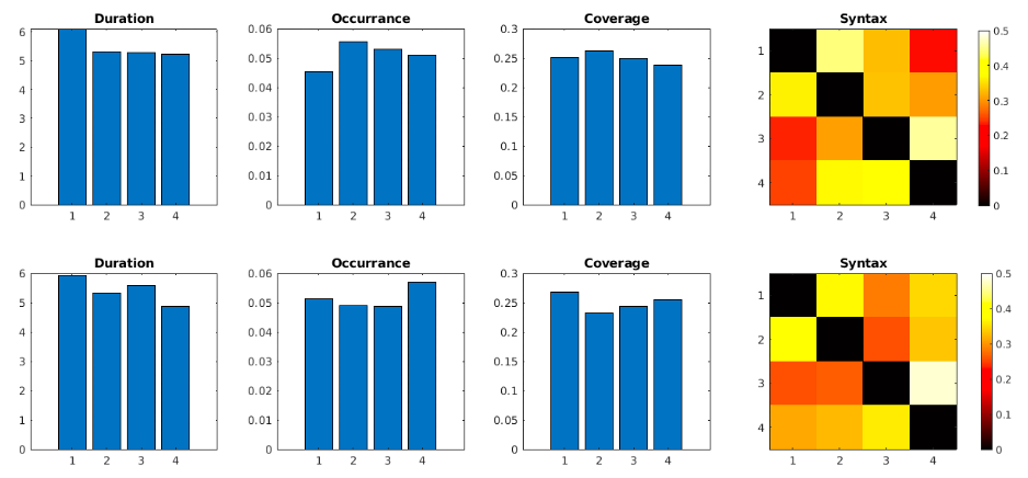
Results: We demonstrate that this novel approach can identify distinct network states, their average duration, frequency of occurrence, total coverage of entire scan and transition probabilities between states.
Impact: We provide proof-of-concept that commonly used EEG-microstate analysis can be adapted to characterize dynamic changes in brain network states in fMRI data. This novel analysis could provide novel insights in alterations of brain functionality in various disorders.
Background
Dynamic network analysis of resting-state fMRI (rs-fMRI) time series has been performed using approaches such as sliding window functional connectivity [1], co-activation patterns [2] or Hidden-Markov models [3]. All these approaches aim at identifying distinct brain modes that have a distinct spatial topography, recurring over time and potentially show some systematic pattern in their transition probabilities. In EEG literature, there is a well-established notion of “microstates” that possess these characteristics [4]. In this preliminary study, we investigated the feasibility to implement an equivalent approach for detecting microstates in resting-state fMRI data.Methods
We leveraged data from the Human Connectome Project. Data were acquired on a 3T Siemens Skyra scanner including an anatomical scan and rs-fMRI scans. We chose to use two scans from the first imaging session with opposing left-right (LR/RL) phase-encoding directions. Data preprocessing included motion-realignment, regression of motion parameters and CSF/WM signal variations, global-signal regression and normalization to canonical space. Subjects with one scan with excessive motion (frame wise displacement > 0.2mm) were excluded.Preprocessed data then underwent frame-wise spatial standardization, resulting in each time frame possessing voxels that are above the spatial mean or below the spatial mean (This step is similar to the EEG-processing step that re-references the data to an average reference). Using k-means clustering (euclidean distance, K=4, 10000 iterations) on each scan of each subject identified four brain modes/networks which we will name “macrostates”. These four macrostates (for LR and RL scans separately) were then matched across subjects by randomly selecting four “template maps” and fit data in an iterative process by repeatedly assigning all individual maps to the most similar template map, and then updating the template maps by the first principal component of the assigned individual maps. The results of this process are four macrostates representing the best match across all subjects for each session. To compute, average duration, frequency occurrence, percentage of temporal coverage and transition probability (syntax) of macrostates, we submitted the data to the microstate+ toolbox [5] which was developed to compute these characteristics for EEG data. In our case each gray matter voxel was treated as equivalent to an EEG channel.Finally, we computed average duration, occurrence, coverage, and syntax as well as the test-retest reliability (Intraclass correlation coefficient ICC) of these characteristics across the two scan sessions.Results
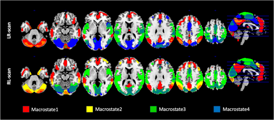
From the HCP900 dataset, 870 subjects with two complete scan sessions were included in the analysis. For each scan session four macrostates (MS) approximately resembling the default-mode network (DMN), a posterior part of the DMN, the visual network and a visual+motor network (Figure1). While test-retest reliability for duration and occurrence was significant (MS1 0.40(p<0.000001), MS2 0.26(p<0.000003), MS3 0.37(p<0.000001), MS4 0.38(p<0.000001) but moderate, ICC for coverage was not significant (MS1 0.08(p=0.120), MS2 0.00(p=0.878), MS3 0.03(p=0.355), MS4 0.02(p=0.384)). Furthermore, MS1, resembling the DMN, showed highest repeatability while MS2, posteriorDMN, was least repeatable. Interestingly, there was a clear pattern of transition probabilities between MS1& MS2 versus MS3 & MS4. Therefore, based on the similarity we also calculated the dynamic characteristics and ICC after combining MS1-2 and MS3-4 which improved reliability for duration (MS1&2 0.44p<0.000001 MS3&4 0.42p<0.000001) and occurrence (MS1&2 0.51(p<0.000001), MS3&4 0.38p<0.000001) and now showed low but significant repeatability for coverage in MS1&2 (ICC= 0.18(p<0.002)) but not for MS3&4 (p=0.95). Finally, MS1&2 lasted significantly longer than MS3&4 but MS3&4 occurred more frequently than MS1&2 in both LR and RL scans (Duration; LR t=8.73(p<0.00001), RL t=7.07(p<0.00001) / Occurrance: LR t=-2.20(p<0.03), RL t=-4.14(p<0.00001).Discussion and Conclusion
In this proof-of-concept study, we successfully translated the well-established microstate analysis form EEG to a rs-fMRI macrostate approach. We identified at least two alternating macrostates that show similar topographies, repeatable dynamic characteristics and represent default mode network and visuo-motor areas, respectively. Future development requires matching and combining macrostates across scans/groups using an additional iterative fitting approach, which potentially will improve test-retest reliability. While EEG-microstates have been shown to be sensitive to inter-individual and disease specific deviations (ref), The fMRI macrostate approach might provide similar insights into distinct dynamic behaviors in brain networks of subjects with psychiatric or neurodegenerative disordersAcknowledgements
This project was funded by NIH R01AG066711 (Jann/Wang).References
[1] Allen et al 2014 Cereb Cortex 24(3):663-676
[2] Liu et al. 2018 Neuroimage 180(Pt B):485-494
[3] Viduarre et al. 2017 PNAS 114(48):12827-12832
[4] Michel & Koenig 2018 Neuroimage 180(Pt B):577:593
[5] Toit & Zhang 2022 Neuroimage 258:119349
Figures