3278
Enhanced neural activity detection in the fMRI using polar Fourier transform1Biomedical Engineering, Amirkabir University of Technology, Tehran, Iran (Islamic Republic of), 2Institute for Research in Fundamental Sciences (IPM), Tehran, Iran (Islamic Republic of), 3Amirkabir University of Technology, Tehran, Iran (Islamic Republic of)
Synopsis
Keywords: fMRI Analysis, fMRI
Motivation: Alternative distortion-reducing methods are needed since NUFFT reconstruction of modestly undersampled radial k-space creates streaking artifacts that influence the entire image and fMRI study's ROI.
Goal(s): Our objective is to demonstrate that the ROI in our fMRI study is less exposed to aliasing artifacts when PFT reconstruction is used as opposed to NUFFT reconstruction.
Approach: Radial-based fMRI images with 2x undersampling were reconstructed using PFT. The neural activity map was then compared to NUFFT-based reconstruction.
Results: Compared to the NUFFT, our proposed approach achieved a qualitatively and quantitatively improved activation map thanks to the distinct artifact characteristic of the PFT.
Impact: Global streaking artifacts in reconstructed images from undersampled radial k-space may seriously affect fMRI study ROI and lead to incorrect brain activity maps. This work used the PFT approach to reduce ROI aliasing artifact in fMRI studies.
Introduction
Using bSSFP with radial acquisition and PFT reconstruction1,2, brain activity analysis demonstrated greater spatial specificity3. Opposite to NUFFT, PFT does not require frequency interpolation. PFT reconstruction of undersampled radial data has different artifact characteristics, unlike NUFFT reconstruction, which shows global streaking. Given that the ROI in the fMRI study is a small sub-region of the brain, we hypothesize that PFT rather than NUFFT may deliver a more accurate image reconstruction from the undersampled radially acquired data, particularly in the ROI where activity is expected. As a result, it may enable the detection of activity in undersampled radial data more effectively than NUFFT. To demonstrate this, we compare the PFT method to the well-known NUFFT in this study.Method
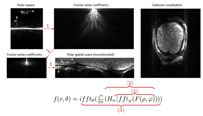
PFT image reconstruction. The PFT formulation and its four sequential stages are illustrated in Figure 1. Using nearest neighbor interpolation, polar images were transformed into cartesian representations to facilitate visualization.Data acquisition. 10 healthy-volunteers performed a visual task in our task-based fMRI experiment. All participants signed consent forms once the local Institutional Review Board (IRB) approved the experiment. According to block-design, subjects observed a checkerboard for each of the six rest-active blocks (24s-24s). Data was collected using bSSPF with radial-readout at a Siemens TIM Trio 3T machine. To cover the occipital lobe, four coronal slices were selected with the following parameters: TR/TE = 6.12/3.06 ms, squared FOV = 224×224 mm2, #spokes = 112, flip angle = 30°, slice thickness = 3 mm, and volume TR = 4s (total 72 measurements).
Setup. Using the PFT and NUFFT algorithms, the original raw-data and 2x retrospectively undersampled data were reconstructed. To analyze the activity of the final images, we used FSL software and performed brain extraction on reconstructed images of both methods under identical conditions after completing preprocessing routines such as removing the first two volumes and applying high pass filtering. In addition to assessing the qualitative activity map, we also computed the Dice coefficient to assess the overlap between the active regions in the brain of the undersampled-reconstructed images and the original activity map derived from the raw k-space data without any undersampling.
Results
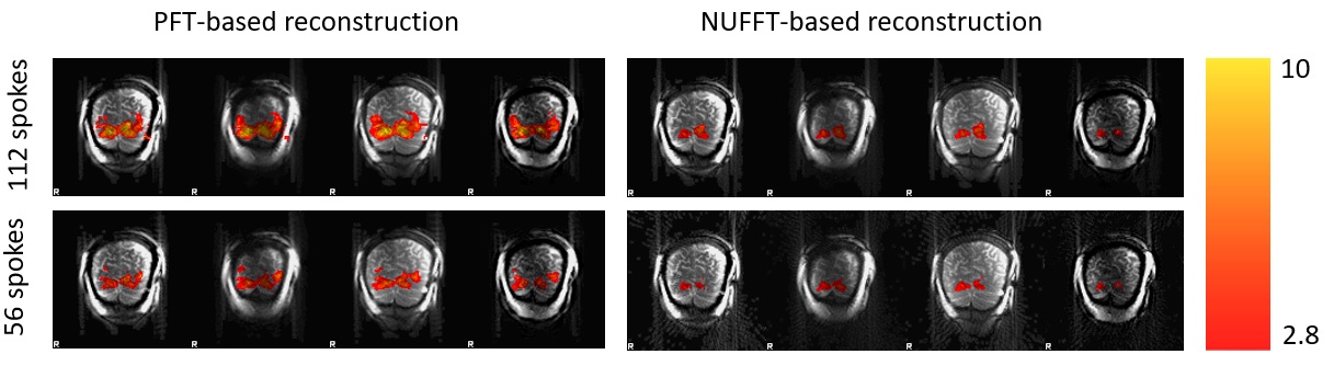
The qualitative comparison between the neural activity derived using PFT reconstruction and the reconstruction based on NUFFT is illustrated in Figure 2. Upon making a deeper examination of the activity generated by both methodologies, it becomes evident that PFT demonstrated significantly elevated Z-score values in the anticipated regions of the visual cortex for the case with 112 radial spokes. In contrast to the PFT, the activate area generated by the NUFFT-based approach would be terminated if the threshold value was increased, owing to its low Z-score. When comparing the case with 56 radial spokes to the reconstructed case with 112 radial spokes, it becomes apparent that the NUFFT-based approach identified some active regions that were absent in the case with 112 spokes, whereas the PFT approach did not introduce any such additions. The mean Dice score (± standard deviation) for the active areas obtained from the undersampled (56 spokes) and highly-sampled (112 spokes) data using the PFT and NUFFT-based reconstruction methods are 0.84 ± 0.07 and 0.72 ± 0.11, respectively. This implies that the use of the PFT results in a more robust identification of brain activity from the undersampled radial data compared to the strategy based on the NUFFT.Discussion
This study conducted a comparison between the PFT-based reconstruction and the NUFFT-based reconstruction methods within the framework of fMRI. Our findings demonstrate an increase in Z-score values in the active regions when employing the PFT method as opposed to the NUFFT-based technique. The use of PFT may result in a more confident identification of the active regions compared to the NUFFT-based methodology. Additionally, our findings demonstrate that the active regions obtained from the undersampled k-space (56 spokes) exhibit a greater degree of overlap, as indicated by a higher dice-score, with the active region produced from the highly-sampled k-space (112 spokes) when the PFT is employed as the reconstruction method. This may be associated with the unique attribute of the aliasing artifact that is observed in the reconstructed image from the undersampled kspace. When utilizing NUFFT to reconstruct the undersampled radial kspace, aliasing artifacts manifest as global streaming artifacts, which have the potential to impact the region of interest (ROI) in an fMRI study. Conversely, PFT reduces the impact of aliasing on the central region of the image. Given that the fMRI study permits the adjustment of the image's center to encompass the region of interest (ROI), neural activity mapping may be enhanced.Acknowledgements
No acknowledgement found.References
1. Higgins WE, Munson DC. A Hankel transform approach to tomographic image reconstruction. IEEE Transactions on Medical Imaging, vol. 7, no. 1, pp. 59-72, March 1988, doi: 10.1109/42.3929.
2. Guo H, Song AW. MRI image reconstruction by polar Fourier trans-form. In: Proceedings of the 12th Annual Meeting of ISMRM, Kyoto, Japan, 2004. Abstract 350.
3. Malekian, V, Rastegar, F, Shafieizargar, B, et al. SSFP fMRI at 3 tesla: Efficiency of polar acquisition-reconstruction technique. Magnetic Resonance Imaging. 2020; 74: 171-180. https://doi.org/10.1016/j.mri.2020.09.005.
Figures