3273
Introducing a task design independent customized cost function for DNN to denoise task-based fMRI data.1Radiology, UZ Brussel (VUB), Brussels, Belgium, 2Radiology, UZ Brussel, Brussels, Belgium
Synopsis
Keywords: fMRI Analysis, fMRI, Denoising, DNN
Motivation: We expected that using a fMRI denoising neural network (DNN) that requires the denoised signal to correlate with a task design matrix in combination with a GLM analysis, can lead to biased results.
Goal(s): To find a DNN cost function that is independent of the task design.
Approach: We suggested a cost function that is based on the preserved frequency content and the correlation with motion regressors, with non-brain signals and with the non-denoised signal.
Results: We found that the DNN with the proposed cost function performed best in reducing the noise while preserving the BOLD signal.
Impact: Our intension with this abstract is to make researchers to think more carefully about the conditions included in their customized cost function in DNN-like denoising models and to check critically the denoised output.
Introduction
To improve the analysis of task-based fMRI, the denoising neural network (DNN) was introduced [1]. DNN is a neural network containing 1D time-dependent fully-connected (tden), convolutional (conv), long short-term memory (LSTM) and time-distributed fully-connected (tdis) layers. The model parameters are estimated per fMRI series by maximizing the difference in the correlation between the denoised signal and the GLM fit signal, between grey matter (GM) and non-GM. The GLM fit signal was the modeled signal (Yfit=b.X with b the fitting parameters) found after fitting the design matrix (X) to the denoised signal (Y). In a latter version [2], for resting-state fMRI, the cost function was changed in minimizing the correlation between GM and non-GM signals.In this study, we hypothesized that including the GLM task design in the DNN cost function can lead to biased results. To avoid this biasing, we suggested a task design matrix independent cost function for DNN in task-based fMRI.
Methodology
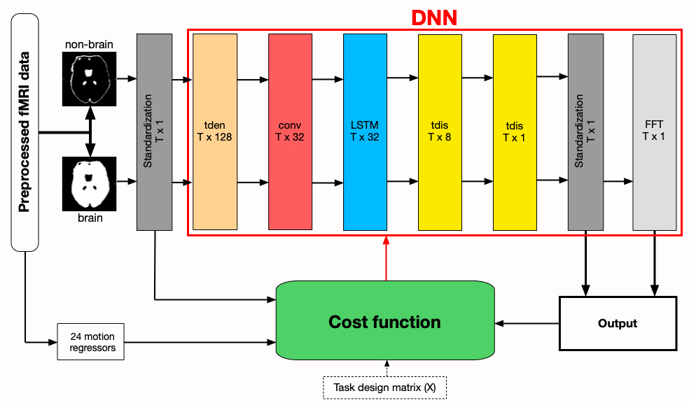
Eight healthy participants underwent a fMRI experiment (3T GE Premier, TE=20/71/121ms, TR=2000ms, 2mm isotropic, 240 volumes). During the experiment, the subjects had to answer a series of episodic (e.g. What will be your next meal?) and general (e.g. What is the color of the sky?) questions.All fMRI data was preprocessed in SPM12 including the combination of the TE timeseries into the averaged series. The used DNN model consisted of tden, conv, LSTM and 2 tdis layers (Fig. 1). The input to the model were the standardized brain (GM + WM (white matter)) and non-brain (head - brain) signals. A Fourier transformation (FFT) was applied on the denoised brain signals.
The model was trained on the first subject for maximal 30 epochs on 50000 voxels randomly chosen in the brain and non-brain masks of which 20% were used for validation. We trained the model with 3 cost functions:
Cost 1: maximal mean difference in the correlation between the denoised signal (Y) and the GLM fit signal (Yfit=b.X), between brain and non-brain areas
Cost 2: minimal mean correlation with noise regressors (motion parameters + temporal derivatives + squared regressors) + minimal mean correlation between brain and non-brain voxels
Cost 3: Cost 2 + maximal mean correlation between the denoised and its non-denoised signals + maximal fractional frequency content within the 0.005-0.12 Hz band
The additional terms in the last cost function were inspired by the hypothesis that the denoised signal should remain similar to the non-denoised signal but contains mainly the task BOLD signals.
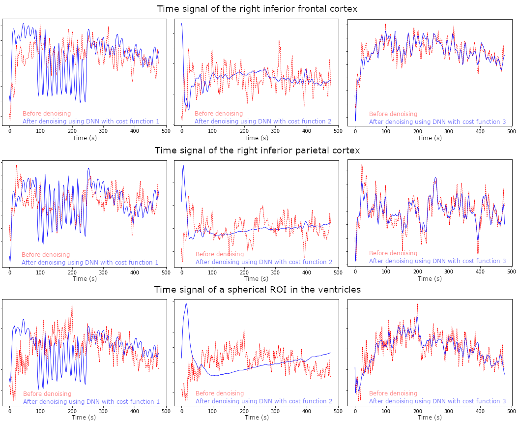
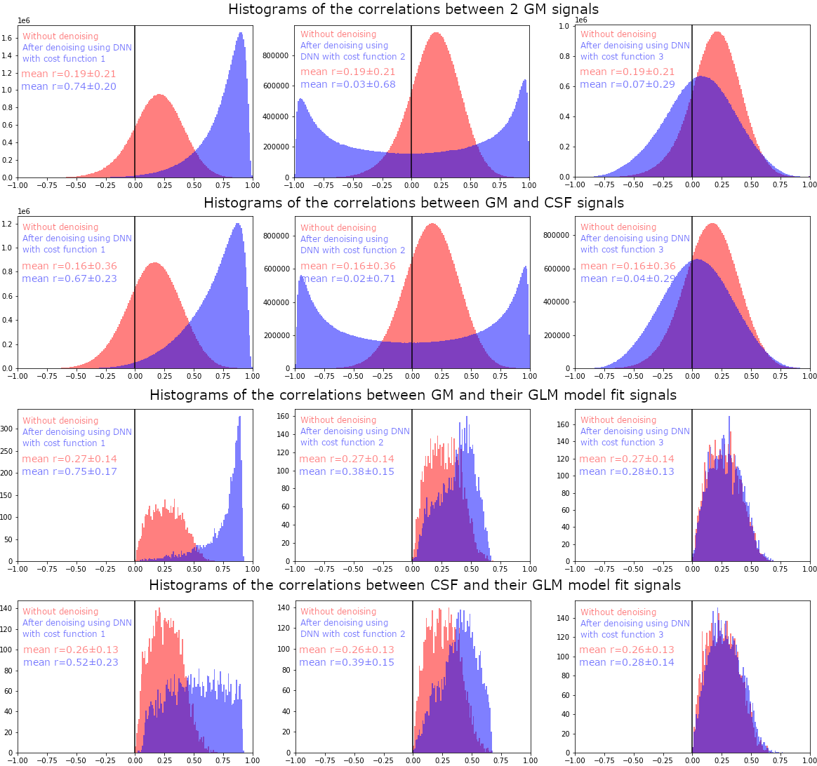
To evaluate the output of the trained models in the first subject, we plotted the signals of the right inferior parietal (IPC) and frontal cortex (IFC), 2 areas in which we expected to find BOLD task activity, and the ventricles. Additionally, in the assumption that the signals of 2 randomly selected voxel will not correlate in the absence of noise or task effects, we looked at the histograms showing the correlations between 2 GM signals, GM and CSF signals, GM and its GLM fit signals and CSF and its GLM fit signals.
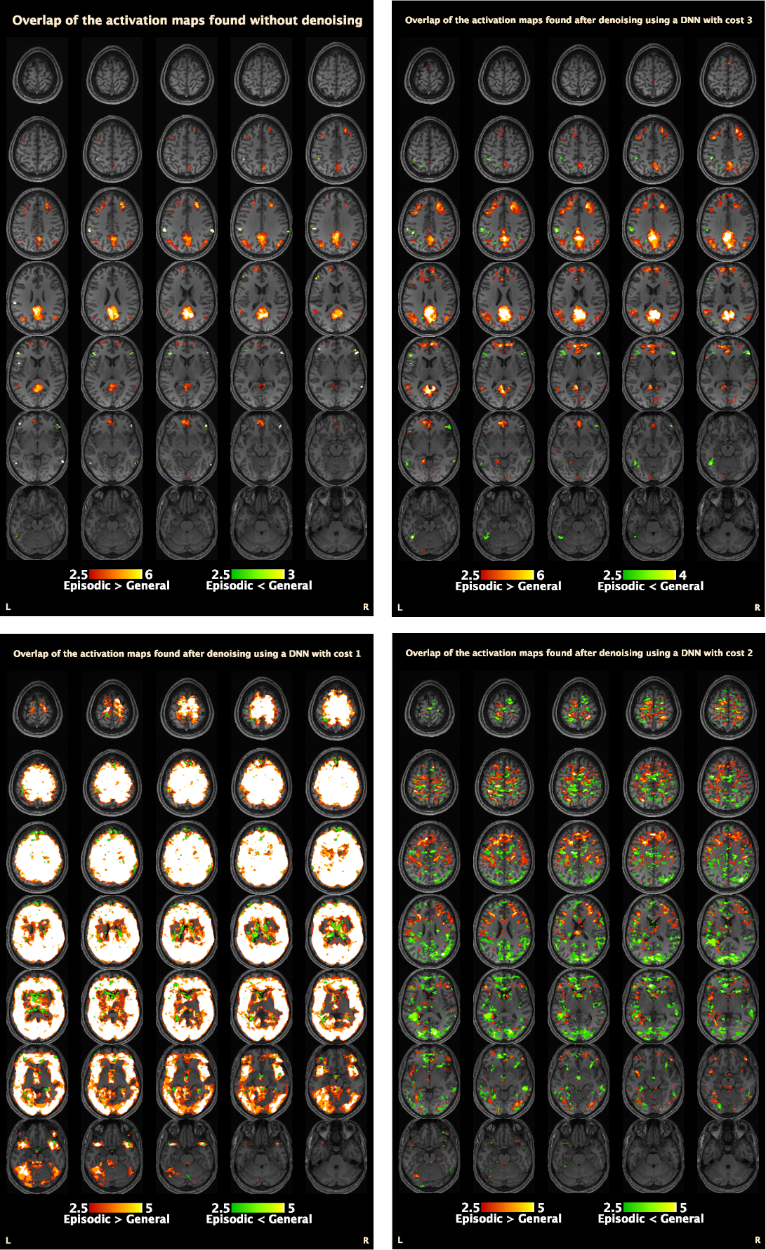
The trained DNN models were used to denoise all fMRI scans. For the resulting activation maps, based on Monte-Carlo simulations, only activations found in at least 3 subjects were considered to reduce the risk for type 1 errors.
Results
The plots of the denoised compared to the non-denoised signals in 3 regions of interest are presented in figure 2.The histograms of the correlations between 2 GM signals, GM and CSF signals, GM and its GLM fit signals and CSF and its GLM fit signals are shown in figure 3.
Only the activation maps found after denoising using a DNN with cost function 3 were found to be more extensive but still comparable to the activation maps found without denoised (fig. 4).
Discussion
As was initially hypothesized, our results suggest that using the GLM task design matrix in the cost function can lead to biased results. Additionally, only requiring the minimization of the correlation between brain and non-brain signals was found to be insufficient. Adding the suggested extra conditions to the cost function resulted in a reduction of the noise while the BOLD signals were preserved.Conclusion
Our results illustrate that DNN tend to convert towards and output that meets the conditions specified in the cost function. As such, care should be taken in the specification of the cost function in DNN based denoising, to avoid erroneous or biased results. Using a task design independent cost function is advisable.Acknowledgements
The Hyperband Multi-echo EPI sequence was provided by GE under a research collaboration.References
1. Yang Z., Zhuang X., Sreenivasan K., Mishra V., Curran T., Cordes D. 2020. A robust deep neural network for denoising task-based fMRI data: An application to working memory and episodic memory. Med. Image Anal. 60:101622
2. Yang Z., Zhuang X., Sreenivasan K., Mishra V., Cordes D., 2021. Disentangling time series between brain tissues improves fMRI data quality using a time-dependent deep neural network. NeuroImage 223:117340
Figures