3271
Improving Coil Setup and Data Processing Strategies for Concurrent (f)MRI and Brain-Stimulation Studies1Leibniz Research Centre for Working Environment and Human Factors, Dortmund, Germany
Synopsis
Keywords: Data Processing, fMRI, brain stimulation
Motivation: In concurrent MRI-brain-stimulation studies various coil setups are used which show either limited access or limited spatial signal coverage.
Goal(s): Our goal is to come up with an optimized coil setup and data processing strategy that allow for open access to the head while maintaining whole brain imaging capabilities.
Approach: Three commonly used coil setups (head array, wrapped body array, TMS surface coil) where used and spatial signal homogenization as well as multi-echo fMRI postprocessing strategies were applied.
Results: By comparing various data processing strategies we were able to improve similarity of activation patterns across the different coil setups used.
Impact: Our results will help to improve researchers to chose optimized data acquistion and postprocessing strategies for whole brain and network based fMRI studies in concurrent brain-stimulation and MRI investigations.
Introduction
Transcranial magnetic stimulation (TMS) is an established non-invasive method for stimulating the human brain. However, its neurophysiological and behavioral effects remain poorly understood. The concurrent application of TMS and functional magnetic resonance imaging (fMRI) provides a robust research approach that merges TMS's causal capabilities with fMRI's high spatial resolution. Here, we conduct a comparative assessment of different TMS-fMRI setups and preprocessing methods. Our primary objective is to elucidate the constraints inherent in current procedures and thereby define optimal strategies for probing the impact of brain stimulation on both behavior and neural activity. This investigation holds significant promise for advancing future research employing this sophisticated technique.Methods
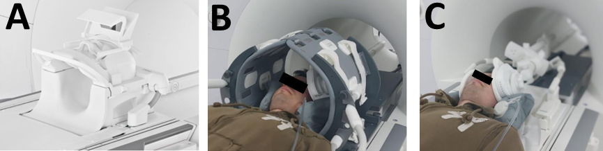
1. Acquisition (rsfMRI)a) Testing three MRI head coils: “standard” 64-Channel coil (Fig1 A), Custom-made coil setup combining two 18ch body array coils wrapped around the head using a home-made holder for maximum accessibility and space for TMS brain stimulation equipment (Fig1 B), commercially available MRI-TMS coil(1) (Fig1 C) consisting of two 7ch coils, one of the two 7ch coils has a MR compatible TMS stimulation coil attached, for MR acquisition.
b) MRI sessions included resting-state fMRI (rsfMRI), anatomical MRI, task-based working memory fMRI, spectroscopy, resting-state perfusion (ASL), and multi-shell diffusion-weighted imaging (DWI) at our 3T Prisma scanner (Siemens Healthiners, Erlangen, Germany). For rsfMRI (10 min), participants were asked to keep their eyes closed. EPI sequence: TR=1250 ms, multiband factor=3, and 3 TE times (13, 35, 56 ms).
2. Preprocessing
a) Homogenization: Spatial signal intensity homogenization was done by applying a signal intensity correction profile obtained from two images acquired with the respective receive coil and with the scanners integrated body coil.
b) Multi echo fMRI data were used to calculate T2*-maps by fitting an exponential decay curve. S0 maps were calculated for each EPI image and time courses with increased SNR were extracted from these S0 maps(2) for further analysis.
3. Postprocessing
a) Independent component analysis (ICA) was performed to obtain brain networks from rsfMRI data using FSL's MELODIC.
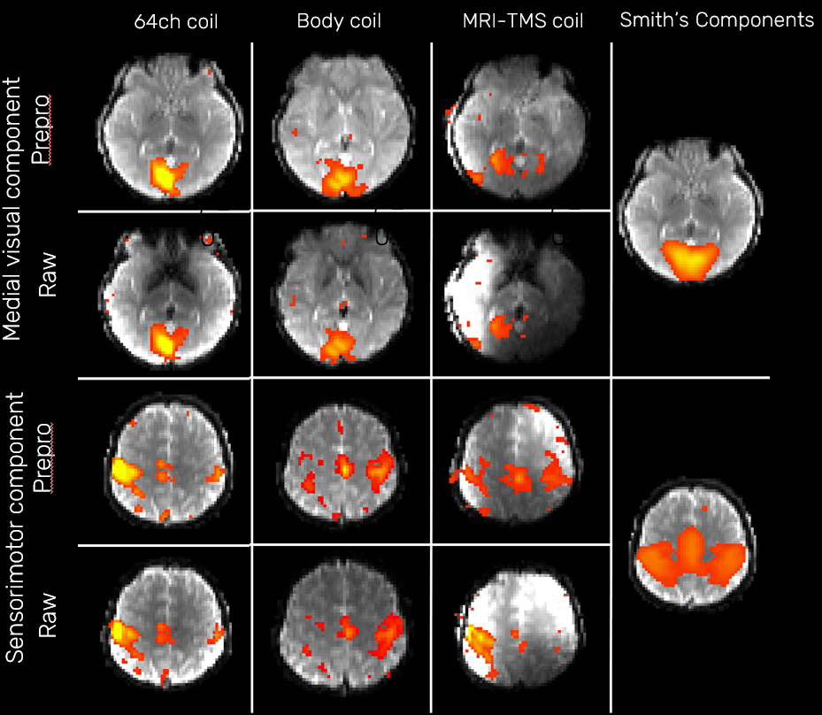
b) Motor and visual ICA networks were identified based on cross-correlation analyses with a set of major brain networks as described by Smith et al. (3).
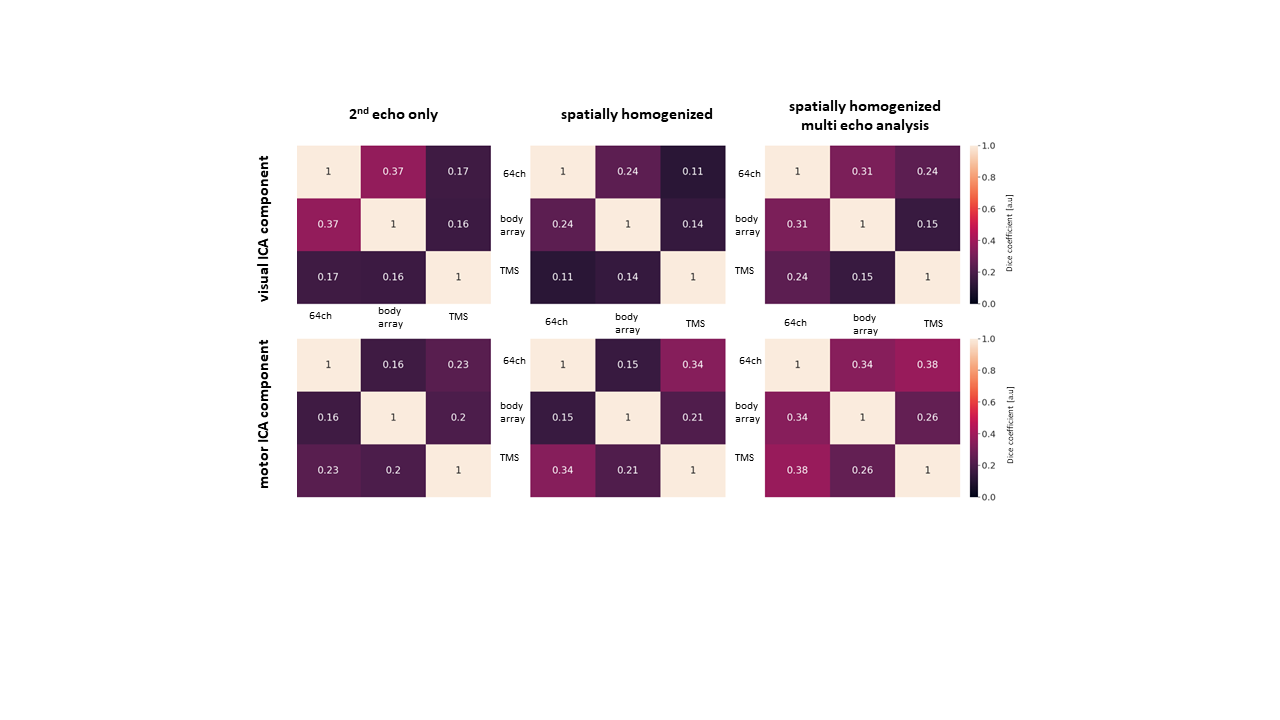
c) Spatial similarities of visual and motor ICAs were compared using the Sørensen–Dice coefficient (0: no similarity, 1: identical spatial overlap of components).
Results
Correlation of ICA components with visual component as identified by Smith (3) increased with data preprocessing (from r=0.48 to r=0.59, 64ch,r=0.59 to r=0.61 for body array and r=0.33 to r=0.42 MRI-TMS-coil) and slightly increased for motor components (r=0.33 to r=0.37 for 64ch, r=0.26 to r=0.32 for body array, and remained unchanged for the MRI-TMS coil).Sørensen–Dice coefficient showed higher similarity of components obtained with 64-channel coil and body array coil setup. However, the spatial similarity of components obtained with MRI-TMS coil vs. 64-channel coil or body array coil was lower for visual and motor networks identified by ICA.
Conclusion
Brain networks identified from resting-state fMRI data spatially varied depending on the coil setup and data preprocessing strategy used. Results obtained from wrapped around body array coil setup closer resembled the findings obtained with the 64-channel coil. The optimal coil configuration differs depending on whether brain networks or only local cortical activity in proximity to the coil are under investigation. On the other hand, the MRI-TMS coil provides best acessibility for brain stimulation whereas no brain TMS brain stimulation can be performed using the 64ch coil, the best compromise with respect to access and with improved data preprocessing strategies is the body array setup for deep brain and brains network studies. With the enhanced coil setup we will be able to improve concurrent brain networks studies during brain stimulation.Acknowledgements
No acknowledgement found.References
(1) Navarro de Lara, L. I. et al. A Novel Coil Array for Combined TMS/fMRIExperiments at 3 T, Magnetic Resonance in Medicine 74:1492–1501 (2015)
(2) Ahmed, Z. et al. ME-ICA/tedana: 23.0.1. (2023) doi:10.5281/ZENODO.1250561
(3) Smith, S. M. et al. Correspondence of the brain’s functional
architecture during activation and rest. Proc. Natl. Acad. Sci. U.S.A. 106,
13040–13045 (2009).
Figures