3264
Optimization of the power independent of number of slices presaturated ultrashort echo time (PINS-UTE) pulse sequence1Department of Computer Science, Mathematics, Physics and Statistics, University of British Columbia, Kelowna, BC, Canada, 2UBC MRI Research, Department of Radiology, Faculty of Medicine, University of British Columbia, Vancouver, BC, Canada, 3Biomedical Engineering and Imaging Institute, Icahn School of Medicine at Mount Sinai, New York, NY, United States
Synopsis
Keywords: Pulse Sequence Design, Pulse Sequence Design
Motivation: Ultrashort echo time (UTE) sequences are essential for short T2 imaging, but have resulted in long scan times. Simultaneous multi-slice techniques can reduce scan times, but until the recent development of the PINS-UTE sequence, they been incompatible with UTE sequences.
Goal(s): We aim to optimize the slice profiles of the PINS-UTE sequence.
Approach: We investigate the effects of minimum radiofrequency subpulse duration, repetition time (TR), and gradient areas on in-vivo slice profiles.
Results: A minimum subpulse duration of 153.6 µs, TR of 250 ms with crusher gradients, and spoiler gradients with areas of 11.7 ms*mT/m on in-plane axes resulted in optimal slice profiles.
Impact: Power independent of number of slices (PINS) presaturation enables simultaneous multi-slice ultrashort echo time acquisitions and can reduce scan times for short T2 imaging. In this work, out-of-slice signal artifacts are reduced by optimizing elements of the PINS-UTE sequence.
Introduction
Tissues and chemical species that exhibit short T2 decay (1-10 ms) such as cortical bone1,2 and sodium3,4 do not appear in conventional MR images5. Conventional sequences require rephasing gradients that limit minimum echo times (TEs).Centre-out half radiofrequency (RF) pulses and two-dimensional (2D) acquisitions1,2, or whole-volume excitations and three-dimensional (3D) acquisitions3,4 have achieved ultrashort echo times (UTEs) of 80-220 µs1-4. Half RF pulses require two excitations per line of k-space6, while whole-volume excitations require an increased number of projections to satisfy the Nyquist criterion7. UTE scan times have ranged from 10-20 mins1-4 – too long for routine clinical implementation.
Simultaneous multi-slice (SMS) pulse sequences have reduced scan times with minimal loss in signal-to-noise ratio8, but also require rephasing gradients. Spatial presaturation techniques9 may be used to achieve SMS excitation without rephasing gradients. We have previously demonstrated the feasibility of a power independent of number of slices (PINS) presaturated UTE (PINS-UTE) sequence that enables SMS excitation with UTE acquisition to reduce scan times for short T2 imaging10. In this study, we have investigated the effects of various sequence elements on the slice profiles of the PINS-UTE sequence to minimize artifacts from out-of-slice signal contamination.
Methods
PINS-UTE saturates wide regions spoiled by gradients, followed by a whole-volume excitation of the narrow slices of remaining longitudinal magnetization10 (Figure 1). Data acquisition from the simultaneously excited slices begins after the transmit-receive switching delay.A modified Multiband RF toolbox11 was used to design a 90º PINS prepulse with a time bandwidth product of 20.68 that saturates 47 mm wide regions leaving 3 mm gaps of longitudinal magnetization. The PINS prepulse and PINS-UTE sequence were simulated with 0.1 mm resolution using the modified Multiband RF toolbox.
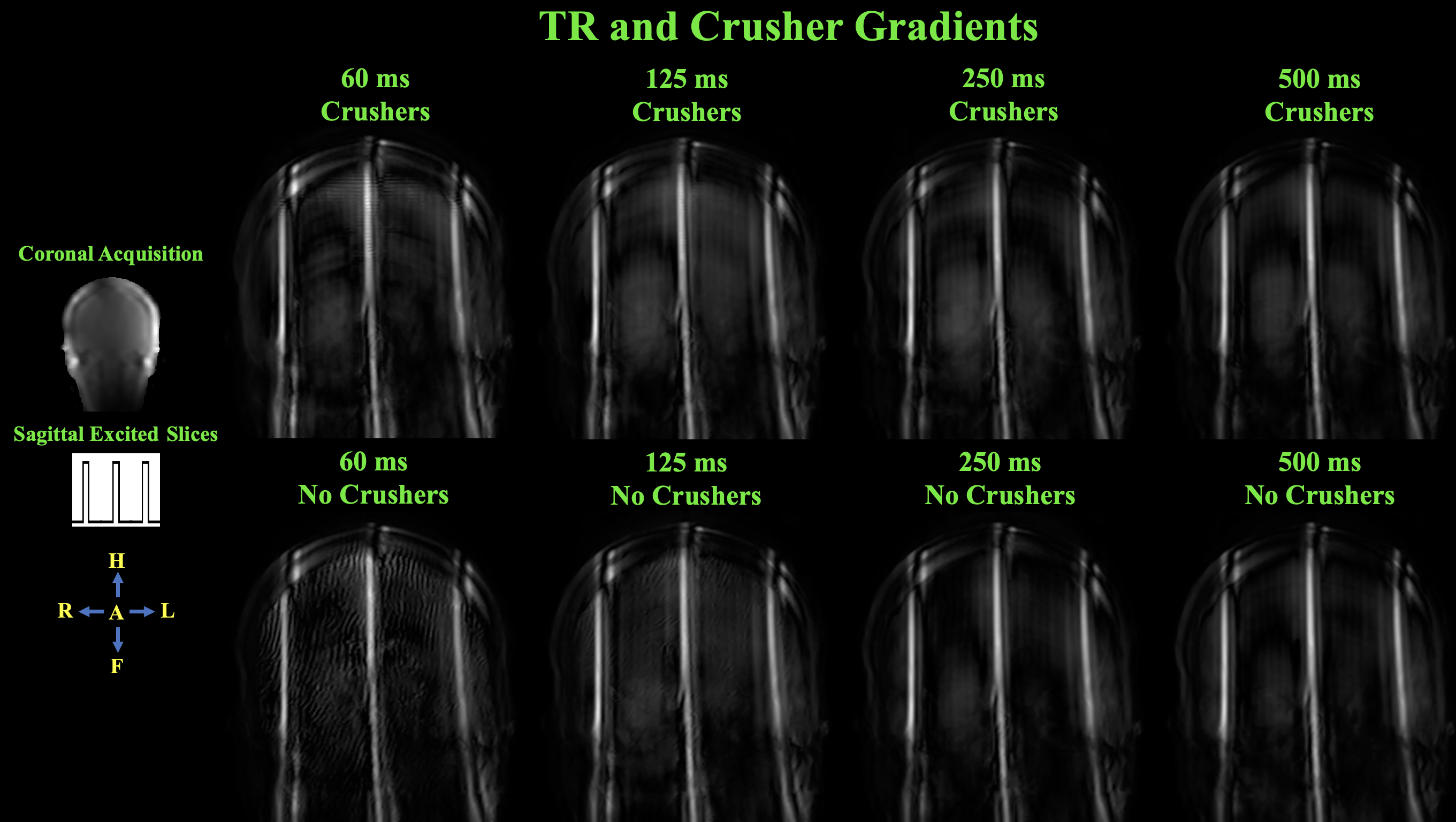
The PINS-UTE sequence was used to investigate slice profiles in-vivo using a 3T Philips Ingenia Elition X with a 32 channel head coil. Unless otherwise mentioned, a minimum PINS subpulse duration of 153.6 µs, a TR of 250 ms, crusher gradients, and spoiler gradients with areas of 23.5 ms*mT/m on all axes were used. Slice projection images were acquired using: (1) minimum PINS subpulse durations of 12.8, 51.2, 102.4, 153.6, 204.8, and 256.0 µs, (2) spoiler gradients with areas of 23.5, 11.7, 5.9, and 2.9 ms*mT/m (to cause 2π dephasing over 1, 2, 4, and 8 mm, respectively) on no axes, the axis normal to excited slices, the axes in-plane with excited slices, and all axes, and (3) TRs of 60, 125, 250, and 500 ms with and without crusher gradients.
Results
The PINS RF and gradient waveforms, and simulated transverse magnetization profiles after the prepulse and at the end of the PINS-UTE sequence are shown in Figure 2. The results of varying minimum PINS subpulse durations in-vivo are shown in Figure 3. The results of varying spoiler gradient areas and orientations in-vivo are shown in Figure 4. The results of varying TR with and without crusher gradients in-vivo are shown in Figure 5.Discussion
For the same field of view (FOV), resolution, and TR as current 2D1,2 and 3D3,4 UTE sequences, the PINS-UTE sequence with a centre-out radial acquisition is expected to reduce scan times by a factor of 2 times the number of simultaneously excited slices (Nsms) and 0.84 times Nsms, respectively. In addition, the PINS-UTE sequence is able to match the minimum TE of current 2D1,2 and 3D3,4 UTE sequences.Minimizing out-of-slice signal is required to reduce artifacts. Figure 3 shows a minimum PINS subpulse duration of 153.6 µs minimizes out-of-slice signal, as desired subpulse flip angles are reached. Figure 4 shows a minimum TR of 250 ms minimizes out-of-slice signal; however, short T2 application will enable further reduction in TR. Figure 5 shows spoiler gradients with areas of 11.7 ms*mT/m on in-plane axes minimizes out-of-slice signal. The striping between slices seen in Figures 4 and 5 results from transverse magnetization remaining in phase between the PINS prepulse and whole-volume excitation and between sequence repetitions, respectively. To minimize out-of-slice signal, the PINS-UTE sequence should use a minimum subpulse duration of 153.6 µs, 250 ms TR, crusher gradients, and spoiler gradients with areas of 11.7 ms*mT/m on in-plane axes.
Conclusion
The PINS-UTE sequence is expected to reduce scan times for short T2 imaging. Compared to current 3D UTE3,4 sequences, the PINS-UTE sequence also offers increased flexibility in resolution and the ability to manually select the number of acquired slices. To minimize artifacts, the PINS-UTE sequence should use a minimum PINS subpulse duration of 153.6 µs, 250 ms TR, crusher gradients, and spoiler gradients with areas of 11.7 ms*mT/m on in-plane axes.Acknowledgements
We acknowledge the support of the Natural Sciences and Engineering Research Council of Canada, Canadian Foundation for Innovation, University of British Columbia, and Walter C. Sumner Memorial Foundation. Special thanks to the Technologists at UBC MRI Research. Rebecca Feldman and Erin MacMillan contributed equally to supervising this research.References
- Du J, Carl M, Bydder M, et al. Qualitative and quantitative ultrashort echo time (UTE) imaging of cortical bone. J Magn Reson. 2010;207(2):304-311.
- Chang EY, Bae WC, Shao H, et al. Ultrashort echo time magnetization transfer (UTE-MT) imaging of cortical bone. NMR Biomed. 2015;28(7):873-880.
- Eisele P, Kraemer M, Dabrinhaus, et al. Characterization of chronic active multiple sclerosis lesions with sodium (23Na) magnetic resonance imaging—preliminary observations. Eur J Neurol. 2021;28(7):2392-2395.
- Riemer F, Solanky BS, Stehning C, et al. Sodium (23Na) ultra-short echo time imaging in the human brain using a 3D-Cones trajectory. MAGMA. 2014;27(1):35-46.
- Bernstein MA, King KF, and Zhou XJ. Handbook of MRI pulse sequences. Elsevier. 2004.
- Tyler DJ, Robson MD, Henkelman RM, et al. Magnetic resonance imaging with ultrashort TE (UTE) PULSE sequences: Technical considerations. J Magn Reson Imaging. 2007;25(2):279-289.
- Konstandin S, Nagel AM, Heiler PM, et al. Two-dimensional radial acquisition technique with density adaption in sodium MRI. Magn Reson Med. 2010;65(4):1090-1096.
- Barth M, Breuer F, Koopmans PJ, et al. Simultaneous mutlislice imaging techniques. Magn Reson Med. 2015;75(1):63-81.
- Felmlee JP and Ehman RL.Spatial presaturation: A method for suppressing flow artifacts and improving depiction of vascular anatomy in MR imaging. Radiology. 1987;164(2):559-564.
- Reich JA, MacMillan E and Feldman R. Design of a Two-dimensional Ultrashort Echo Time Simultaneous Multi-slice Pulse Sequence. Proc Intl Soc Mag Reson Med. 2023;31:2229.
- Seada SA, Price AN, Schneider T, et al. Multiband RF pulse design for realistic gradient performance Magn Reson Med. 2019;81(1):362-376.
Figures
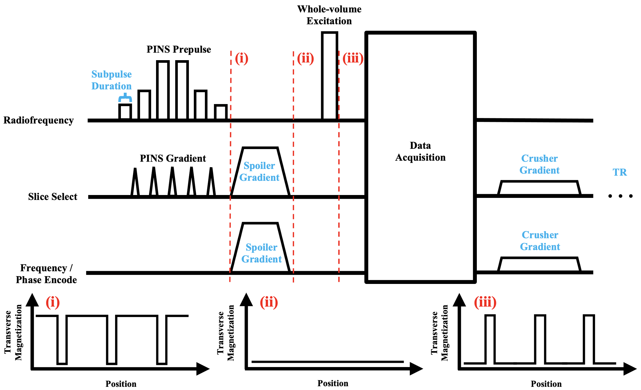
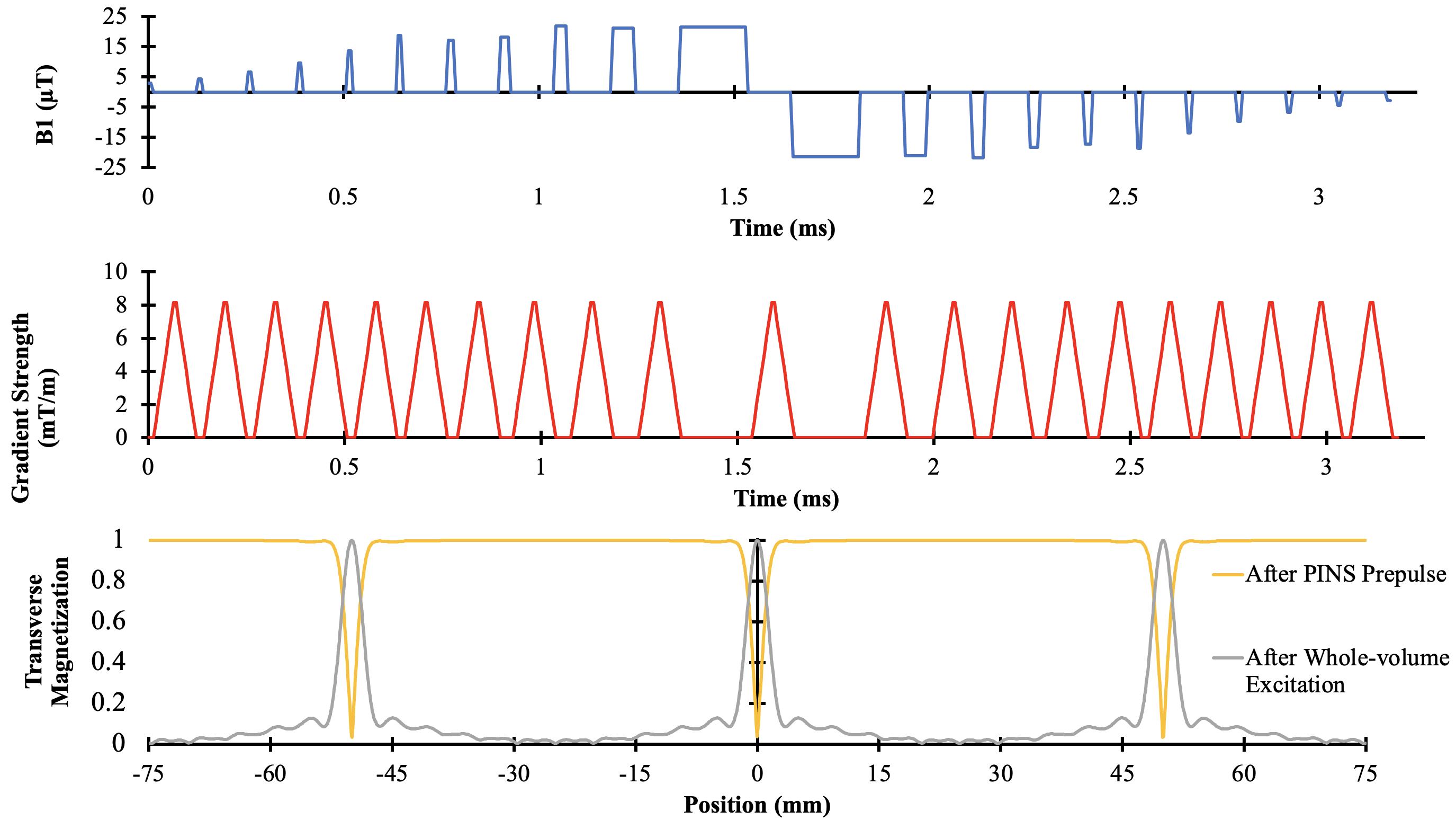
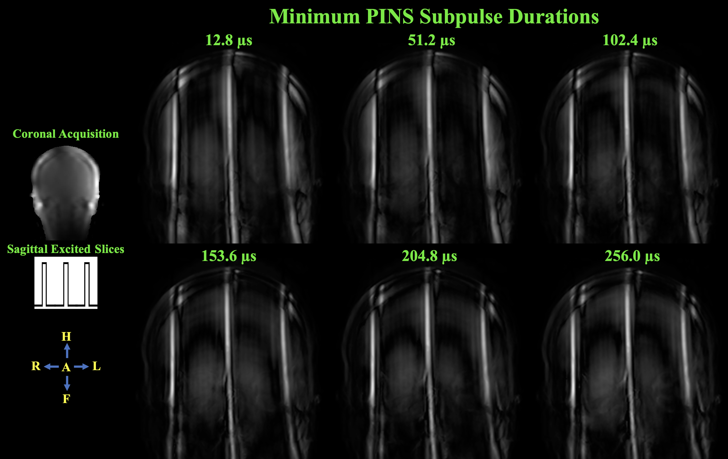
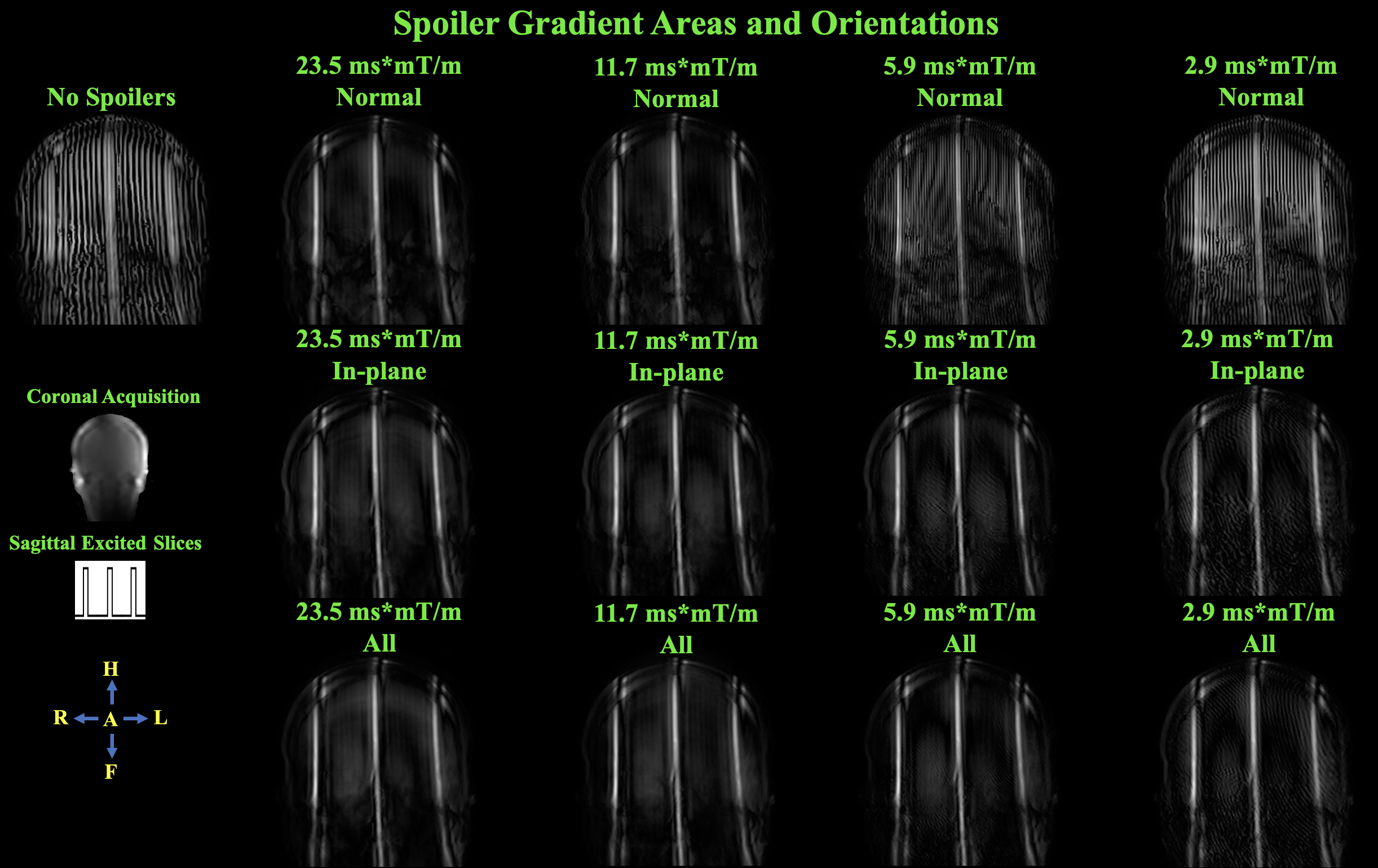
Figure 4. In-vivo slice projection images (coronal acquisition with sagittal excited slices) acquired with spoiler gradient areas of 23.5, 11.7, 5.9, and 2.9 ms*mT/m. For each spoiler gradient area, slice projection images were acquired with a spoiler gradients normal to the excited slices, in-plane with the excited slices, and on all axes. A slice projection image was also acquired with no spoiler gradients. A minimum PINS subpulse duration of 153.6 µs and TR of 250 ms were used.