3260
Convex Gradient Optimization for the Free Running Framework1Universite de Lausanne, Lausanne, Switzerland, 2universite de Lausanne, Lausanne, Switzerland
Synopsis
Keywords: Pulse Sequence Design, Cardiovascular
Motivation: Designing optimal gradient waveforms is a convex problem. They must satisfy gradient hardware constraints, physiological constraints, and being time-optimal.
Goal(s): By shortening TE, we are more robust to motion artifact and less vulnerable to field inhomogeneities. By shortening TR and consequently TA, we can potentially minimize banding artifacts and obtain shorter scan time, increasing motion artifact robustness.
Approach: The gradient optimized method “GrOpt” was integrated directly into free-running 3D-radial-bSSFP and GRE acquisitions at 3T
Results: We achieved 14.0 %, 5.7% and 5.4% reduction in TE, TR and TA for bSSFP and 25.1%, 6.6% and 6.5% reduction in TE, TR and TA for GRE, .
Impact: In 3D-radial-bSSFP, CVX achieves 14.0%, 5.7% and 5.4% reduction in TE, TR and TA, respectively. In 3D-radial-GRE, 25.1%, 6.6% and 6.5% reduction in TE, TR and TA was obtained, reducing the effect of inhomogeneities, motions, eddy current and scan time.
INTRODUCTION
Typically, gradient waveforms are created by combining analytical solutions and timely activation of the gradients separately on each axis. This approach complicates the process of programming pulse sequences and is also time-consuming. As a result, most commercial MRI pulse sequences do not exploit the available gradient hardware or efficiently address physiological interference. Consequently, MRI inherent hardware capabilities are not always fully leveraged. The problem to design gradient waveforms which satisfy gradient hardware constraints is convex (CVX), physiological constrains (Peripheral nerve stimulation (PNS), SAR) do exist, while the requirement for being time-optimal and sustaining desired imaging parameters (e.g. without eddy current artefacts) needs to be satisfied. GrOpt [1–3], a gradient optimizer function, can solve this convex problem. To this day, this technique has not been implemented either in 3D-radial-bSSFP or GRE sequences in the free-running framework [4,5]. Such sequence provide cardiac and respiratory motion-resolved whole-heart 5D imaging for the assessment of anatomy, function [5,6], and blood-flow [7,8]. A first insight on how CVX optimization could benefit a free-running sequence through TE, TR, TA, PNS, and eddy current minimization is presented for GRE and bSFFP.METHODS
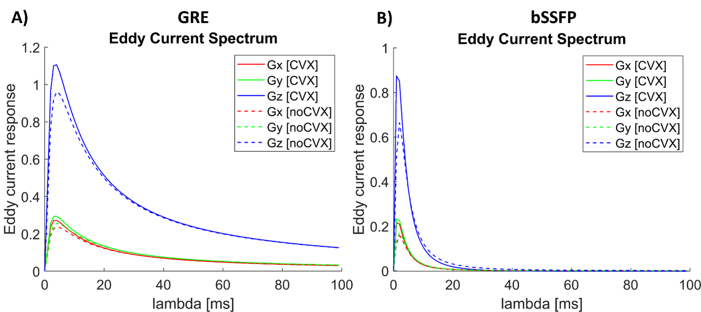
Eddy currents can be simulated according to a time constant λ as [9]:Eddy(λ) = \integral_{Texcite}^{TE} dG/dt * e^(-t/λ) dt (1)
where the derivative of the gradient G, the slew rate, is integrated from the RF pulse excitation to the TE. PNS constraints was determined from the SAFE model [10]:
SAFE(G(t)) = α1 |e^(-t/τ1)*dG/dt|+ α2 |e^(-t/τ2)*dG/dt|+ α3 |e^(-t/τ3)*dG/dt| (2)
where αi represent the weight on each slew rate axis and τi are their respective time constants. According to [2], they can be approximated to α1=0.898ms, α2=0.097ms, α3=0.74ms and τ1=0.16ms, τ1=0.74ms and τ1=0.16ms. The slew rate and PNS were compared, based on their maximum allowed limit, by simulating the equations (1 and 2) into the gradient activation timeline with and without optimization.
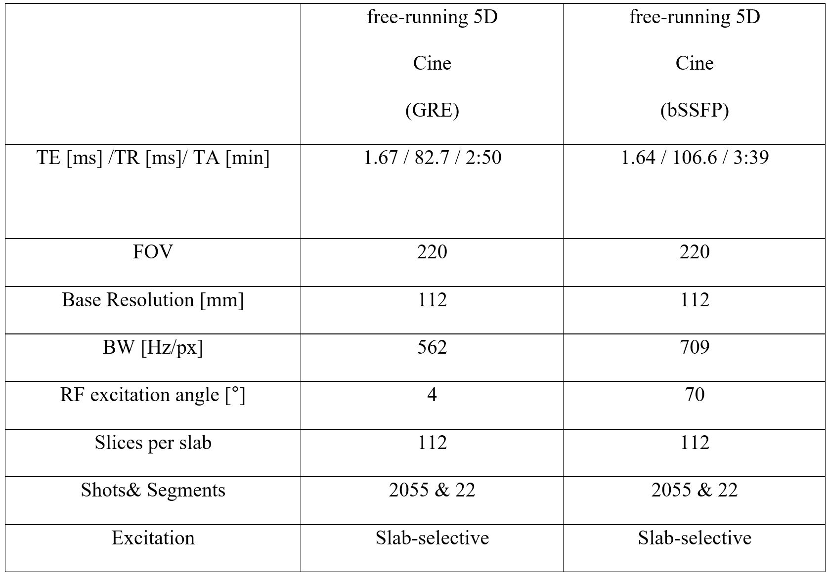
Two volunteers were imaged at 3T (Prisma) with free-running bSSFP and GRE sequences with and without CVX optimization. Table 1 presents the acquisition parameters for these sequences, where TR represents the cumulative TR from all the minTR at each phase encoding steps. For non-optimized sequences, the “normal" gradient mode (34mT/m) was selected. For optimized sequences, the CVX gradients in the entire sequence were designed to account for maximum gradient amplitude (80mT/m*95%), slew rate available (80/(80*5.3)*1000*95%= 179 mT/m/ms) and raster time (10us). PNS threshold was 1. A 95% factor was included to allow gradients to start. Finally, we aim to quantify the gain in TE, TR and TA for each sequence and qualitatively assess image differences.
RESULTS
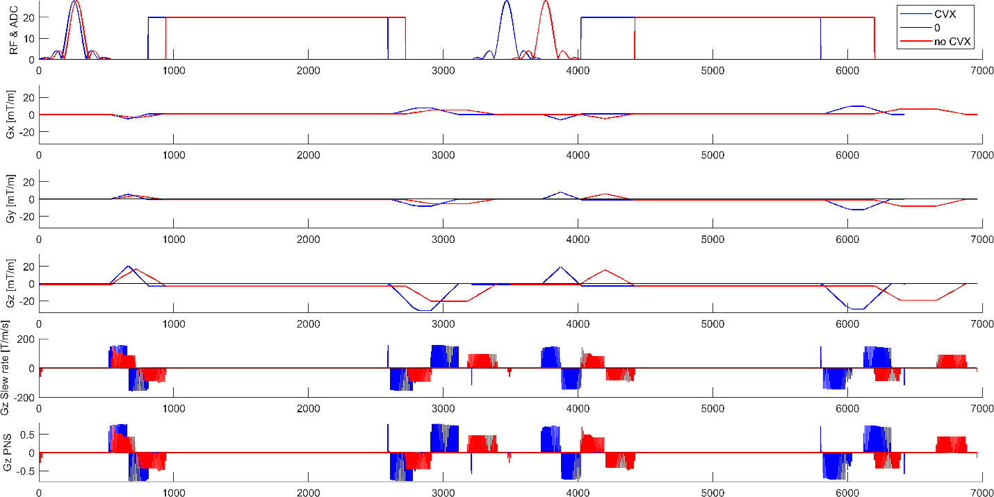
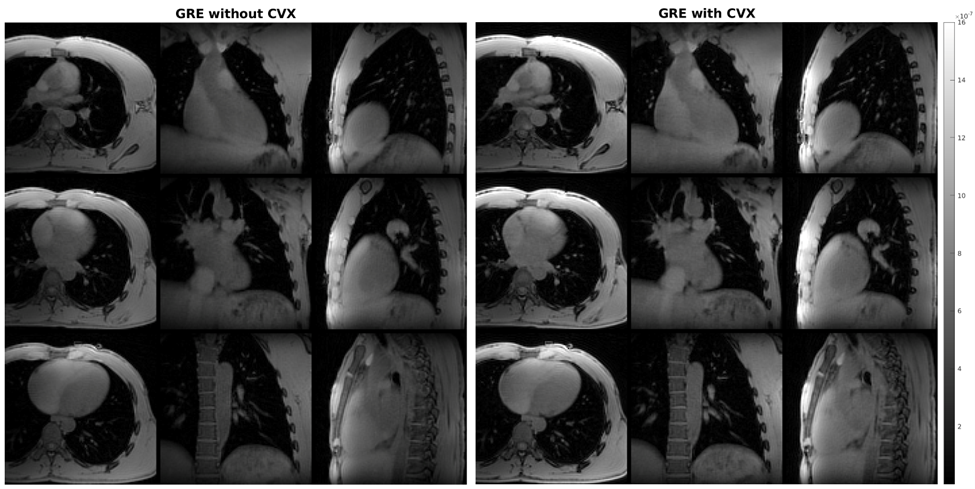
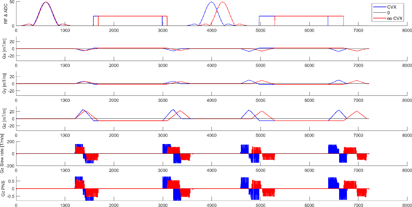
For GRE, we were able to decrease the TE, TR, and TA by 25.1%, 6.7% and 6.5%, respectively (TE=1.25ms, TR=77.2ms, TA=2:39min). In bSSFP, we were able to decrease the TE, TR, and TA by 14.0%, 5.7% and 5.4%, respectively (TE=1.41ms, TR=100.5ms, TA=3:27min). During ramp time in the GRE sequence (Figure 1), the slew rate and PNS clipped to the maximum allowed limit using CVX. Without CVX all the gradients were derated if one of them reached the limit. Although CVX showed a TE and TR decrease by 25.14% and 6.65%, it did not allow to improve blood to myocardium contrast on GRE images(Figure 2). Despite a 14% gain in TE and a 5.7% improvement in TR for bSSFP after CVX(Figure 3), the tradeoff between the required high RF excitation angle of 70° for SNR in bSSFP led to incompatible SAR, which forced the TR to increase to 250ms. As a result, bSSFP showed severe banding artefacts (images not shown). The acquisitions were slab-selective resulting in increased eddy current response on the Gz axis(Figure 4).Discussion
After CVX, TE was reduced by 25% in GRE and inhomogeneities were reduced. The absence of changes in blood to myocardium contrast on GRE CVX images is attributable to the Ernst angle (FA=4° at T1=1471ms and T2=45ms at 3T) that maximizes SNR but not CNR. TE could be further reduced by 0.7ms(70% shorter) using the mpVERSE pulse [2], which would lead to reduced T2*dephasing and SNR gain. Further improvements could be made by nulling eddy current at a desired time constant (during readout). TR and TA were also reduced to some extent (6%),which may reduce motion artefacts, and could be advantageous for long scan time such as in 5D flow imaging.Conclusion
Convex optimization of the gradients with integrated GrOpt toolbox directly implemented in the free-running sequence allows to fully exploit the MRI gradient hardware, thereby reducing the effect of field inhomogeneities,motion,and eddy currents. It helps decrease scan time while respecting safety constraints.Acknowledgements
No acknowledgement found.References
[1] M.J. Middione, M. Loecher, K. Moulin, D.B. Ennis, Optimization methods for magnetic resonance imaging gradient waveform design, NMR Biomed. 33 (2020) e4308. https://doi.org/10.1002/NBM.4308.
[2] M. Loecher, P. Magrath, E. Aliotta, D.B. Ennis, Time-optimized 4D phase contrast MRI with real-time convex optimization of gradient waveforms and fast excitation methods, Magn. Reson. Med. 82 (2019) 213–224. https://doi.org/10.1002/MRM.27716.
[3] M. Loecher, M.J. Middione, D.B. Ennis, A gradient optimization toolbox for general purpose time-optimal MRI gradient waveform design, Magn. Reson. Med. 84 (2020) 3234–3245. https://doi.org/10.1002/MRM.28384.
[4] S. Coppo, D. Piccini, G. Bonanno, J. Chaptinel, G. Vincenti, H. Feliciano, R.B. Van Heeswijk, J. Schwitter, M. Stuber, Free-running 4D whole-heart self-navigated golden angle MRI: Initial results, Magn. Reson. Med. 74 (2015) 1306–1316. https://doi.org/10.1002/mrm.25523.
[5] L. Di Sopra, D. Piccini, S. Coppo, M. Stuber, J. Yerly, An automated approach to fully self-gated free-running cardiac and respiratory motion-resolved 5D whole-heart MRI, Magn. Reson. Med. 82 (2019) 2118–2132. https://doi.org/10.1002/mrm.27898.
[6] C.W. Roy, J. Heerfordt, D. Piccini, G. Rossi, A.G. Pavon, J. Schwitter, M. Stuber, Motion compensated whole-heart coronary cardiovascular magnetic resonance angiography using focused navigation (fNAV), J. Cardiovasc. Magn. Reson. 23 (2021) 1–17. https://doi.org/10.1186/s12968-021-00717-4.
[7] S. Ma, C.T. Nguyen, F. Han, N. Wang, Z. Deng, N. Binesh, F.G. Moser, A.G. Christodoulou, D. Li, Three-dimensional simultaneous brain T1, T2, and ADC mapping with MR Multitasking, Magn. Reson. Med. 84 (2020) 72–88. https://doi.org/10.1002/MRM.28092.
[8] M.B.L. Falcão, L. Di Sopra, L. Ma, M. Bacher, J. Yerly, P. Speier, T. Rutz, M. Prša, M. Markl, M. Stuber, C.W. Roy, Pilot tone navigation for respiratory and cardiac motion-resolved free-running 5D flow MRI, Magn. Reson. Med. 87 (2022) 718–732. https://doi.org/10.1002/MRM.29023.
[9] E. Aliotta, K. Moulin, D.B. Ennis, Eddy current-nulled convex optimized diffusion encoding (EN-CODE) for distortion-free diffusion tensor imaging with short echo times, Magn. Reson. Med. (2017). https://doi.org/10.1002/mrm.26709. [10] F.X. Hebrank, M. Gebhardt, SAFE-Model -- A new method for predicting peripheral nerve stimulations in MRI, Proc Intl Soc Mag Reson Med. 8 (2000) 2007.
Figures