3257
Technical Advances of T1 Relaxation-Enhanced Steady-State (T1RESS) Sequences with Radial Acquisition for Contrast-Enhanced Examination1The Bernard and Irene Schwartz Center for Biomedical Imaging, Department of Radiology, New York University Grossman School of Medicine, New York, NY, United States, 2Center for Advanced Imaging Innovation and Research (CAI2R), Department of Radiology, New York University Grossman School of Medicine, New York, NY, United States, 3Vilcek Institute of Graduate Biomedical Sciences, New York University Grossman School of Medicine, New York, NY, United States, 4Radiology, Northshore University HealthSystem, Evanston, IL, United States, 5Siemens Medical Solutions, New York, NY, United States
Synopsis
Keywords: Pulse Sequence Design, Tumor, Stack-of-stars Acquisition
Motivation: Sequences from the T1RESS family offer increased lesion conspicuity in contrast-enhanced brain examinations. Recently, radial variants have been developed to improve motion robustness.
Goal(s): To enable broader clinical utilization of radial T1RESS sequences.
Approach: Dynamic imaging is enabled through combination with GRASP reconstruction. Fat/water separation is achieved by integrating the multi-echo Dixon technique. Further scan acceleration is achieved by applying 1D GRAPPA along the kz dimension.
Results: The proposed extensions are demonstrated for brain imaging at 3T in volunteers and patients, revealing dynamic lesion-enhancement patterns, successful fat/water separation, and supplementary scan acceleration with comparable quality and lesion conspicuity.
Impact: The proposed technical extensions of radial stack-of-stars T1RESS sequences, including dynamic reconstruction, fat/water separation, and parallel imaging, will enable broader clinical utilization of this novel sequence family and may result in noticeably improved sensitivity for lesion detection throughout the body.
Introduction
Contrast-enhanced T1-weighted imaging is an important component of many clinical protocols, such as for detection of brain tumors and metastases. A widely used acquisition is the magnetization-prepared rapid gradient-echo (MP-RAGE) sequence, which provides high signal intensity in lesions with impaired blood-brain barrier and in blood vessels after gadolinium injection. As an alternative, a novel family of sequences has been proposed, named T1 relaxation-enhanced steady-state (T1RESS), which offer improved conspicuity of enhancing lesions and an option for suppression of flowing blood1,2 to increase visibility of small metastases near vessels. Recently, T1RESS variants with radial stack-of-stars sampling have been developed (Figure 1) that provide higher motion robustness compared to Cartesian implementations.To broaden the clinical applicability, this work presents three technical extensions of the stack-of-stars T1RESS sequences, including the combination with GRASP3 for dynamic contrast-enhanced imaging (DCE-MRI), an option for fat/water separation using the Dixon principle4, and an option for further acceleration by applying GRAPPA parallel imaging5 along the Cartesian direction of the stack-of-stars trajectory. All extensions are demonstrated for brain imaging in healthy volunteers and patients at 3T.
Methods
Dynamic ImagingThe stack-of-stars T1RESS sequences have been implemented with golden-angle ordering (Figure 1). Partitions are acquired as inner loop and radial views as outer loop. This scheme allows utilizing advanced GRASP reconstruction, which exploits temporal correlations by applying a total-variation constraint to recover images from undersampled data. The number of radial views was chosen to balance temporal resolution and image quality. In this work, we used 13 radial views/frame, corresponding to a temporal resolution of 10.14 seconds/frame. To capture sufficient pre-contrast information, the acquisition was started 20sec before injection of a gadolinium-based contrast agent (flowrate 4.0 ml/sec).
Dixon Fat/Water Separation
Although bright fat signal may not be a major concern in brain imaging, providing an option for fat suppression is essential for using the sequences in the neck and other body areas. Therefore, a multi-echo readout was integrated into the uT1RESS sequences with weak spoiler gradients at the end of each TR. The three-point Dixon method was implemented to generate fat and water images.4
GRAPPA Acceleration
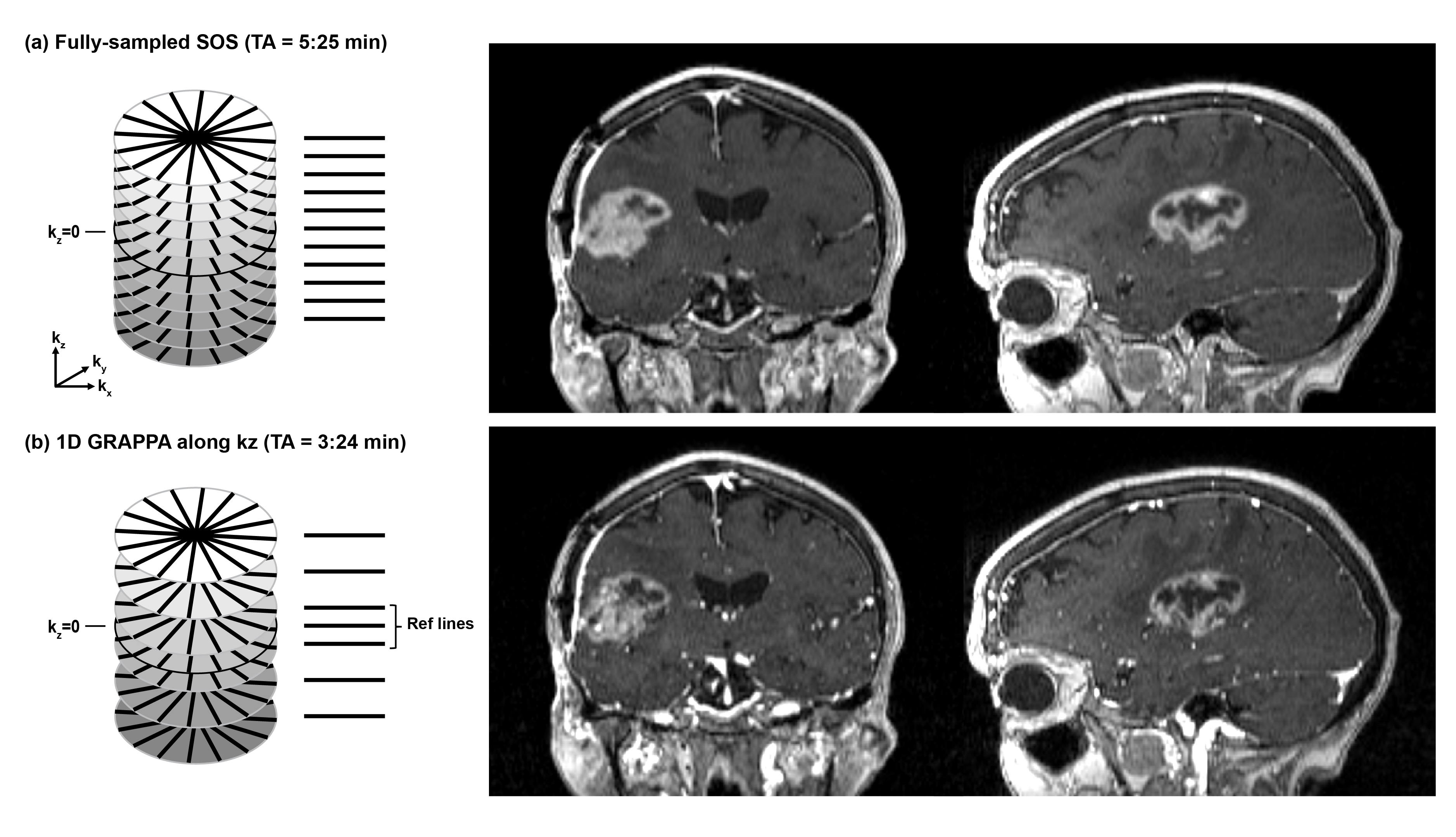
To further accelerate the scans and achieve clinically feasible durations for isotropic resolution, 1D GRAPPA was incorporated in the Cartesian direction of the stack-of-stars geometry. Encoding steps along kz are skipped as shown in Figure 5. The GRAPPA algorithm has been implemented as first reconstruction step, followed by an FFT along kz to enable subsequent slice-by-slice processing.
Results
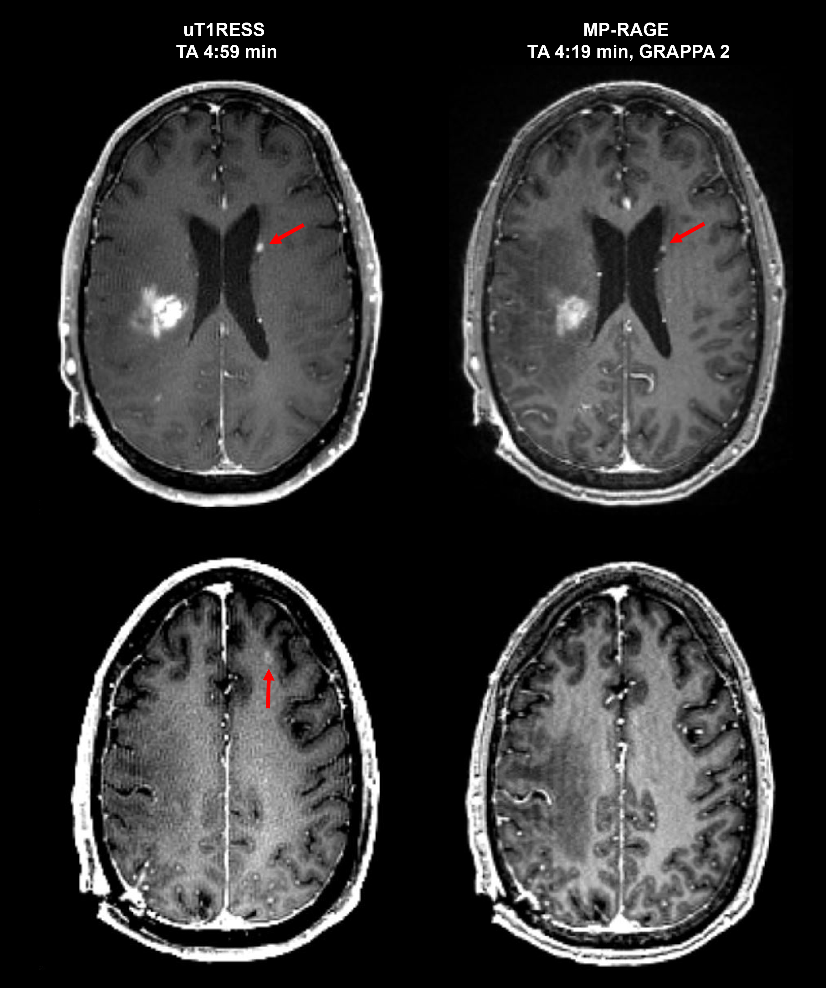
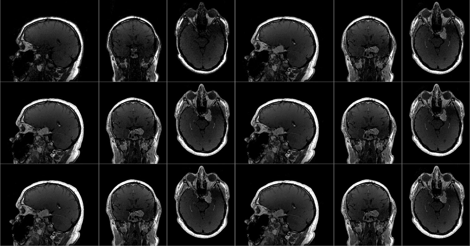
Figure 2 shows a comparison between uT1RESS and MP-RAGE for a patient with brain lesions. The lesions are better noticeable due to the darker tissue background, especially for small lesions (red arrows). Motion-related artifacts are visible in the MP-RAGE image and absent in the uT1RESS image due to the higher robustness of radial sampling. Moreover, blood vessels are attenuated compared to the bright signal seen in the MP-RAGE images.Figure 3 demonstrates the feasibility of generating dynamic images from the radial data. Gradual lesion enhancement is observed in the dynamic series. Blood vessels remain dark, which is a property of the echo-uT1RESS variant.
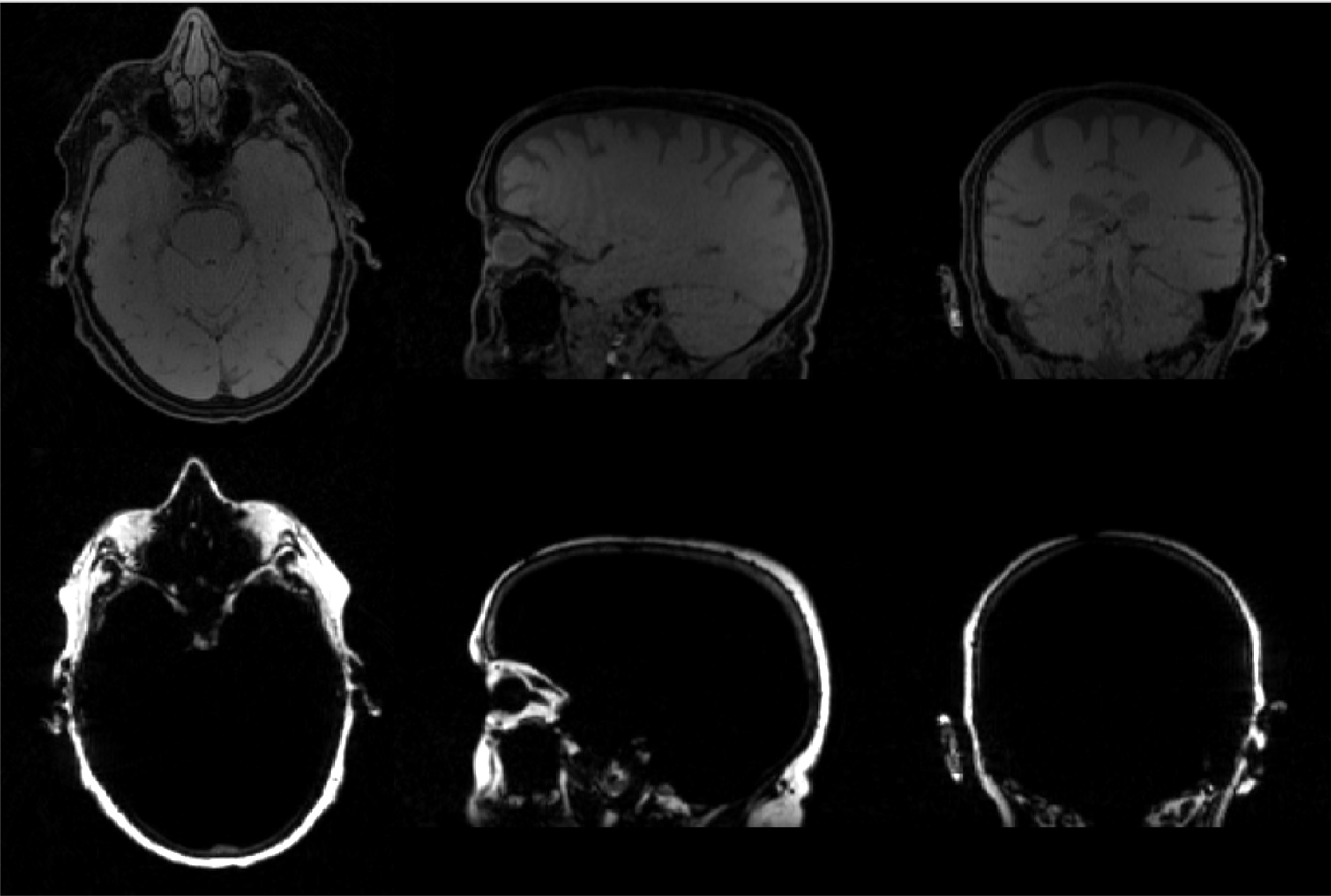
Figure 4 shows fat and water images obtained by performing three-echo Dixon of the uT1RESS variant.
Figure 5 compares images without and with 1D GRAPPA acceleration of the uT1RESS variant. Although accelerated images show slightly lower SNR, the visibility of the lesion remains unaffected. The lesion-to-background contrast and signal of blood vessels differ slightly because of the shorter repetition time between the CM modules with GRAPPA.
Discussion
Here, we present three technical extensions of radial stack-of-stars T1RESS sequences, aiming to make this novel class of sequences more widely applicable in clinical practice. The combination with the GRASP reconstruction technique allows obtaining dynamic information from a single T1RESS acquisition and may be used as input for pharmacokinetical modeling to derive biomarkers. The incorporation of Dixon fat/water separation allows translating the T1RESS technique outside of the brain with the goal of achieving a similar improvement in lesion conspicuity in other body regions, such as for prostate examination. Lastly, the addition of GRAPPA parallel imaging in the kz direction as supplemental acceleration mechanism to the undersampling capabilities of radial sampling make the radial T1RESS variants competitive with 2D GRAPPA-accelerated Cartesian variants while providing better motion robustness.Future work involves optimizing acquisition parameters for the accelerated uT1RESS protocols. Moreover, the clinical performance of 1D GRAPPA for echo-uT1RESS acquisition needs to be validated, as the reduced recovery time leads to slightly altered contrast properties. Furthermore, we will investigate the combination of Dixon and GRAPPA, which would be desirable for various clinical protocols.
Acknowledgements
This work was supported by NIH R01 CA263091 and R21 CA273280. It was performed under the rubric of the Center for Advanced Imaging Innovation and Research (CAI2R, www.cai2r.net), an NIBIB National Center for Biomedical Imaging and Bioengineering (NIH P41 EB017183).References
1. Edelman R, Leloudas N, Pang J, Bailes J, Merrell R, Koktzoglou I. Twofold improved tumor-to-brain contrast using a novel T1 relaxation-enhanced steady-state (T1RESS) MRI technique. Science advances. 2020 Oct 28;6(44):eabd1635.
2. Edelman RR, Koktzoglou I, Leloudas N, Pang J. T1 Relaxation-Enhanced Steady-State (T1RESS): An Improved Three-Dimensional Method for Contrast-Enhanced Imaging of Brain Tumors.
3. Feng L, Grimm R, Block KT, Chandarana H, Kim S, Xu J, Axel L, Sodickson DK, Otazo R. Golden-angle radial sparse parallel MRI: combination of compressed sensing, parallel imaging, and golden-angle radial sampling for fast and flexible dynamic volumetric MRI. Magn Reson Med. 2014 Sep;72(3):707-17.
4. Berglund J, Johansson L, Ahlström H, Kullberg J. Three‐point Dixon method enables whole‐body water and fat imaging of obese subjects. Magn Reson Med. 2010 Jun;63(6):1659-68.
5. Griswold MA, Jakob PM, Heidemann RM, Nittka M, Jellus V, Wang J, Kiefer B, Haase A. Generalized autocalibrating partially parallel acquisitions (GRAPPA). Magn Reson Med. 2002 Jun;47(6):1202-10.
Figures
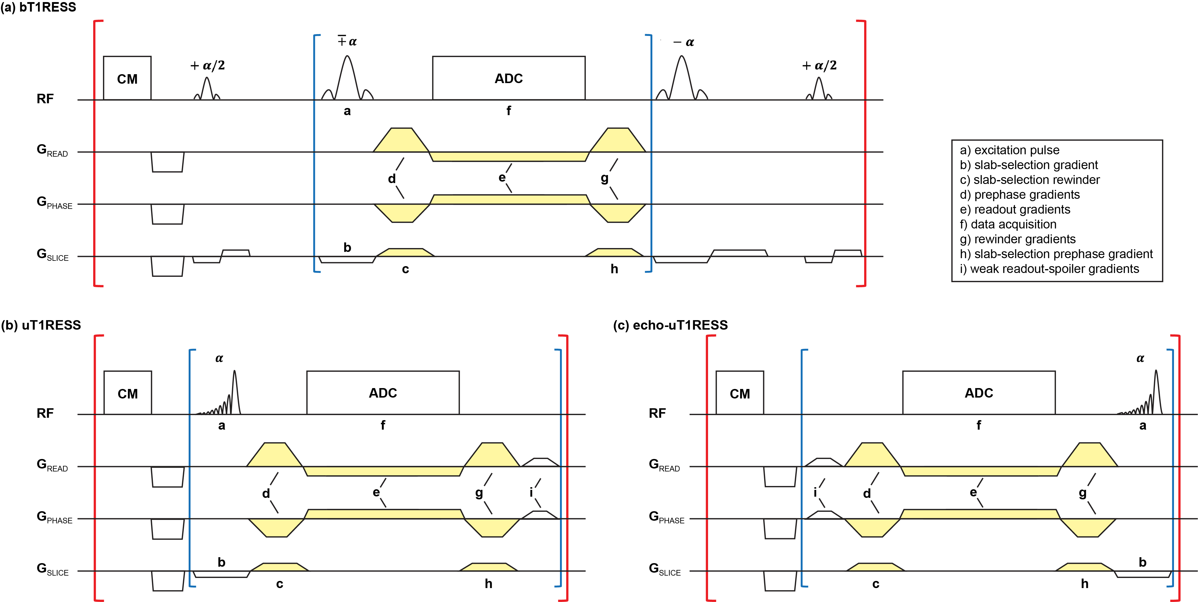
Fig 1: Timing diagrams of the three radial stack-of-stars variants of T1RESS sequences. (a) Balanced T1RESS with bSSFP readout. (b) Unbalanced T1RESS with weakly spoiled GRE readout. (c) Echo-uT1RESS with PSIF readout.
Red backets indicate the outer loops over radial views, blue brackets show the inner loop over partitions. CM is a three-composite pulse saturation recovery module. Gradients d, e, g are modulated according to the radial views. Gradients c, h are modulated by the partition number. Gradient i is a weak spoiler with fixed direction during the entire acquisition.