3256
gSlider-TSE for high-resolution isotropic T2-weighted imaging with high contrast and high SNR1State Key Laboratory of Brain and Cognitive Science, Institute of Biophysics, Chinese Academy of Sciences, Beijing, China, 2University of Chinese Academy of Sciences, Beijing, China, 3The Innovation Center of Excellence on Brain Science, Chinese Academy of Sciences, Beijing, China, 4Institute of Artificial Intelligence, Hefei Comprehensive National Science Center, Hefei, China
Synopsis
Keywords: Pulse Sequence Design, Pulse Sequence Design
Motivation: Conventional 2D-TSE offers superior tissue contrast compared to 3D-TSE, but its slice thickness is restricted by SNR and slice profile of RF pulse.
Goal(s): Our objective was to achieve 0.4mm isotropic T2-weighted imaging with high contrast and high SNR.
Approach: Amplitude and phase modulated gSlider pulses were developed and incorporated into 2D-TSE. Their performances were evaluated in phantom and in vivo.
Results: The gSlider-TSE achieved 0.4mm isotropic imaging with high accuracy. The SNR was comparable to 8 times averaged conventional 2D-TSE image.
Impact: The gSlider-TSE provides high-resolution isotropic imaging with submillimeter slice thickness and high SNR, while preserving the high contrast of conventional 2D-TSE.
Introduction
Conventional 2D-TSE (Turbo Spin-Echo, TSE) typically provides better tissue contrast than 3D-TSE. However, the slice thickness in 2D-TSE cannot be pushed to submillimeter because of limited Signal-to-Noise Ratio (SNR) and achievable slice profile of excitation pulses. Therefore, high-resolution isotropic imaging is challenging for conventional 2D-TSE. The generalized Slice Dithered Enhanced Resolution (gSlider) technique was introduced to obtain thin slices from phase-modulated RF encoded thick slabs, achieving sub-millimeter isotropic resolution in diffusion imaging1. In this study, we introduced the gSlider technique to conventional 2D-TSE to enhance the slice thickness. In addition to the conventional phase-modulated gSlider (PM-gSlider) pulses, we designed and evaluated amplitude-modulated gSlider (AM-gSlider) pulses. The gSlider-TSE sequence was implemented on a 7T MRI scanner to demonstrate its effectiveness for 0.40mm isotropic imaging on a phantom and in vivo.Methods
The PM-gSlider pulses were generated by the sigpy2 toolbox, with the magnetization of each composing slice sequentially flipped to negative y-axis. The AM-gSlider pulses were designed to sequentially preserve each composing slice in z-axis. A combined approach was used for the AM-gSlider pulse design, which employed one or more Shinnar-LeRoux (SLR) 90° excitation RF pulses with frequency shift. The PM/AM-gSlider pulses with time-bandwidth product (TBWP) of 12 were used for excitation. The pulses and corresponding simulated slab profiles were shown in Figure 1. The phantom and in vivo data were acquired on a MAGNETOM 7T plus MR scanner (Siemens Healthcare, Erlangen, Germany). The reconstruction of PM-gSlider involves two methods: one utilizing only magnitude and the other one combining both magnitude and phase (denoted as “PM-gSlider (phase)”). The reconstruction of AM-gSlider only utilized magnitude images.Based on our previous experience, the performance of the refocusing pulses dramatically impacts the slice profile of gSlider-TSE images. We firstly compared three different refocusing pulses: pure Gaussian pulse (usually used in thin-slice 2D-TSE imaging), SE18012A2_2 pulse (180° optimized amplitude modulated rf pulse in Siemens’ library) and SLR pulse (TBWP=6) to identify the most effective refocusing pulse (Figure 2).
After clarifying the refocusing pulse that best preserved the gSlider-TSE profile, transversal data with the proposed PM/AM-gSlider-TSE and conventional 2D-TSE were acquired to demonstrate the feasibility of high-resolution isotropic imaging. We compared PM/AM-gSlider-TSE with the 2D-TSE in terms of SNR and reconstruction accuracy. All images were normalized and the PM/AM-gSlider images were subtracted from the 8-average 2D-TSE images to obtain the difference maps.
In in vivo experiments, slides were collected in an oblique coronal position perpendicular to the long axis of the hippocampus body. Scanning parameters of the sequences were listed in Table 1.
Results
In Figure 2, we present the performance of three types of refocusing pulses in gSlider-TSE. Notably, the PM/AM-gSlider-TSE using SLR refocusing provides appreciable slice profiles with consistent brightness across all the reconstructed slices. As shown in Figure 3, when reconstructed with phase information, the images of PM-gSlider-TSE match well with the TSE references, and are comparable to that of AM-gSlider-TSE. Despite some reconstruction error (pointed by yellow arrows), PM-gSlider reconstructed with only magnitude provides the highest SNR equivalent to that of 8 averages of 2D-TSE. In Figure 4, PM-gSlider reconstructed using magnitude alone produced the most appreciable results with the highest SNR and the least artifacts. Dramatic reconstruction artifacts appear in PM-gSlider with phase reconstruction, indicated by the blue arrow. The locations of the image corruptions are consistent with that of multi-channel phase combination errors in Phase images. Images reconstructed from AM-gSlider exhibit poor SNR, and are sensitive to motion artifacts.Discussion
With the proposed gSlider-TSE approach, high-contrast TSE imaging with 0.4 mm isotropic resolution was achieved with high accuracy. PM-gSlider-TSE with magnitude-only reconstruction has almost 3 times SNR than conventional 2D-TSE, with acceptable reconstruction error in single slices. Incorporating phase information is proved to boost the reconstruction accuracy of PM-gSlider-TSE, but suffers from the errors originating from multi-channel phase combination. AM-gSlider-TSE has similar accuracy to PM-gSlider-TSE phase reconstruction, but SNR is lower as its signal comes from 4 of 5 slices than PM-gSlider-TSE.The limitation of gSlider-TSE is that the imaging slab will be repeated excited in every acquisition, as opposed to the sequential excitation of individual slices in 2D-TSE. Therefore, the interval of adjacent excitations cannot be as short as conventional 2D-TSE (1s in our case), making the acquisition time longer than 2D-TSE. In the future work, spiral trajectory3 may be introduced to improve the acquisition efficiency and SNR compared to conventional cartesian sampling.
Conclusion
We developed gSlider-TSE for 0.40mm isotropic imaging with high contrast and SNR. Our experiments demonstrate PM-gSlider with magnitude-only reconstruction provides highest SNR and smaller artifacts.Acknowledgements
This work was supported in part by National Natural Science Foundation of China (82271985, 82001804, 81961128030), Youth Innovation Promotion Association CAS (2022093), National Science and Technology Innovation 2030 Major Program (2022ZD0211900, 2022ZD0211901), Ministry of Science and Technology of China grant (2019YFA0707103), and National Nature Science Foundation of China grant (31730039).
References
1. Setsompop, K., Fan, Q., Stockmann, J., et al. High-resolution in vivo diffusion imaging of the human brain with generalized slice dithered enhanced resolution: Simultaneous multislice (gSlider-SMS). Magnetic Resonance in Medicine. 2018;79(1):141–151.
2. Martin, J., Ong, F., Ma, J., et al. SigPy.RF: Comprehensive Open-Source RF Pulse Design Tools for Reproducible Research. Proc. ISMRM Annu. Meet. Exhib. (2020).
3. Wang, Z., Allen, S. P., Feng, X., et al. SPRING-RIO TSE: 2D T2-Weighted Turbo Spin-Echo brain imaging using SPiral RINGs with retraced in/out trajectories. Magnetic Resonance in Medicine. 2022;88(2):601–616.
Figures
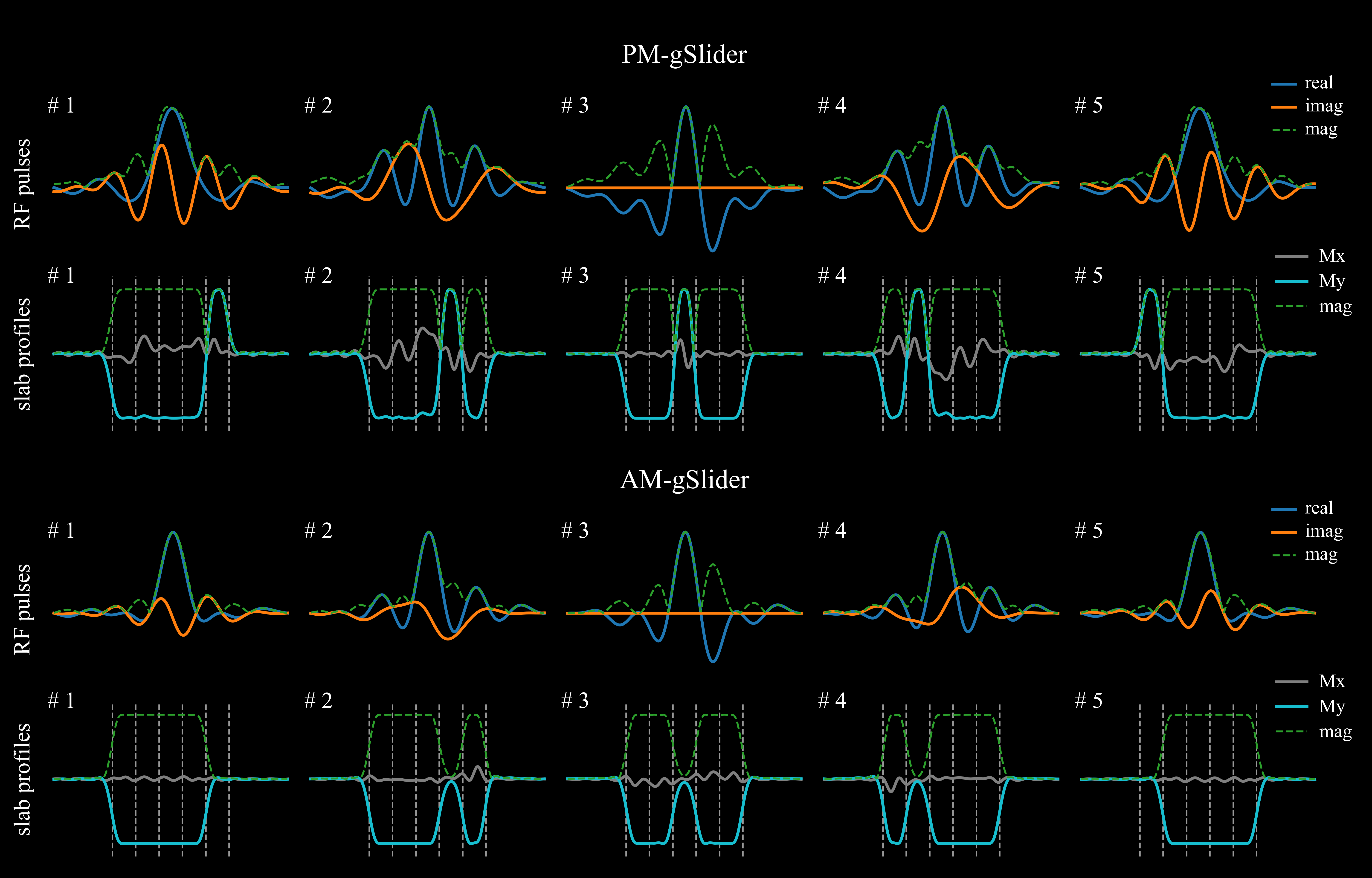
Figure 1. The illustration of the pulse shapes and slab profiles of PM-gSlider and AM-gSlider pulses.
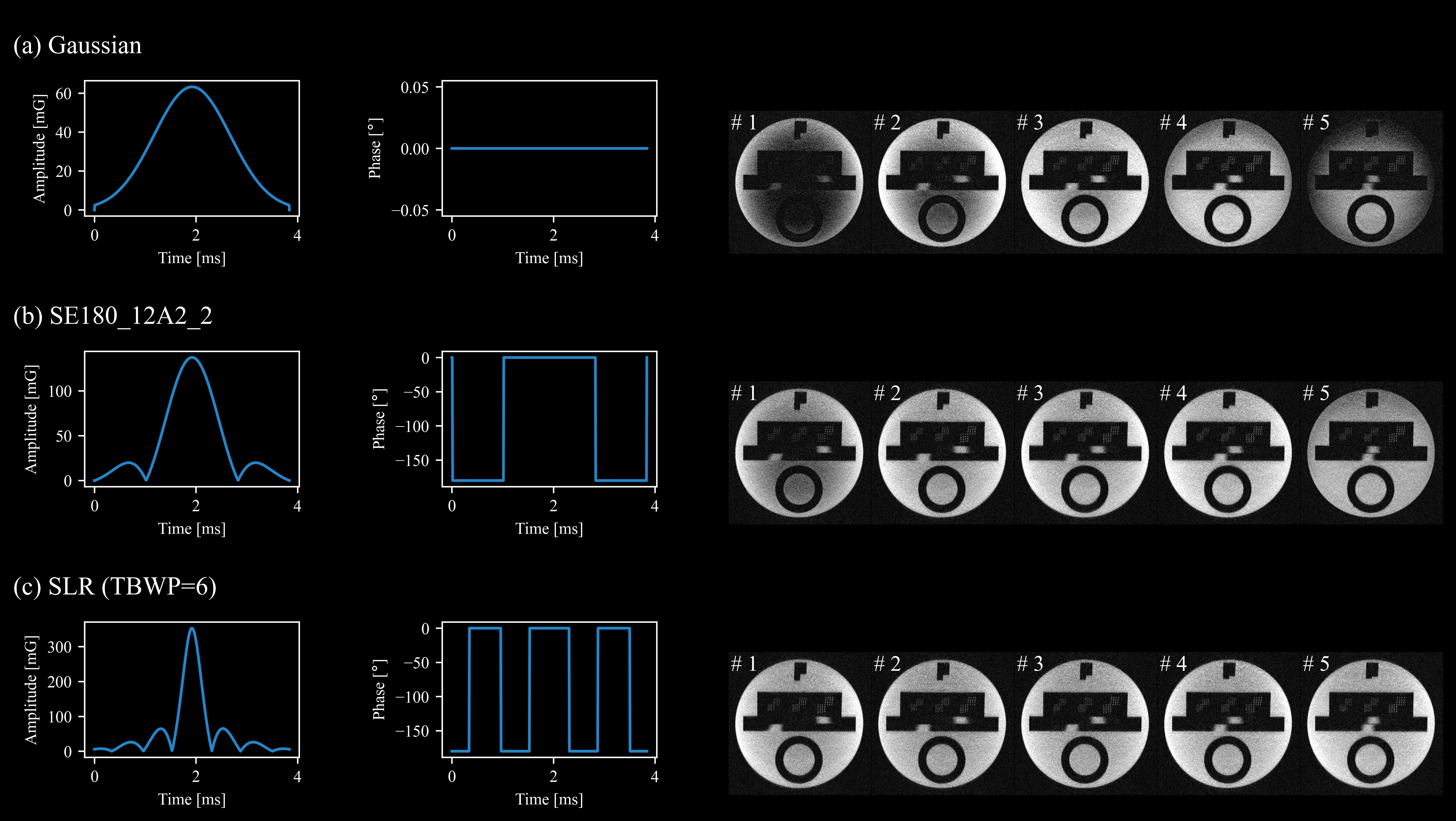
Figure 2. The illustration of three different refocusing pulses and corresponding phantom images. (a) pure Gaussian pulse. (b) SE18012A2_2 pulse (180° optimized amplitude modulated rf pulse in Siemens’ library). (c) SLR pulse (TBWP=6). When employing SLR refocusing, the PM/AM-gSlider-TSE provides appreciable slice profiles with consistent brightness across all the reconstructed slices.
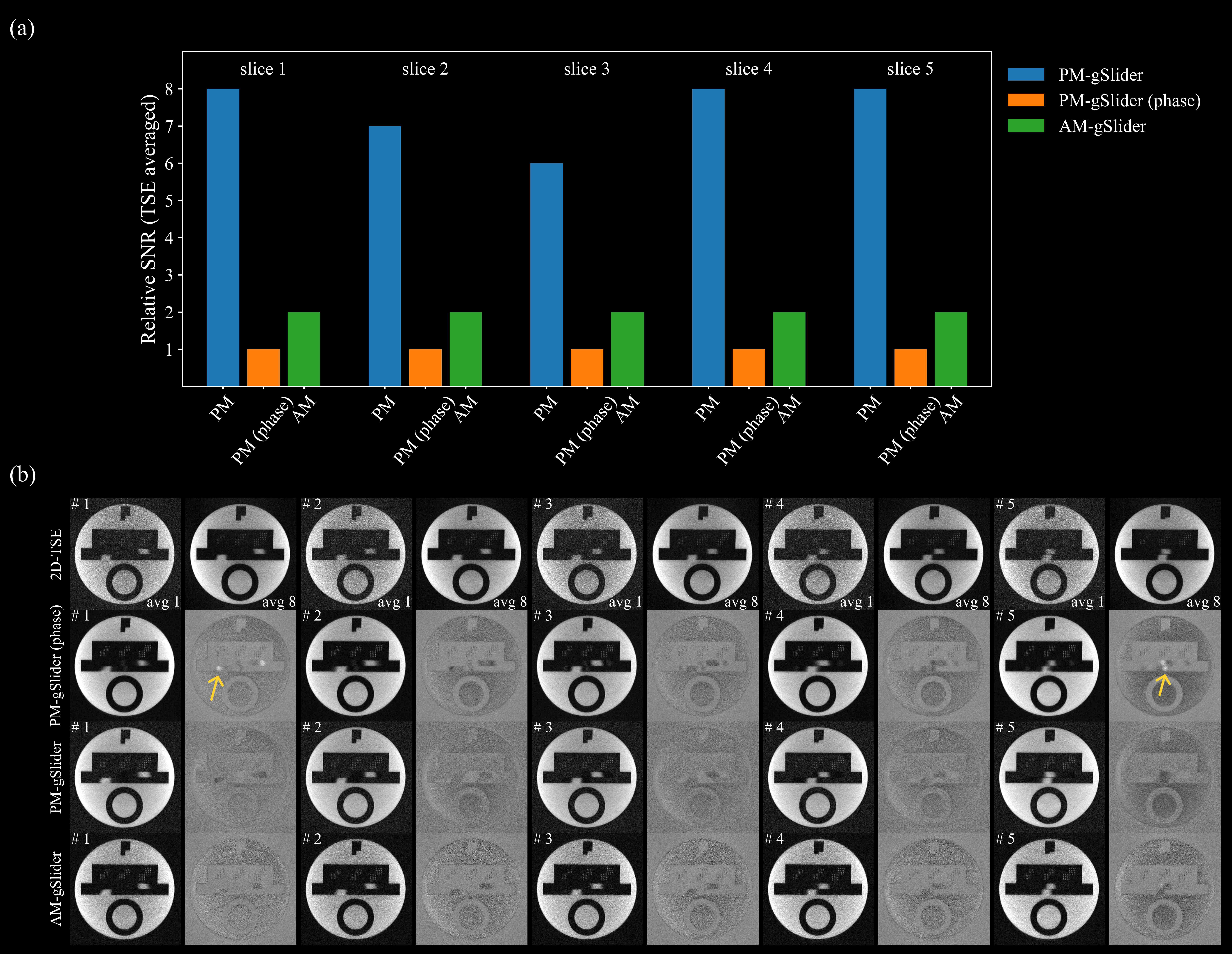
Figure 3. Comparison of PM/AM-gSlider and conventional 2D-TSE in phantom study. (a) relative SNR of PM/AM-gSlider compared to 2D-TSE with averaged in multi-times. (b) phantom images with 0.4mm isotropic resolution and corresponding difference maps. The PM-gSlider reconstructed using magnitude alone achieved the highest SNR, but a few errors were present, as indicated by the yellow arrows.
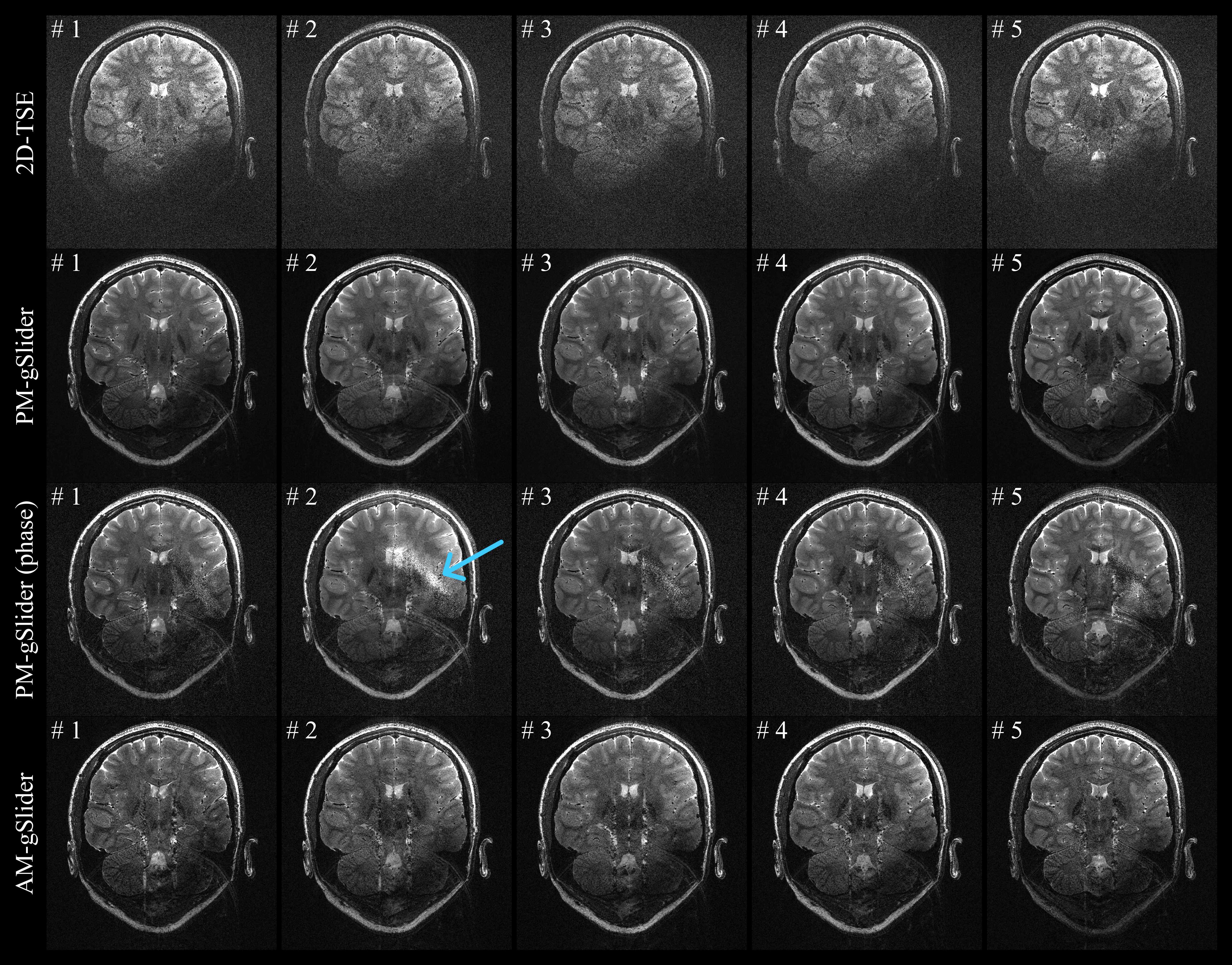
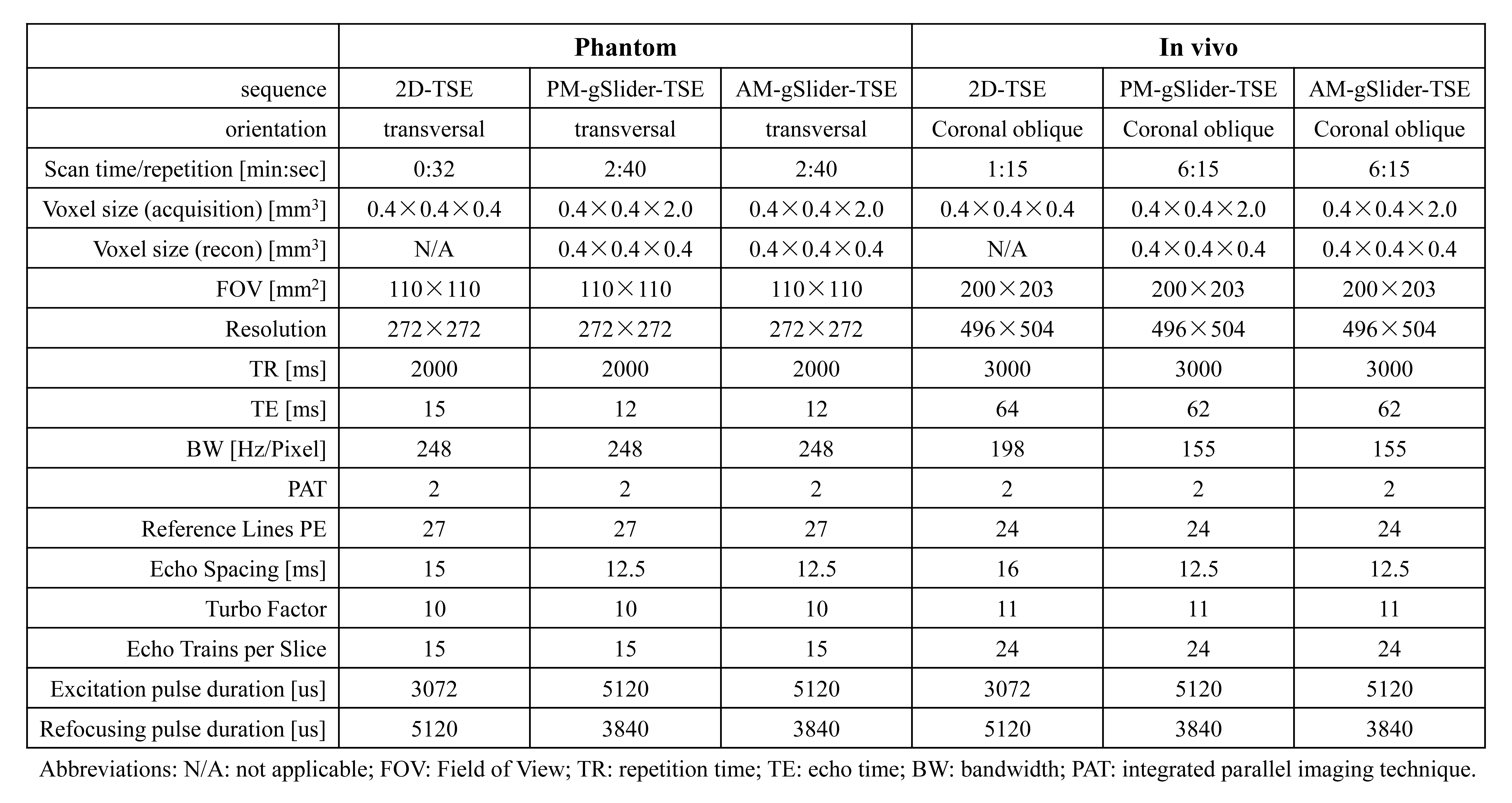
Table 1. Sequence parameters for phantom and in vivo imaging.