3254
Modified Three-dimensional Double-echo Steady-state Echo-planar Imaging with Fly-back Gradients for Quantitative Susceptibility Mapping1Biomedical Engineering Department, The University of Arizona, Tucson, AZ, United States
Synopsis
Keywords: Pulse Sequence Design, Pulse Sequence Design
Motivation: The selection of cerebrospinal fluid (CSF) as a zero-reference can yield significant and interpretable susceptibility measurements in quantitative susceptibility mapping (QSM).
Goal(s): Our goal is to develop a 3D dual-pathway sequence capable of achieving acquisition of multi-contrast images with a reduced scan time and improving image quality. These images will be used in selecting the zero reference in QSM.
Approach: We implement a 3D double-echo steady-state echo-planar imaging (DESS-EPI) sequence with fly-back gradients, designed to generate multi-contrast images while eliminating Nyquist artifact.
Results: Our preliminary findings indicate the feasibility of utilizing multi-echo images in QSM and global CSF segmentation.
Impact: The use of global CSF as a reference in QSM can enhance susceptibility measurements. The development of our modified 3D DESS-EPI sequence improves image quality, reduces scan time, and shows promise for global CSF segmentation in QSM.
Introduction
Quantitative Susceptibility Mapping (QSM) is an advanced magnetic resonance imaging (MRI) technique for quantification of magnetic susceptibility in various tissues within the human body1. Magnetic susceptibility is a fundamental property of tissue that characterizes its response to a magnetic field. In QSM, measurements compared to a chosen reference (a process known as "zero-referencing”) is essential since the susceptibility is determined up to a constant2,3. To address this challenge, a common reference tissue is selected, and its susceptibility is subtracted from the susceptibility measurements of other tissues.In brain QSM, cerebrospinal fluid (CSF) is frequently employed as the reference tissue due to its similarity to pure water in terms of magnetic susceptibility. However, the nonuniformity of ventricular CSF may introduce uncertainty in the reference susceptibility2,3. To address this variability and enhance the accuracy of QSM, an approach is suggested, involving the utilization of a global CSF flow as a reference2,3. In this study, we develop a novel multi-contrast imaging sequence, which can be applied in QSM with global CSF zero reference.
Methods
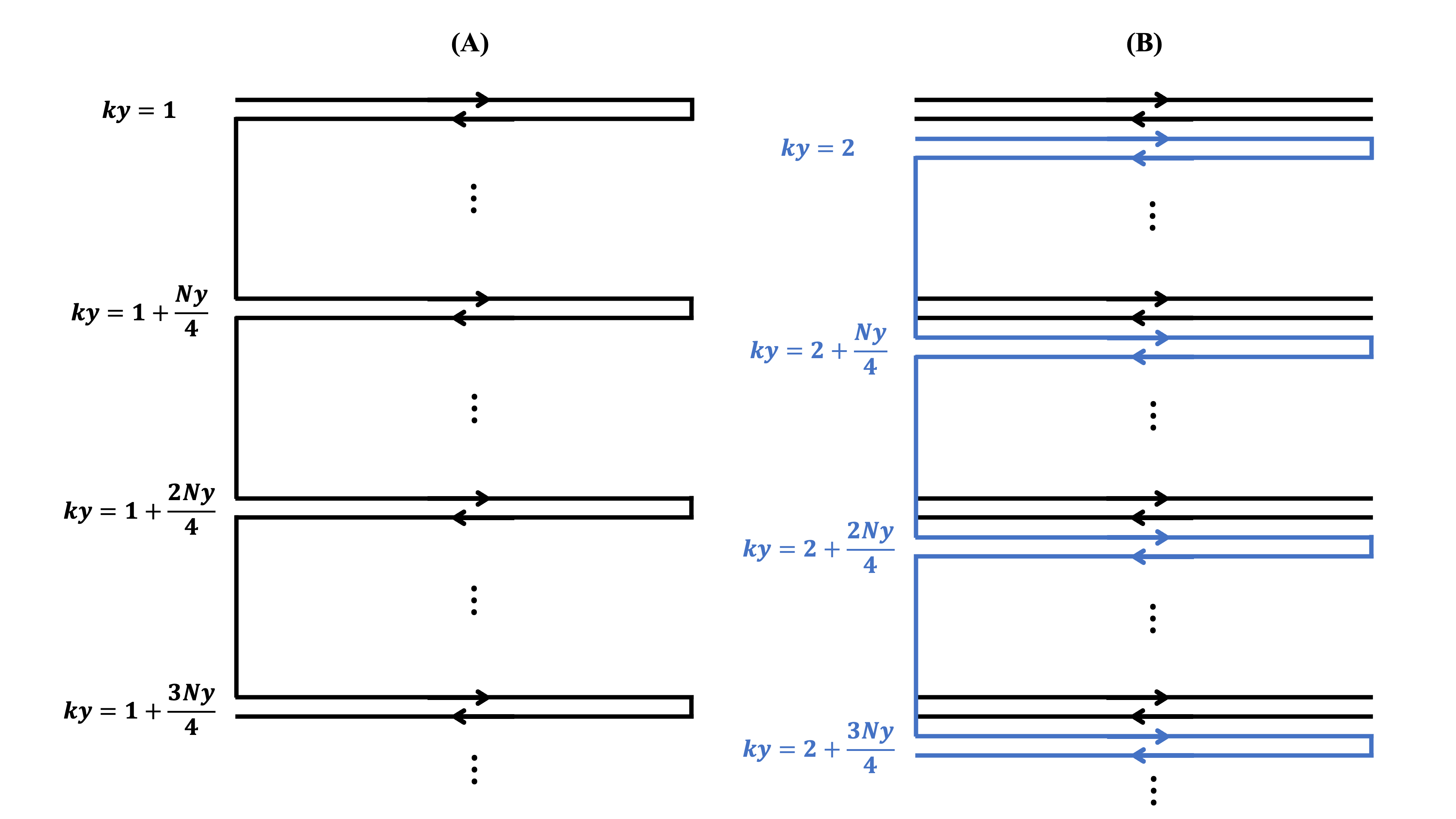
Traditional DESS imaging technique acquires dual echoes within each repetition time (TR), consisting of fast imaging with steady-state precession (FISP) and inverse fast imaging with steady-state precession (PSIF)4,5. To enhance the scan time efficiency and eliminate Nyquist artifact, we develop a modified 3D DESS-EPI sequence with fly-back gradients. Figure 1 shows the readout gradients of 3D fly-back DESS-EPI and Figure 2 illustrates the corresponding k-space acquisition trajectory. The k-space data generated from the first radiofrequency (RF) pulse in Figure 1 corresponds to specific ky lines = $$$1$$$, $$$1+\frac{N_y}{4}$$$, $$$1+\frac{2N_y}{4}$$$, $$$1+\frac{3N_y}{4}$$$ in Figure 2.A, where Ny represents the matrix size along phase-encoding direction. Each ky line acquires k-space data twice using fly-back gradients, resulting in two echoes (FISP and PSIF) for each acquisition. The second RF pulse in Figure 1 generates k-space data corresponding to ky lines = $$$2$$$, $$$2+\frac{N_y}{4}$$$, $$$2+\frac{2N_y}{4}$$$, $$$2+\frac{3N_y}{4}$$$in Figure 2.B. With the use of fly-back gradients, a total of four echo images with different TE are acquired, ensuring that the images are free from Nyquist artifacts.In our study, we implement our 3D fly-back DESS-EPI sequence using PulSeq framework6 and acquire data at a 3T Siemens scanner equipped with 32-channel RF coils. Scan parameters included: FOV = 240 mm x 240 mm, spatial resolution = 1.5 mm x 1.5 mm x 1 mm and flip angle = 8o. TR and echo time (TE) settings are as follows: TR = 64 msec, TEFISP of two FISP echo (T2* time of FISP) = 27 msec, 34 msec and TEPSIF of two PSIF echo (T2* time of PSIF) = 30 msec, 33 msec. Notably, the PSIF echo not only undergoes T2* relaxation but also includes T2 relaxation, resulting in T2 time = 91 msec, 94 msec and providing enhanced T2 contrast. The FISP images with different TE values are used for QSM using STI Suite Software Package7, while the PSIF images are utilized for CSF flow segmentation by applying a threshold.
Results and Discussion
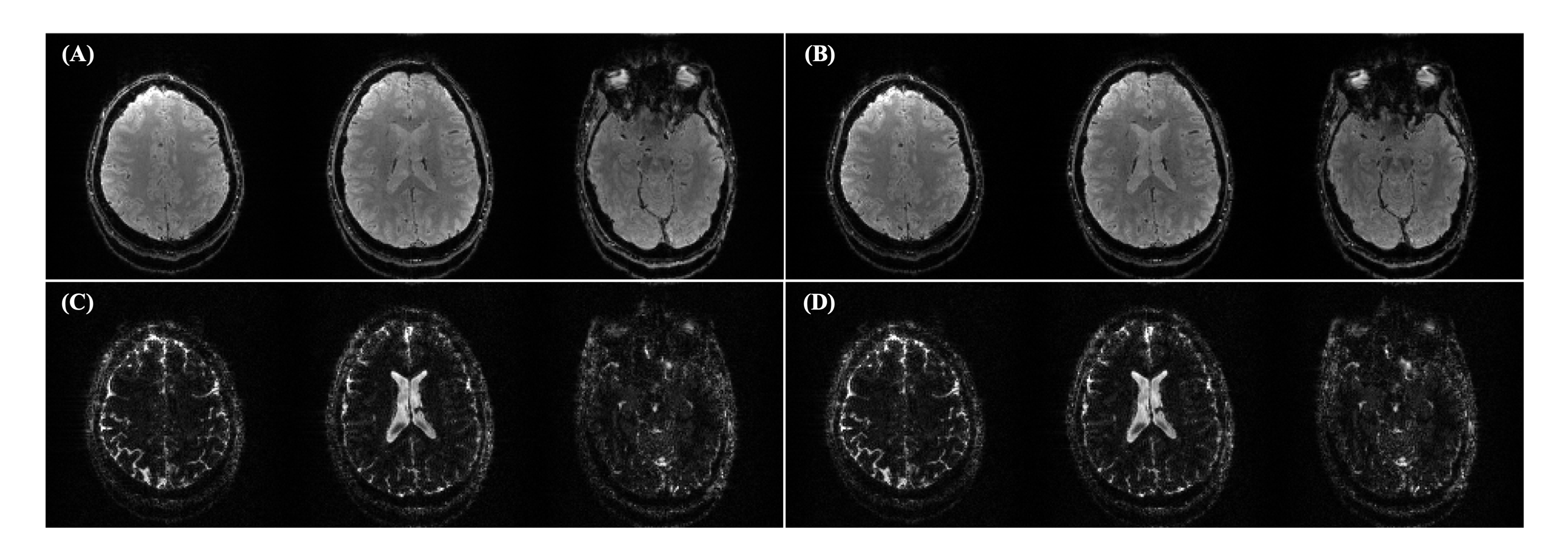
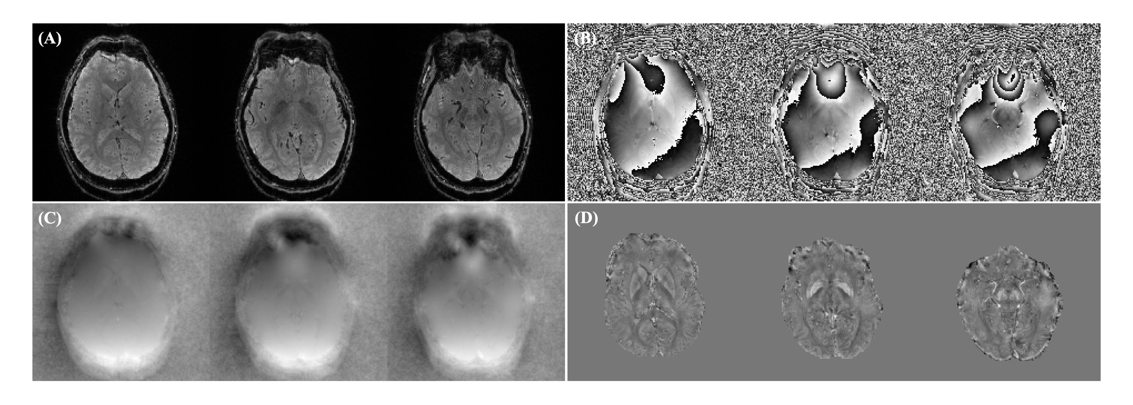
Figure 3 shows magnitude images obtained using our 3D fly-back DESS-EPI sequence, showing the FISP and PSIF images with different contrast. It can be seen that the PSIF images exhibit a higher SNR within CSF, making it feasible for enhancing the accuracy of CSF segmentation and a critical component of zero reference in QSM.The results from STI Suite Toolbox are presented in Figure 4, including: FISP magnitude image with TE = 27 msec (Figure 4.A), its corresponding FISP phase image (Figure 4.B), unwrapped phase image (Figure 4.C) and susceptibility mapping (Figure 4.D). These images provide valuable insights into the tissue properties and susceptibility variations within the imaged region like midbrain nuclei.
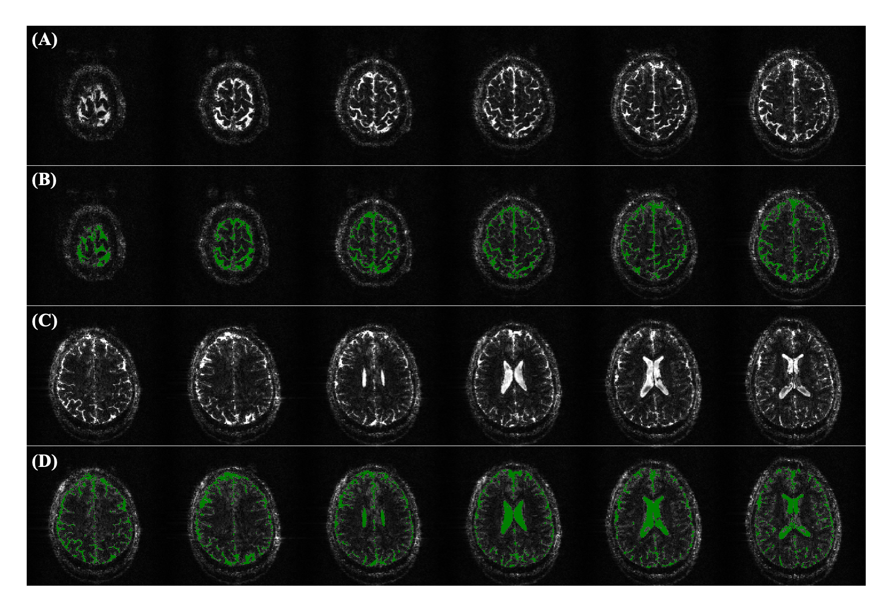
Furthermore, the PSIF magnitude images, as demonstrated in Figure 5, play a pivotal role in the segmentation process. In this figure, segmentation results are shown through a straightforward thresholding technique, simplifying the process of distinguishing CSF from other tissue types. It can be noted that our proposed multi-contrast sequence can provide a precise global CSF segmentation, which can be incorporated as a reference point to further enhance the accuracy and robustness of susceptibility mapping in our future studies.
Conclusions
We develop an efficient 3D fly-back DESS-EPI with dual pathways for multi-contrast imaging. Our preliminary results demonstrate its utility for susceptibility mapping generation and its potential application in global CSF segmentation, serving as a zero-reference regularization in brain QSM.Acknowledgements
No acknowledgement found.References
1. Ruetten PPR, Gillard JH, Graves MJ. Introduction to Quantitative Susceptibility Mapping and Susceptibility Weighted Imaging. Br J Radiol. 2019;92(1101):20181016.
2. Liu Z, Spincemaille P, Yao Y, et al. MEDI+0: Morphology enabled dipole inversion with automatic uniform cerebrospinal fluid zero reference for quantitative susceptibility mapping. Magn Reson Med. 2018;79(5):2795-2803.
3. Dimov AV, Nguyen TD, Spincemaille P, et al. Global cerebrospinal fluid as a zero-reference regularization for brain quantitative susceptibility mapping. J Neuroimaging. 2022;32(1):141-147.
4. Bruder H, Fischer H, Graumann R, et al. A new steady-state imaging sequence for simultaneous acquisition of two MR images with clearly different contrasts. Magn Reson Med. 1988;7(1):35-42.
5. Wu ML, Chang HC, Chao TC, et al. Efficient imaging of midbrain nuclei using inverse double-echo steady-state acquisition. Med Phys. 2015;42(7):4367-4374.
6. Layton KJ, Kroboth S, Jia F, et al. Pulseq: A rapid and hardware-independent pulse sequence prototyping framework. Magn Reson Med. 2017;77(4):1544-1552.
7. Li W, Avram AV, Wu B, et al. Integrated Laplacian-based phase unwrapping and background phase removal for quantitative susceptibility mapping. NMR Biomed. 2014;27(2):219-227.
Figures