3253
Multi-contrast Imaging with High Quality and High Scan Efficiency using Three-dimensional Dual-pathway Echo-planar Imaging Sequence1Biomedical Engineering Department, The University of Arizona, Tucson, AZ, United States
Synopsis
Keywords: Pulse Sequence Design, Pulse Sequence Design
Motivation: While current multi-contrast imaging technique spoiled gradient recalled echo (SPGR) requires a long acquisition time and with a relatively low signal-to-noise ratio (SNR), an efficient three-dimensional (3D) dual-pathway sequence may improve image quality with a reduced scan time.
Goal(s): Our goal is to demonstrate an image quality improvement in multi-contrast imaging with our proposed 3D double-echo steady-state echo-planar imaging (DESS-EPI) sequence.
Approach: We design and implement a 3D DESS-EPI sequence and evaluate SNR and contrast-to-noise ratio (CNR) of acquired images.
Results: Our results demonstrate the potential advantages of the 3D DESS-EPI sequence in terms of imaging quality and efficiency.
Impact: The improvement in time efficiency and image quality using our 3D DESS-EPI sequence illustrates the prospective benefits in multi-contrast imaging, which is appropriate for temperature mapping, quantitative mapping, field mapping, susceptibility-weighted imaging (SWI) and quantitative susceptibility mapping (QSM).
Introduction
Multi-contrast imaging provides images with various tissue parameters like T1-, T2-, T2*-relaxation times or proton density, commonly used for diagnosis and monitoring of neurological disease1. However, acquiring a series of images with different timing parameters requires a relatively long scan time with current routine sequence SPGR. Instead, dual-pathway sequences have been proposed to allow a simultaneous acquisition of two images with clearly different contrast, improving acquisition speed over standard SPGR method2,3.Our previous 2D DESS-EPI sequence compared to SPGR, offers certain advantages4: (1) exhibits enhanced signal; (2) provides improved phase contrast. However, the SNR and CNR still remains low in 2D acquisition mode. To further improve the SNR and CNR, we introduce our 3D DESS-EPI sequence for multi-contrast imaging.
Theory
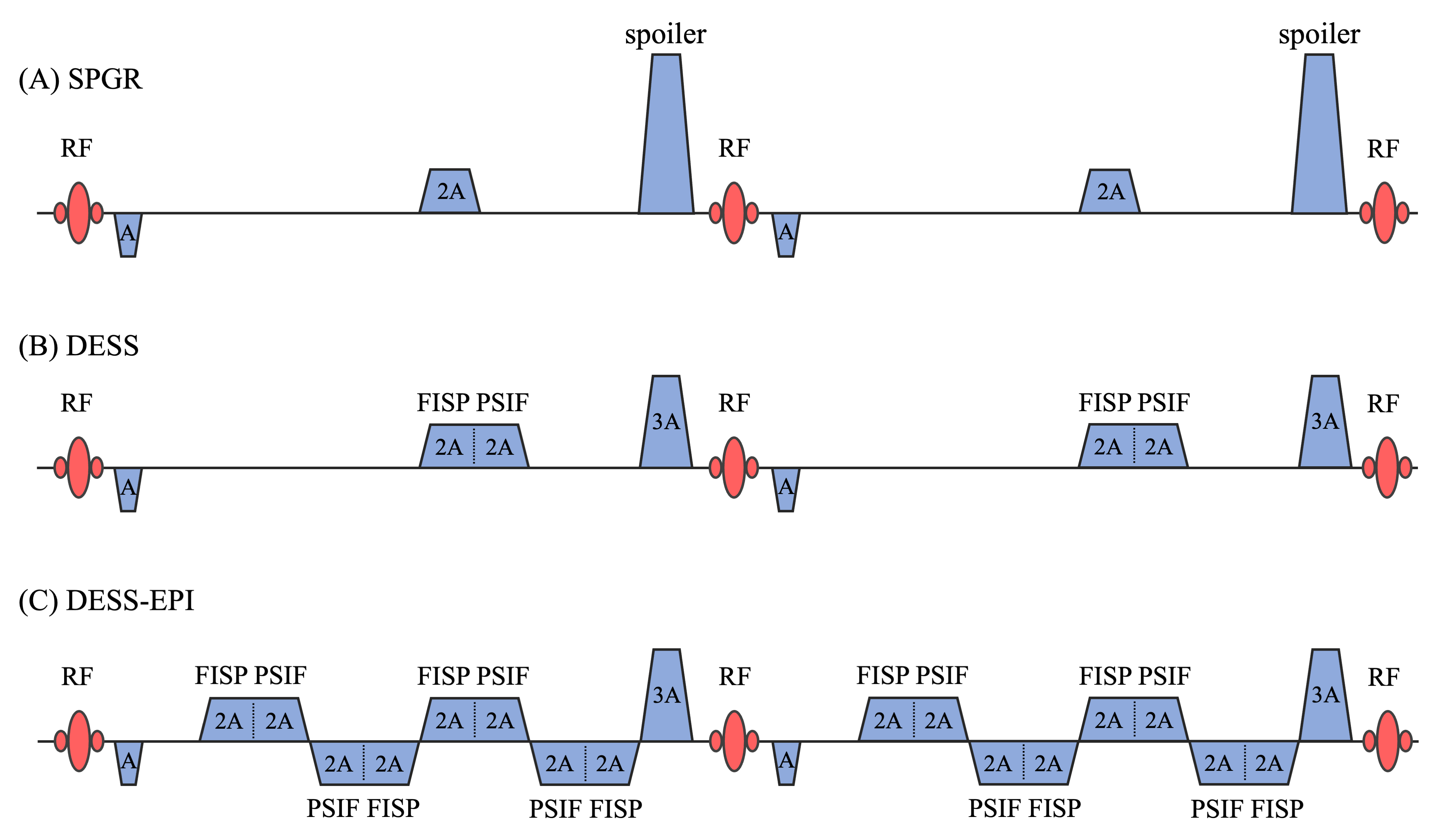
Figure 1 shows the read-out gradient of SPGR, DESS and DESS-EPI sequence. SPGR contains a spoiling gradient at the end of each cycle, spoiling the transverse magnetization and causing a low SNR. Instead, DESS sequence acquires dual echoes with balanced gradients within each repetition time (TR): fast imaging with steady-state precession (FISP) and inverse fast imaging with steady-state precession (PSIF). Compared to SPGR, DESS imaging technique: (1) acquires dual echoes with both T2 and T2* contrast; (2) enables a higher SNR since the steady-state FISP echo is acquired with a relatively long spin history from previous TR's contribution.Methods
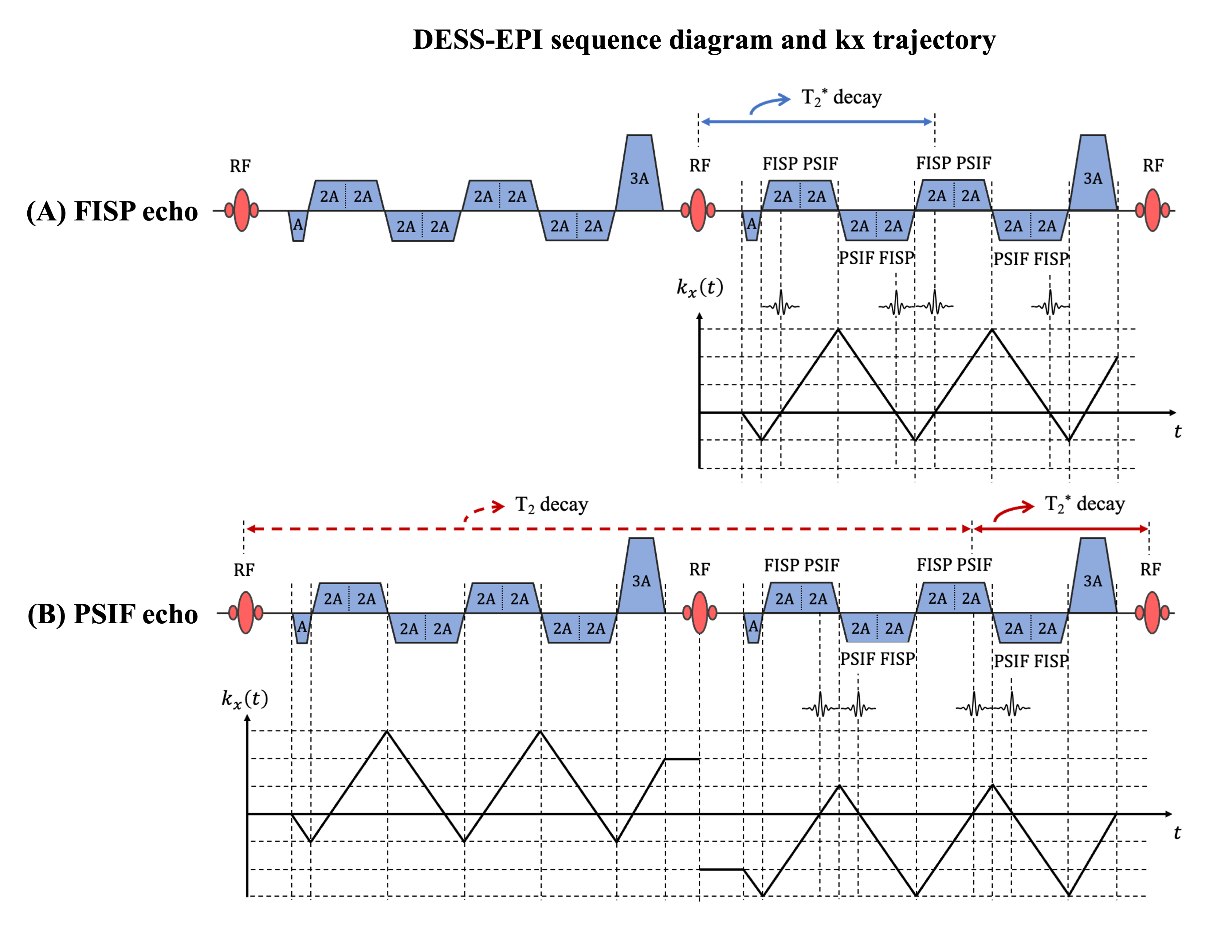
To further improve the time efficiency, DESS-EPI is developed to acquire eight echoes within each TR. Figure 2 shows the kx trajectory for FISP and PSIF echoes in DESS-EPI sequence. The first echo FISP consists of free induction decay excited from current TR and magnetization refocused from previous TR, contributing to increased signal intensities compared to SPGR since no spoiling gradients are added. FISP echo experiences a T2* contrast accumulation, illustrated with blue double arrow in Figure 2.A. The second echo PSIF excited from previous TR is partially refocused at current TR. PSIF experiences a T2 relaxation plus a T2* decaying, indicated with red double arrow in Figure 2.B.In our study, we use PulSeq framework5 to implement 3D SPGR and our 3D DESS-EPI sequences and acquire data at a 3T Siemens scanner equipped with 32-channel RF coils. Scan parameters included: FOV = 240 mm x 240 mm, spatial resolution = 1.5 mm x 1.5 mm x 1 mm and flip angle = 30o. For 3D SPGR, two sets of data with different TR and echo time (TE) are acquired: SPGR1, TR/TE = 18 msec/11 msec; SPGR2, TR/TE = 33 msec/26 msec. For 3D DESS-EPI: TR = 38 msec, TEFISP = 11 msec and TEPSIF (T2* time of PSIF) = 26 msec. The scan duration is 9.2 mins, 16.9 mins and 4.9 mins for SPGR1, SPGR2 and DESS-EPI respectively. A composite image of DESS-EPI is generated by summing up both FISP and PSIF image with a weighting factor of 0.5, optimizing image contrast for a midbrain nuclei depiction. SNR is calculated by averaging signal from a ROI in the occipital lobe and comparing it to the background noise. CNR of midbrain (including red nuclei and substantia nigra) and cerebrospinal fluid (CSF) region is evaluated.
Results and Discussion
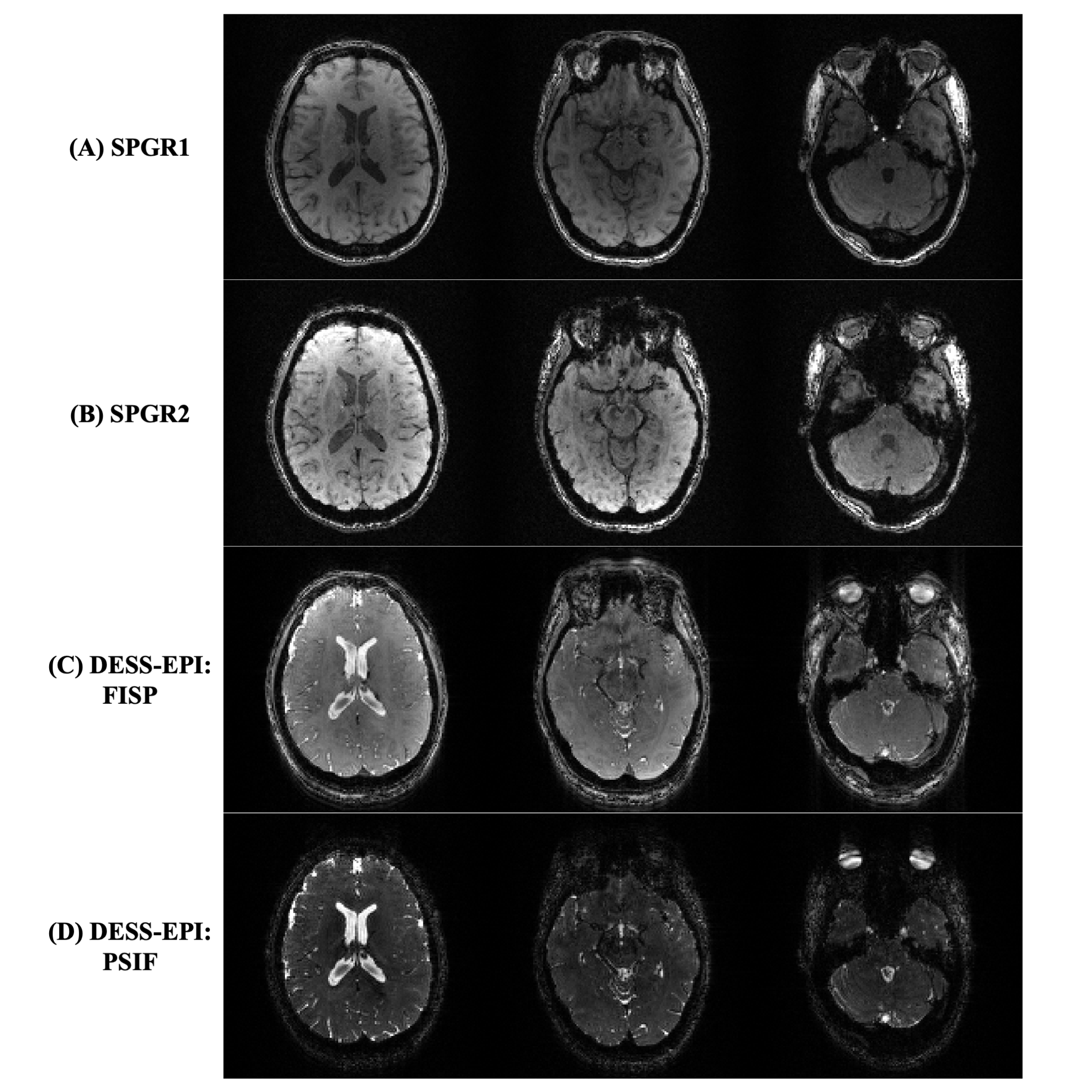
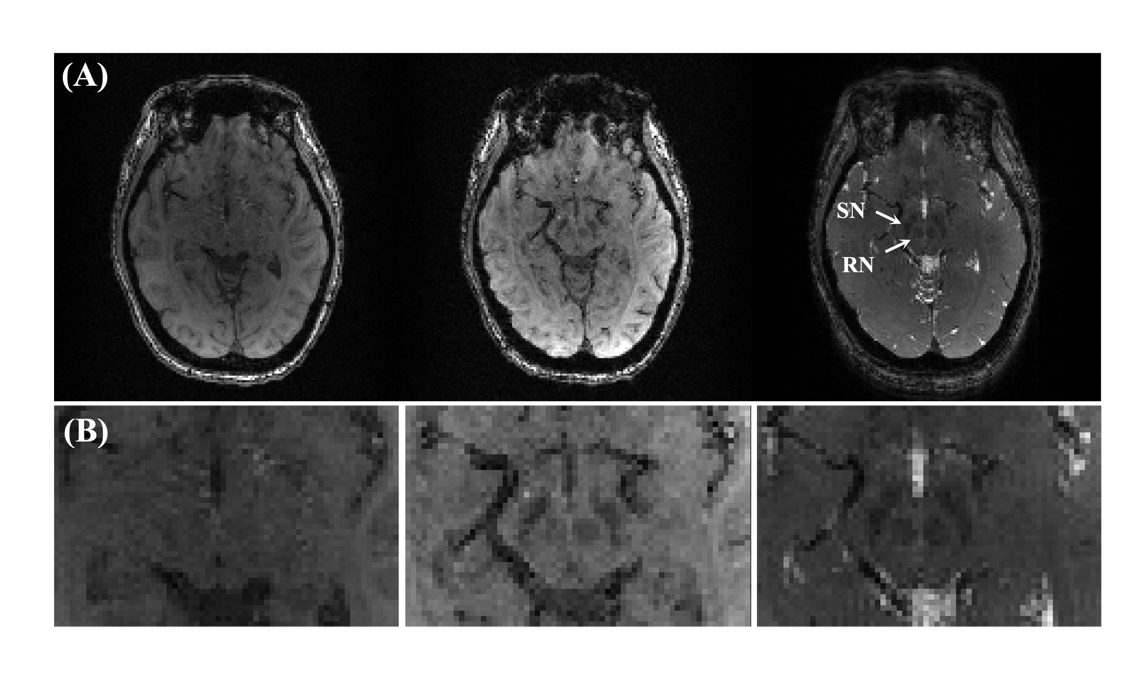
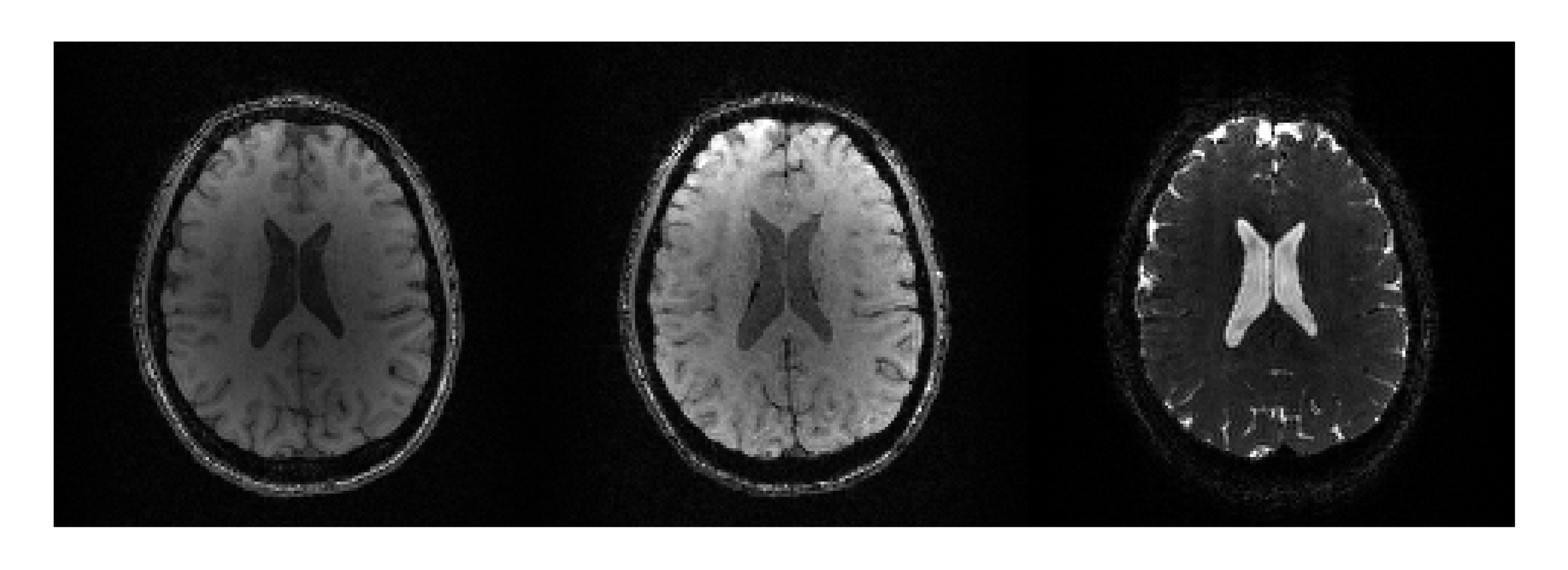
Figure 3 shows the magnitude images of SPGR1, SPGR2 and DESS-EPI at different slice location. The SNR in SPGR1 and SPGR2 image is 19.39 and 25.28, respectively and increases to 73.04 (FISP) and 38.76 (PSIF) in DESS-EPI.The magnitude images in Figure 4 shows red nuclei and substantia nigra (indicated with arrows): from left to right are SPGR1, SPGR2 and a composite image of DESS-EPI. Midbrain nuclei can be observed in images with a TE of at least 25 msec, as shown in Figure 4 (column 2 and 3). The CNR of midbrain nuclei from SPGR1, SPGR2 and DESS-EPI is 0.65, 7.01 and 16.60, respectively. A significant improvement in midbrain nuclei CNR is shown with our proposed sequence.
Figure 5.A shows the magnitude images of SPGR1, SPGR2 and PSIF from DESS-EPI. The CNR of CSF from SPGR1, SPGR2 and DESS-EPI is 22.06, 25.36 and 117.36, respectively. The PSIF image from DESS-EPI sequence offers a better CNR of CSF, making it a valuable choice for segmentation purpose.
Conclusion
We develop an efficient dual-pathway sequence 3D DESS-EPI. Our experimental results illustrate a remarkable fourfold increase in scan efficiency while maintaining similar contrast. Our multi-echo images exhibit a threefold improvement in SNR. Furthermore, CNR of midbrain nuclei increases from 7.01 to 16.60 and CNR of CSF increases from 25.36 to 117.36.Acknowledgements
No acknowledgement found.References
1. Seiler A, Noth U, Hok P, et al. Multiparametric Quantitative MRI in Neurological Diseases. Front Neurol. 2021;12:640239.
2. Wu ML, Chang HC, Chao TC, et al. Efficient imaging of midbrain nuclei using inverse double-echo steady-state acquisition. Med Phys. 2015;42(7):4367-4374.
3. Bruder H, Fischer H, Graumann R, et al. A new steady-state imaging sequence for simultaneous acquisition of two MR images with clearly different contrasts. Magn Reson Med. 1988;7(1):35-42.
4. Han S, Ramanna S, Chen N-k. Multi-contrast Imaging using Dual-pathway Echo-planar Imaging Sequence. Proc Intl Soc Mag Reson Med 31 (2023).
5. Layton KJ, Kroboth S, Jia F, et al. Pulseq: A rapid and hardware-independent pulse sequence prototyping framework. Magn Reson Med. 2017;77(4):1544-1552.
Figures