3251
Pulseq on Philips MRI Systems: Unlocking and Validating Open-Source Sequences1Department of Radiology, High Field MRI group, University Medical Center Utrecht, Utrecht, Netherlands
Synopsis
Keywords: Pulse Sequence Design, Pulse Sequence Design, Pulseq, reproducable, open, open-source, UHF, 7T, Philips
Motivation: Conventional MRI vendor-specific sequence development limits research transferability and reproducibility. Harmonized frameworks, like Pulseq, overcome these limitations - but Pulseq was not supported yet on Philips MRI scanners.
Goal(s): To develop a fully-compliant Pulseq interpreter for Philips MRI systems, facilitating the use of open-source sequences previously incompatible with these platforms.
Approach: Custom adaptation of Pulseq for the Philips R5.4 platform, followed by validation with standard imaging sequences using a field camera and phantom/in vivo scans, and comparison with native sequences.
Results: Demonstration of the first Philips-compatible Pulseq interpreter, evidenced by successful scans at 7T, marking a leap in cross-vendor research capabilities.
Impact: The successful adaptation of Pulseq sequences to Philips MRI system enables researchers to deploy and disseminate advanced MRI techniques universally, fostering cross-vendor collaboration and accelerating the evolution of MRI technology.
Introduction
MRI sequence development is often constrained by vendor-specific environments, limiting the sharing and reproducibility of research across different MR scanners, potentially impeding scientific progress.Open-source frameworks like Pulseq have emerged to overcome these barriers, enabling hardware- and software-independent sequence development in MATLAB or Python1-6. Pulseq was supported by three major MRI vendors (Siemens1, Bruker1, and GE2) through interpreters or sequence conversion.
Philips MRI scanners, however, utilize a distinct platform that historically restricted runtime flexibility, thus hindering Pulseq integration. However, recent digital Philips hardware has become so fast that we hypothesized that extensive runtime modifications are in reach.
We explored this possibility and developed the first fully-compliant Pulseq interpreter on a Philips MR-system. We demonstrate the functionality of this Pulseq interpreter through field measurements and imaging experiments with a phantom and in-vivo at 7T.
Methods
Pulseq implementation
We ported Pulseq to the R5.4 Philips MRI platform into a spectroscopy sequence that starts with dummy sequence-objects that are internally modified beyond the conventional restrictions, to achieve the flexibility needed for full Pulseq compatibility. Only six source files are modified to prepare the sequence, read Pulseq sequences, and convert those into Philips objects. The customized files will be shared through the MR-Paradise repository for Philips research sites, linked at openmr.nlValidation Experiments
The interpreter's accuracy was validated using three sequences: a gradient echo, a spiral sequence, and a MP-RAGE, adapted from Pulseq's GitHub7,8. Initial validations were through simulations, where Philips' sequence viewer outcomes were juxtaposed with Pulseq's plotted waveforms. Waveform consistency was confirmed using a 16-channel field camera (Skope, Switzerland9). We then conducted phantom and in-vivo scans on a 7T Achieva MRI with 8Tx40Rx head coil (Nova Medical, USA) using Pulseq and matched native sequences, to evaluate imaging performance. Phantom scans are 256x256mm2 2D-GRE at 1mm2 (with TE/TR/α/BW=3ms/15ms/20°/900Hz/px) and the in-vivo scan demonstrated is 3D MP-RAGE with FOV of 256x240x192mm3 at 1mm3 (with TE/TR/α/BW=3ms/9ms/6°/250Hz/px) accelerated by R=2x2 to 3min.Data Reconstruction
Images are reconstructed from k-space data exported from the scanner, read into MATLAB via Reconframe (Gyrotools, Switzerland). Raw data was sorted using acquisition labels from native scans, or directly from the Pulseq sequence files. Phantom scans are reconstructed by a 2D inverse Fourier transform, and RSS coil-combination. The accelerated MP-RAGE scans were reconstructed using the PICS framework from the BART toolbox10.Results
SimulationsSimulations of the Pulseq sequences were visualized with the vendor's tools, showing excellent alignment for the gradient echo sequence, as depicted in Figure 1. However, the spiral waveforms were not accurately represented since the vendor's simulation software does not support the runtime waveform updates performed by the interpreter.
Field Measurements
The camera accurately captured the spiral trajectory up to the gradient crusher's dephasing limit. A comparison of the intended and captured gradients in Figure 2 and 3 reveals minor discrepancies that are typical for spiral sequences11,12.
Image Reconstructions
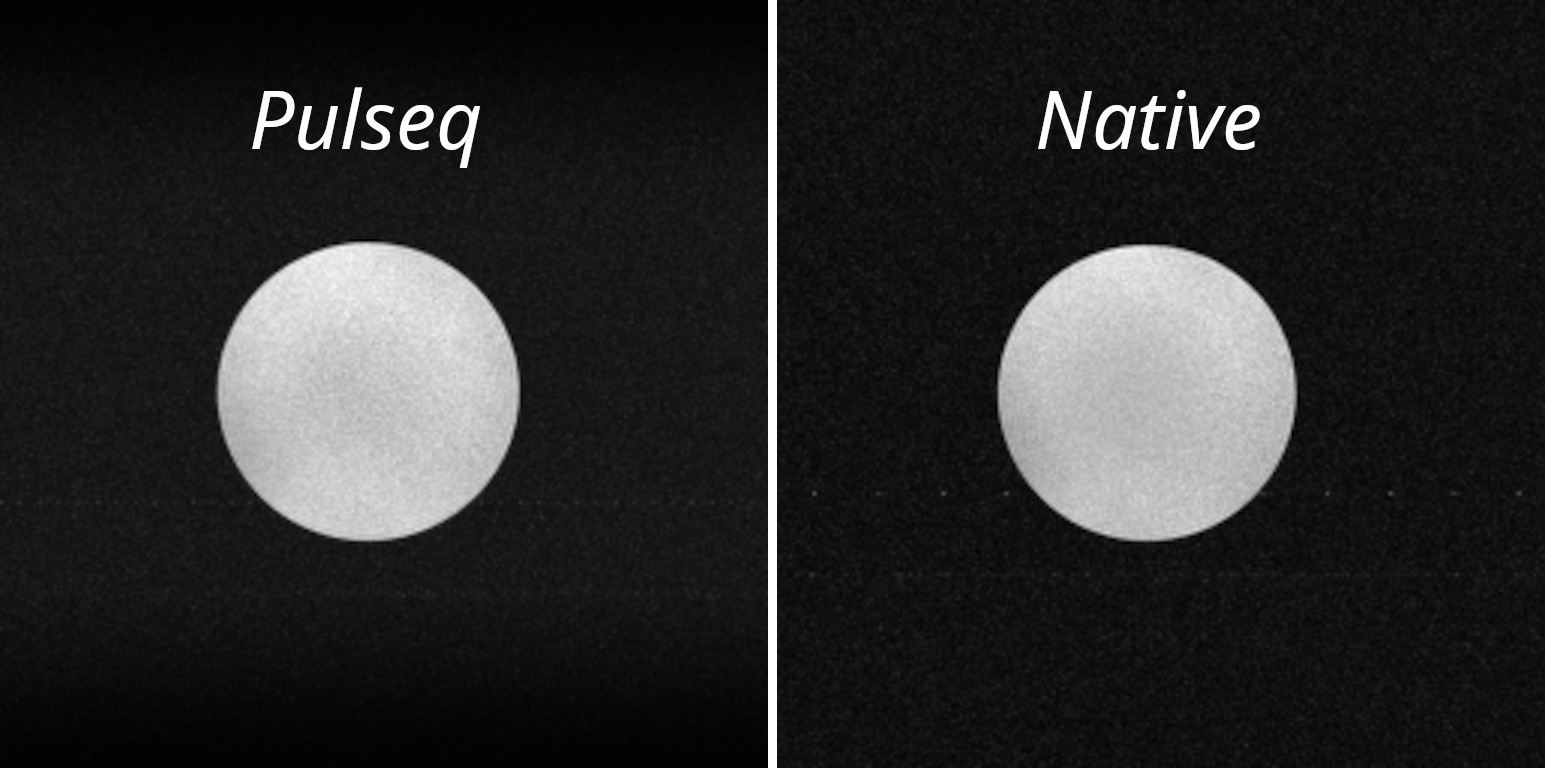
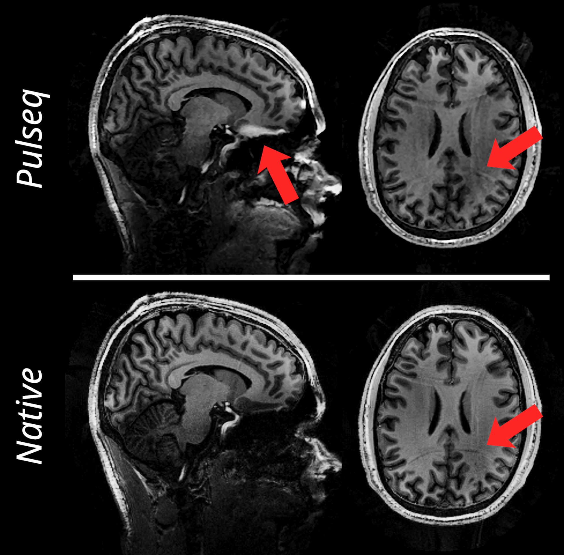
Reconstructed images from both native and Pulseq implementations of 2D-GRE and MP-RAGE are depicted in Figure 4 and 5 respectively. Both images exhibit similar contrast and signal-to-noise ratios (SNR), with observed inhomogeneities attributable to the B1+ field and the receive-coil profile at 7T. The Pulseq MP-RAGE displays slight inversion loss at tissue-boundaries, since the bandwidth was accidentally 50% of the intended bandwidth, unlike the native scan.
Discussion
We implemented a Pulseq interpreter for Philips MRI scanners, and demonstrated imaging with open-source sequences on a Philips 7T system. Reconstructed images from the Pulseq sequences showed similar image quality to the native sequence, with minor differences originating from additional acquisition optimisations in the native sequence.The Pulseq interpreter handles both simple 2D scans and complex, high-resolution in-vivo scans, indicating that the latest Philips hardware offers sufficient speed for significant runtime flexibility. While our implementation was tuned for Pulseq, an almost identical approach is·envisioned for supporting other frameworks.
Despite some hardware timing constraints, such as RF-deadtime, the Philips platform has minimal other timing constraints. RF-dwelltime can be set to popular values in Pulseq sequences (i.e. 1µs), blocks, acquisitions, and trapezoid gradients can align with any time grid, and arbitrary gradients can be upsampled from the common 10µs timing grid.
Conclusion
Enabling Pulseq sequences on Philips MRI systems expands the toolkit for researchers globally. As evidenced by the demonstration of spiral and cartesian readouts, in phantom and in-vivo scans at 7T, our interpreter promises full support for Pulseq sequences to Philips MRI systems.This advancement facilitates the rapid translation and dissemination of cutting-edge innovative MRI techniques across vendors, signifying a leap forward in making MRI research more open, collaborative and reproducible.
Acknowledgements
The authors gratefully acknowledge funding from the Dutch Research Council (NWO),
as this publication is part of the project “Silent MRI with the speed of CT and richer metabolic
information than PET” (with project number 18361) of the research programme “Talent
Programme Vidi TTW 2019” which is (partly) financed by the NWO.
The authors would also like to thank the Spinoza Centre for Neuroimaging (Amsterdam,
The Netherlands) for their provided computational facilities.
References
- Layton, Kelvin J., Stefan Kroboth, Feng Jia, Sebastian Littin, Huijun Yu, Jochen Leupold,Jon‐Fredrik Nielsen, Tony Stöcker, and Maxim Zaitsev. "Pulseq: a rapid andhardware‐independent pulse sequence prototyping framework." Magnetic resonance inmedicine 77, no. 4 (2017): 1544-1552.
- Nielsen, Jon‐Fredrik, and Douglas C. Noll. "TOPPE: A framework for rapid prototypingof MR pulse sequences." Magnetic resonance in medicine 79, no. 6 (2018): 3128-3134
- Jochimsen, Thies H., and Michael Von Mengershausen. "ODIN—object-orienteddevelopment interface for NMR." Journal of Magnetic Resonance 170, no. 1 (2004):67-78
- Magland, Jeremy F., Cheng Li, Michael C. Langham, and Felix W. Wehrli. "Pulsesequence programming in a dynamic visual environment: SequenceTree." Magneticresonance in medicine 75, no. 1 (2016): 257-265
- Cordes, Cristoffer, Simon Konstandin, David Porter, and Matthias Günther. "Portable andplatform‐independent MR pulse sequence programs." Magnetic resonance in medicine83, no. 4 (2020): 1277-1290
- Ravi, Keerthi Sravan, Sneha Potdar, Pavan Poojar, Ashok Kumar Reddy, Stefan Kroboth,Jon-Fredrik Nielsen, Maxim Zaitsev, Ramesh Venkatesan, and Sairam Geethanath."Pulseq-Graphical Programming Interface: Open source visual environment forprototyping pulse sequences and integrated magnetic resonance imaging algorithmdevelopment." Magnetic resonance imaging 52 (2018): 9-15
- "writeGradientEcho - Pulseq." https://pulseq.github.io/writeGradientEcho.html.Accessed 27 Sep. 2023
- "writeSpiral - Pulseq." https://pulseq.github.io/writeSpiral.html. Accessed 27 Sep. 2023
- Wilm, Bertram J., Christoph Barmet, Matteo Pavan, and Klaas P. Pruessmann. "Higherorder reconstruction for MRI in the presence of spatiotemporal field perturbations."Magnetic resonance in medicine 65, no. 6 (2011): 1690-1701
- Uecker M, Ong F, Tamir JI, et al. “Berkeley Advanced Reconstruction Toolbox”. Annual Meeting ISMRM, Toronto 2015, In: Proc. Intl. Soc. Mag.Reson. Med 2015;23:2486
- Papadakis, Nikolaos G., Adam A. Wilkinson, T. Adrian Carpenter, and Laurance D. Hall."A general method for measurement of the time integral of variant magnetic fieldgradients: application to 2D spiral imaging." Magnetic resonance imaging 15, no. 5(1997): 567-578
- Block, K.T. and Frahm, J., 2005. Spiral imaging: a critical appraisal. Journal of MagneticResonance Imaging: An Official Journal of the International Society for MagneticResonance in Medicine, 21(6), pp.657-668.
Figures
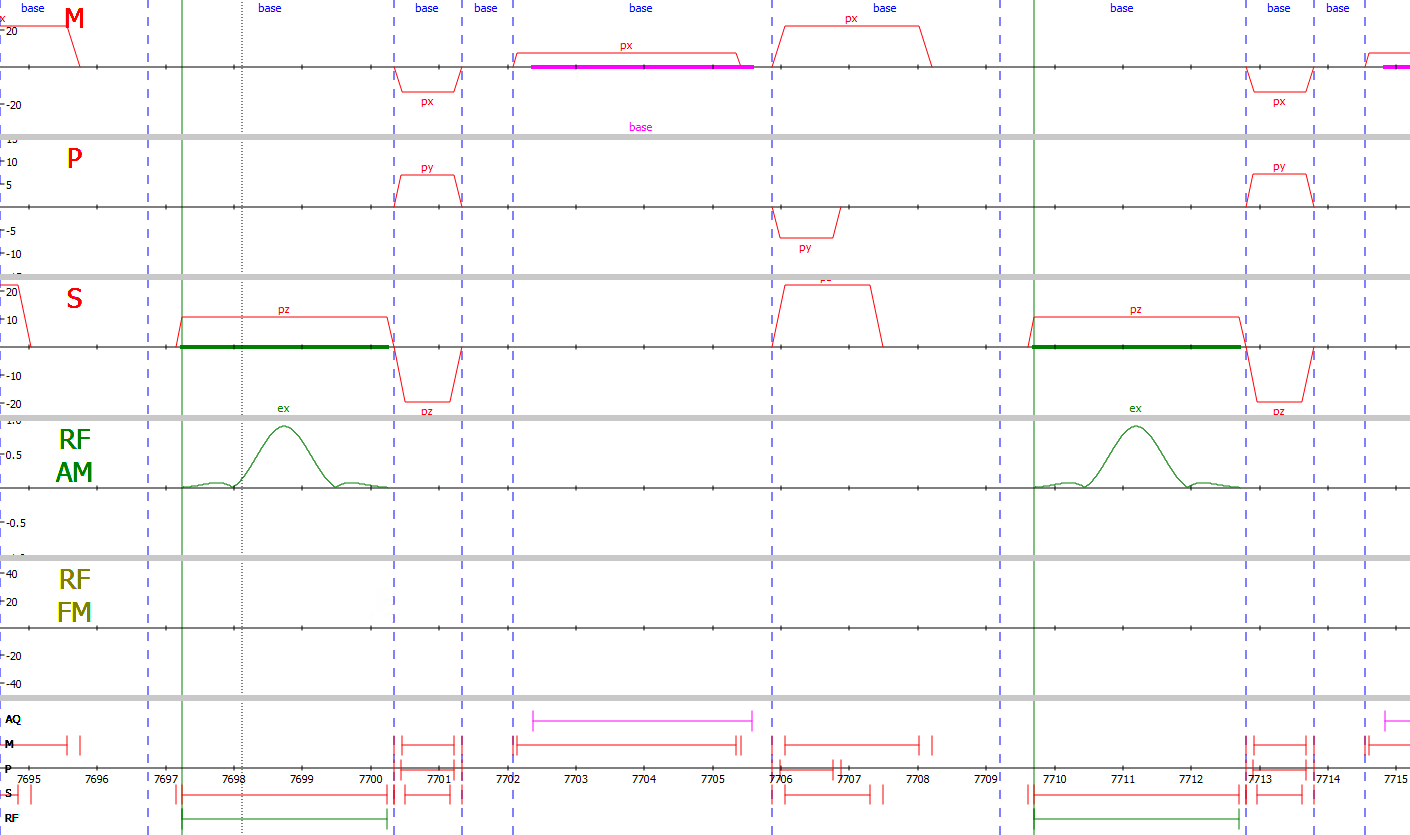
Pulse sequence generated from MRI spectrometer simulation, as visualized by the
vendor-provided graphical viewer, of the Pulseq interpreter performing the gradient echo example sequence file from the Pulseq GitHub7.
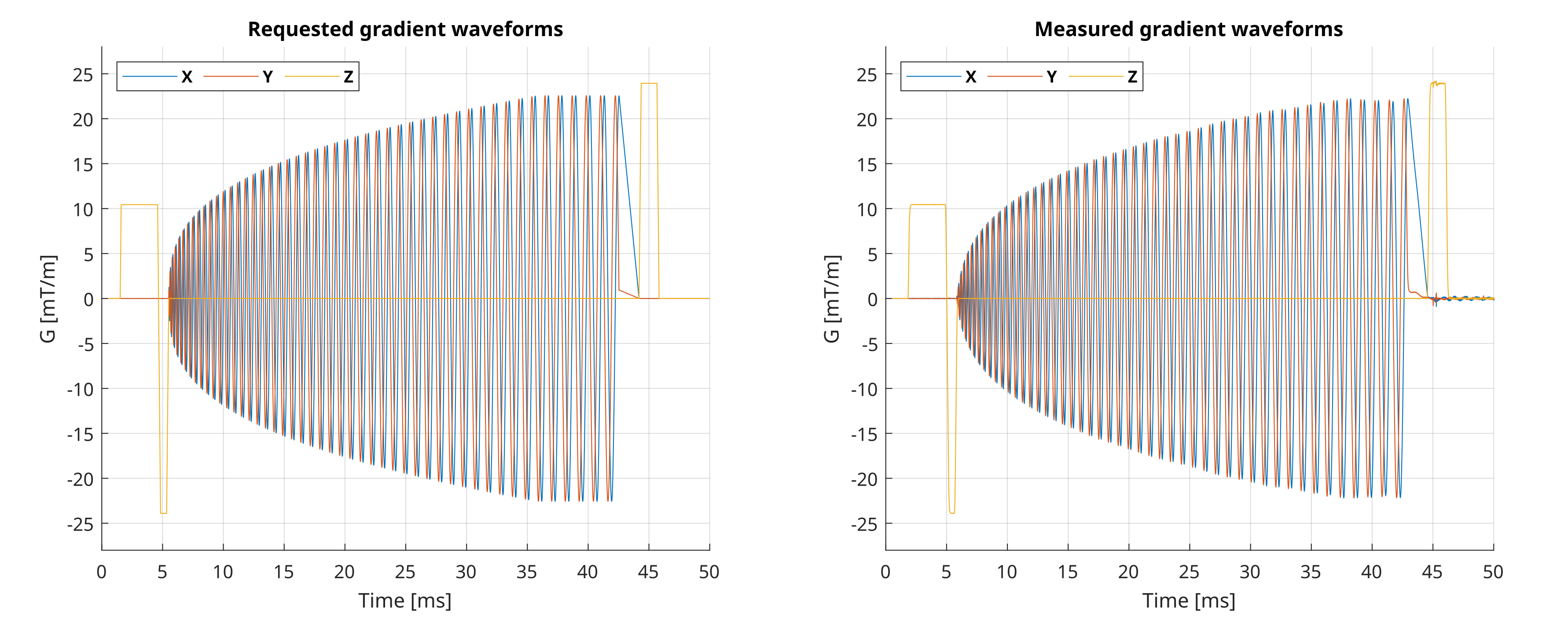
Gradient waveforms of a spiral readout, both as requested in the Pulseq sequence
definition (left) and as measured gradients captured by the field camera (right). This spiral readout is part of the spiral example sequence available from the Pulseq GitHub8.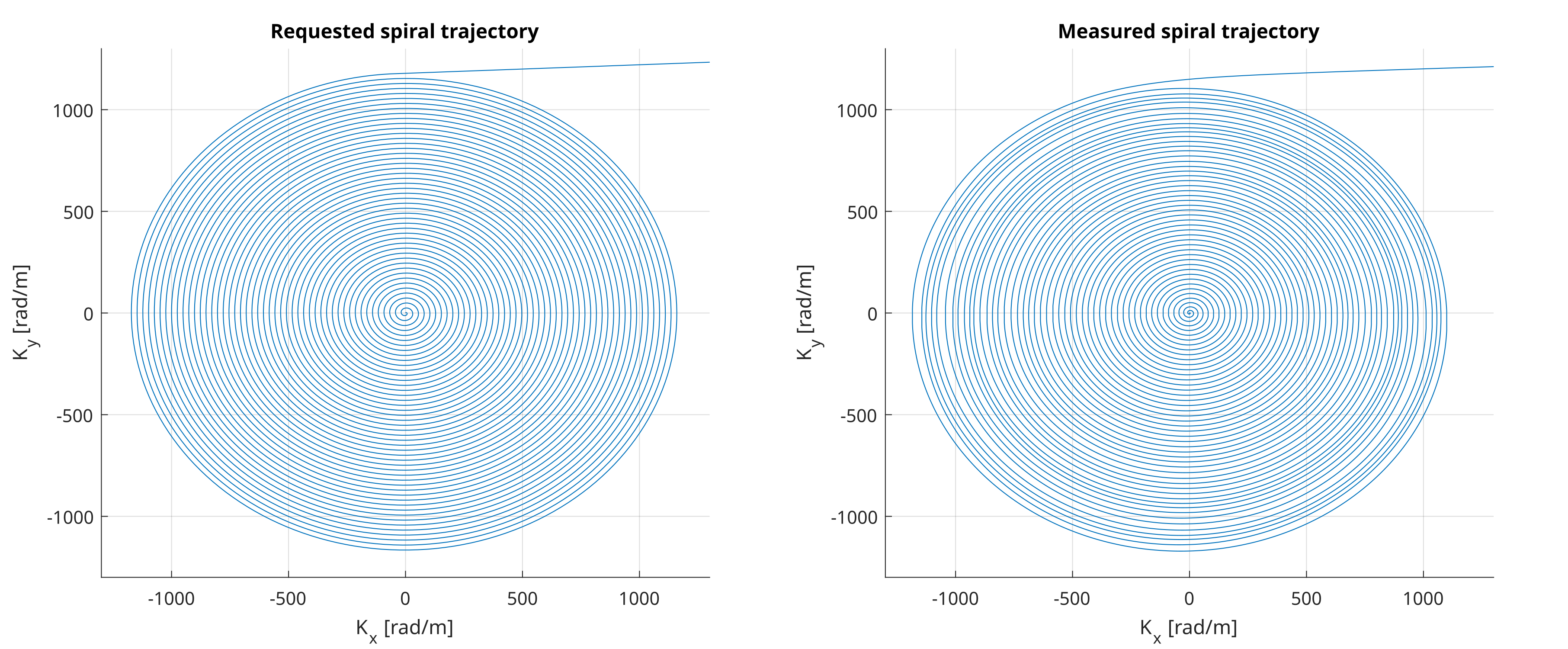
k-space encoding trajectory of a spiral readout, both as requested in the Pulseq
sequence definition (left) and as captured by the field camera (right). This spiral trajectory is part of the spiral example sequence available from the Pulseq GitHub8.