3249
High-Accuracy Ultra-short Inner-Volume Saturation Pulse for 3D Steady-State Imaging1fMRI Laboratory and Applied Physics Program, University of Michigan-Ann Arbor, Ann Arbor, MI, United States, 2fMRI Laboratory and Biomedical Engineering, University of Michigan-Ann Arbor, Ann Arbor, MI, United States
Synopsis
Keywords: Pulse Sequence Design, Software Tools
Motivation: Spatially-tailored excitation is often favorable in MR applications where the region-of-interest only occupies a small portion of the whole FOV.
Goal(s): In this study, we sought to design an inner-volume saturation pulse for steady-state imaging.
Approach: We propose an extension to the AutoDiff tool by Luo et al. Here we use steady-state magnetization error as the design objective instead of one-time excitation error in the original design.
Results: Our proposed pulse design can excite spatially-selective patterns with ultra-short duration(~1.2ms) and high accuracy (error reduced by 50% compared to the original design).
Impact: Our pulse design tool enables highly accurate multi-dimensional spatially-tailored excitations with ultra-short pulses. Such pulses can be plugged into functional MRI sequences, and be played in clinical scenarios where ROI-specific excitations can mitigate motion artifacts and reduce image encoding time.
Introduction
In MRI applications, oftentimes the region-of-interest (ROI) does not occupy the full FOV. For example, in neuroimaging applications cortical regions may be of more importance than the inner-volumetric (IV) regions such as central ventricle. With an ROI-specific excitation, the image quality may be improved for the following reasons: 1) less signal contamination from non-ROI regions, e.g., regions exhibiting motion or physiological noise, 2) higher spatial-temporal resolution, 3) better parallel imaging performance (reduced g-factor).Nevertheless, multi-dimensional spatially selective excitations typically require long pulse durations [1]. Long durations introduce relaxation and off-resonance effects that significantly limit the applications of such pulses. Recently, an RF and gradient joint design tool using AutoDiff was proposed [2], where the design degree-of-freedom is greatly increased and thus much shorter (a few ms) selective pulses are made possible. However, in the original design [2], the pulses are optimized to produce a one-time excitation profile that matches the target while in many cases like steady-state (SS) imaging, the tailored pulses are repeated every TR to achieve better selectivity. To that end, we propose an extension to the original design called steady-state auto-differentiation (SS-AutoDiff) , in which steady-state error is minimized instead. We will present phantom and in-vivo experimental results with an ultrashort (~1.2ms) IV saturation pulse to validate that the proposed design can achieve higher accuracy than the original one.
Method
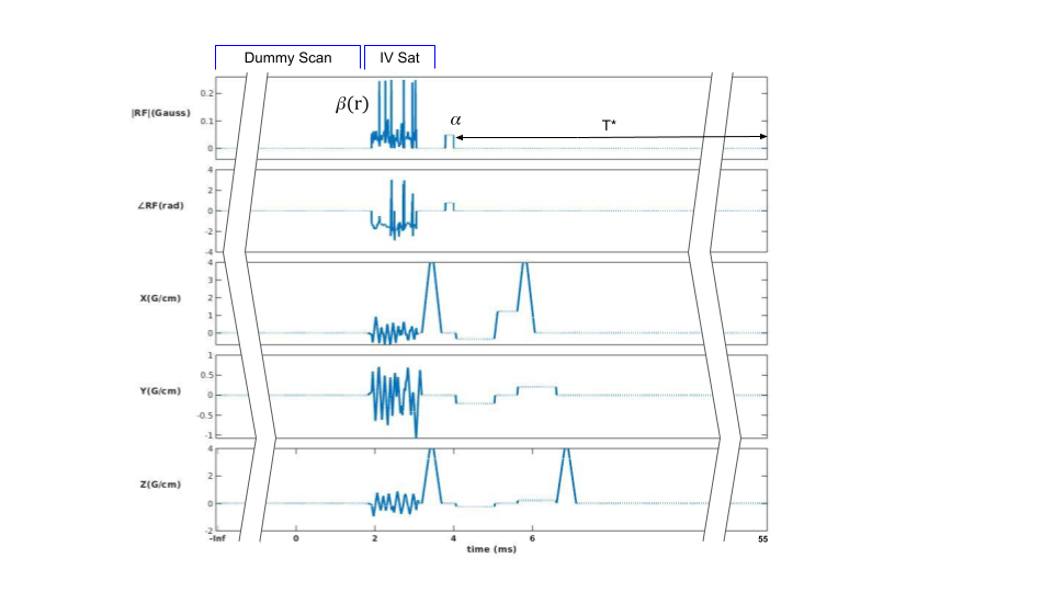
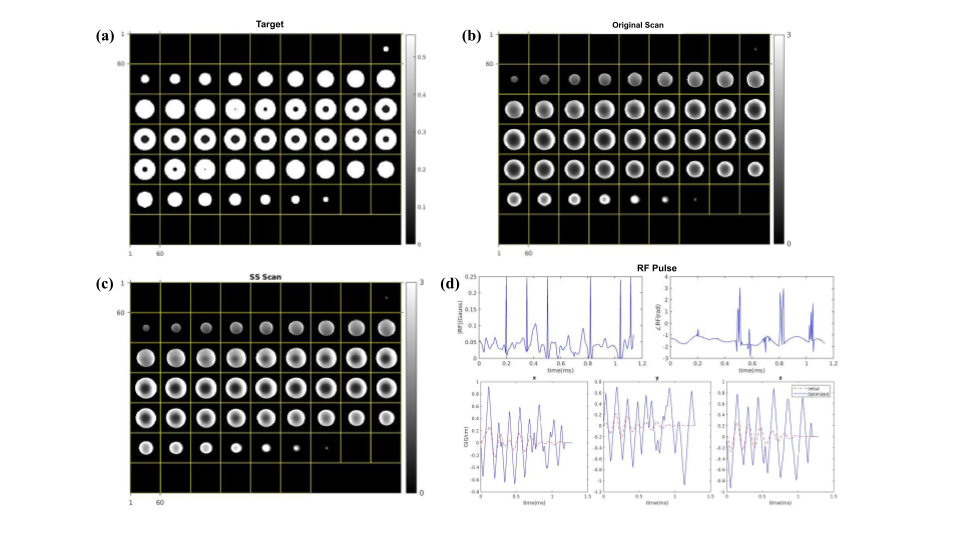
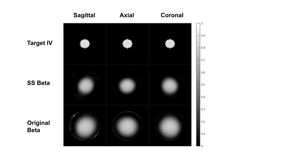
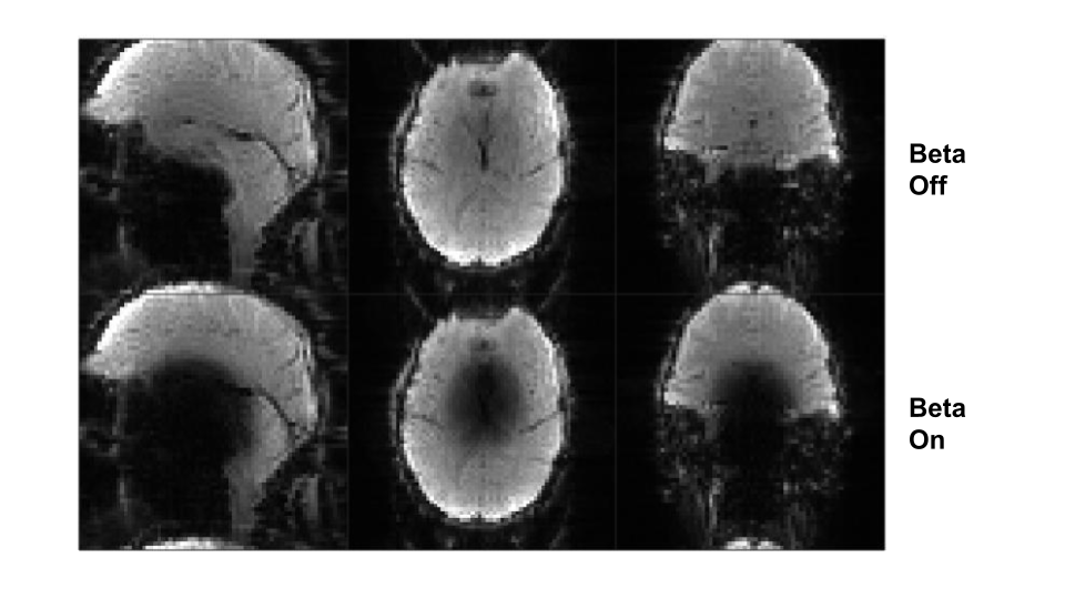
SS_AutoDiff Design: We first derive the expression of steady-state magnetization. The spoiled steady-state $$$M_z$$$ can be expressed as: $$M_z^{SS}(r)=\frac{M_0(1-E_1)}{1-cos\beta(r)cos\alpha E_1}$$ where $$$M_0$$$ is the initial $$$M_z$$$, $$$\beta(r)$$$ and $$$\alpha$$$ are the one-time flip angles of the spatially-tailored pulse and excitation pulse, respectively (Fig.1), and $$$E_1:=exp(-T^*/T_1)$$$. In [2,3], the loss is chosen to be the mean-square-error (MSE) between one-time excitation $$$M_z$$$ (which is $$$M_0cos\beta(r)$$$) and the target $$$M_z$$$. Here, we use an alternative loss to directly match steady-state $$$M_z$$$ with the target: $$L(\beta(r),M_z^d(r))=\frac{1}{N}||M_z^{SS}(r)-M_z^d(r)||_2^2$$ where $$$M_z^d(r)$$$ is the desired pattern. We determine $$$M_z^d(OV)$$$ and $$$M_z^d(IV)$$$ by substituting $$$\beta(OV)=0$$$ and $$$\beta(IV)=60°$$$ into the first equation. We use 1ms SPINS [4] gradients ($$$\alpha=5, \beta=0.5$$$, duration=1ms, $$$u=12\pi, v=2\pi$$$) and a 1ms randomized RF waveform as initializations.Experimental Validation: We first ran a 3D spoiled gradient-echo (SPGR) imaging experiment on a ball phantom (Fig.2, TR=55ms). A spherical IV with 0.3 object radius was placed at the scanner isocenter. 3D spin-warp was used as the readout. We also acquired reference images with beta pulse turned off(Fig.3). We compare the performance of the SS-AutoDiff with the original [2,3] (Fig.2). For the in-vivo experiment, we scanned a volunteer on a GE 3T MR750 scanner with a Nova 32ch head coil under IRB approval. A 3D EPI readout train was used to replace the spin-warp in Fig.1. TR=55ms and resolution was set to be 3mm isotropic (Fig.4).
Results
Figure 2 shows the phantom imaging results and the beta pulse used. Compared to the original design [2,3] (Fig. 2b), the SS-AutoDiff beta pulse has a steady-state $$$M_z$$$ (proportional to the signal strength in Fig.2c) closer to the target (Fig2a). To quantitatively compare their performance and remove unrelated factors such as coil sensitivity gain, we calculate the difference between steady-state signal ($$$M_{xy}$$$) with beta on and off, then normalize the difference with beta-off signal $$$|M_{xy}|$$$. This normalized signal difference is a representation of the beta pulse effect. As Fig.3 shows, the original design has a significantly broader suppression region, resulting in a MSE almost twice as big as that of the SS-AutoDiff design. Fig.4 shows the in-vivo EPI images with and without IV suppression. The IV signal is significantly suppressed while the OV resolution is well preserved compared to the reference (beta off).Discussion
ROI shape and placement: Here we gave a demonstration of isocenter spherical IV suppression. The proposed tool is expected to work equally well for outer-volume (OV) suppression [2]. More customized ROI shape and placement, or even simultaneously exciting multiple regions, should also be possible, although longer pulse duration might be needed.Pulses Initialization: The current choice of pulse initialization is empirical. We have observed that different waveform initializations can lead to significantly different optimization results, which reflects the highly non-convex nature of the design problem. The exploration of better initializations [5] is an open question.
Conclusion
We propose an extension called SS-AutoDiff to the original AutoDiff 3D spatially-tailored pulse design tool. With SS-AutoDiff, we demonstrate that an ultra-short pulse can be inserted into e.g. SPGR or 3D EPI imaging sequences to achieve highly accurate steady-state localized saturation. We believe that these pulses could be useful in a wide range of T1/T2* imaging applications, such as MR corticography and prostate imaging.Acknowledgements
This work was funded by NIH grants R37CA263583 and U24NS120056.References
[1] Stenger VA, Boada FE, Noll DC. Three-dimensional tailored RF pulses for the reduction of susceptibility artifacts in T2*-weighted functional MRI. Magn Reson Med 2000; 44: 525–531.
[2] T. Luo, D. C. Noll, J. A. Fessler and J. -F. Nielsen, "Joint Design of RF and Gradient Waveforms via Auto-differentiation for 3D Tailored Excitation in MRI," in IEEE Transactions on Medical Imaging, vol. 40, no. 12, pp. 3305-3314, Dec. 2021, doi: 10.1109/TMI.2021.3083104.
[3] Nielsen J, Setsompop K. Tailored Multi-Dimensional Partial Saturation Pulses For Inner/Outer-Volume Spoiled Steady-State Imaging. ISMRM, 2023, program number: 2375.
[4] Malik SJ, Keihaninejad S, Hammers A, Hajnal JV. Tailored excitation in 3D with spiral nonselective (SPINS) RF pulses. Magn Reson Med. 2012 May;67(5):1303-15.
[5] Sun H, Fessler JA, Noll D, Nielsen JF. Joint design of continuous excitation k-space trajectory and RF pulse for 3D tailored excitation. Proc Intl Soc Magn Reson Med. 2014:1438.
Figures