3248
Design of a power independent of finite number of slices (PIFINS) pulse for simultaneous multi-slice imaging1Department of Computer Science, Mathematics, Physics and Statistics, University of British Columbia, Kelowna, BC, Canada, 2UBC MRI Research, Department of Radiology, Faculty of Medicine, University of British Columbia, Vancouver, BC, Canada, 3Biomedical Engineering and Imaging Institute, Icahn School of Medicine at Mount Sinai, New York, NY, United States
Synopsis
Keywords: Pulse Sequence Design, RF Pulse Design & Fields
Motivation: Simultaneous mutli-slice (SMS) techniques can reduce scan times. Power deposition from multiband pulses and outer slice signal from power independent of number of slices (PINS) pulses has hampered thoracic and cardiac SMS imaging.
Goal(s): To reduce power deposition in thoracic and cardiac SMS imaging, thereby reducing scan times and motion artifacts.
Approach: A power independent of finite number of slices (PIFINS) pulse was designed to excite any number of slices over a desired field of view with power deposition comparable to single-band pulse.
Results: The PIFINS pulse was used to simultaneously acquire 3 slices in a cylindrical ACR phantom.
Impact: Reducing scan time in thoracic and cardiac imaging reduces motion artifacts. Acceleration with simultaneous multi-slice has been hampered due to high power deposition. The new pulse presented here will enable SMS thoracic and cardiac imaging with reduced power deposition.
Introduction
Simultaneous multi-slice (SMS) techniques have reduced scan times compared to sequential slice acquistions1. Traditional SMS excitation pulses are a superposition of single-band (SB) radiofrequency (RF) pulses to form a multi-band (MB) pulse; however, RF amplifier and power deposition limits are easily exceeded1.SMS excitation with power deposition comparable to a SB pulse can be achieved with the power independent of number of slices (PINS) pulse2. PINS pulses consist of a SB envelope modulated by a comb function producing a series of rectangular subpulses, interleaved with gradient blips2. The resulting slice profiles repeat infinitely with a field of view (FOV) that is determined by the coil size and cannot be reduced. Due to unwanted signal from outer slices, applications of PINS pulses are limited to sagittal orientations in the brain2-4 or refocusing pulses for MB excitations5-7.
By reshaping the subpulses of a PINS pulse with SB subpulses and applying a gradient during the subpulses, the region over which phase-selective excitation is achieved can be windowed, supressing unwanted signal from outer slices. The new pulse – the power independent of finite number of slices (PIFINS) pulse – could excite any number of slices over a defined FOV. In this work, we introduce a PIFINS pulse for flexible SMS imaging with reduced power deposition to ultimately reduce scan times and artifacts for thoracic and cardiac imaging.
Methods
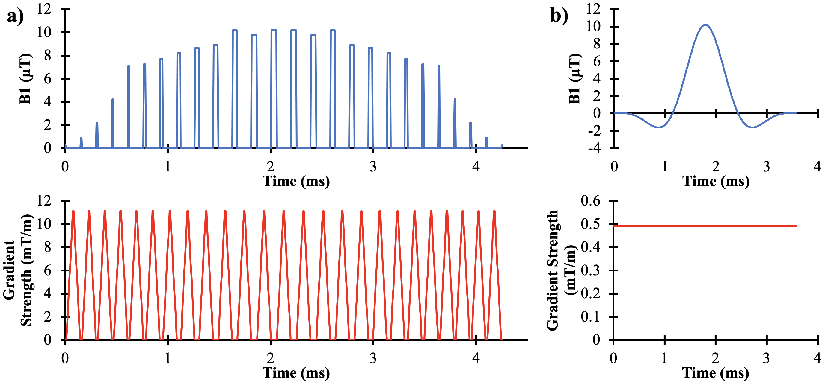
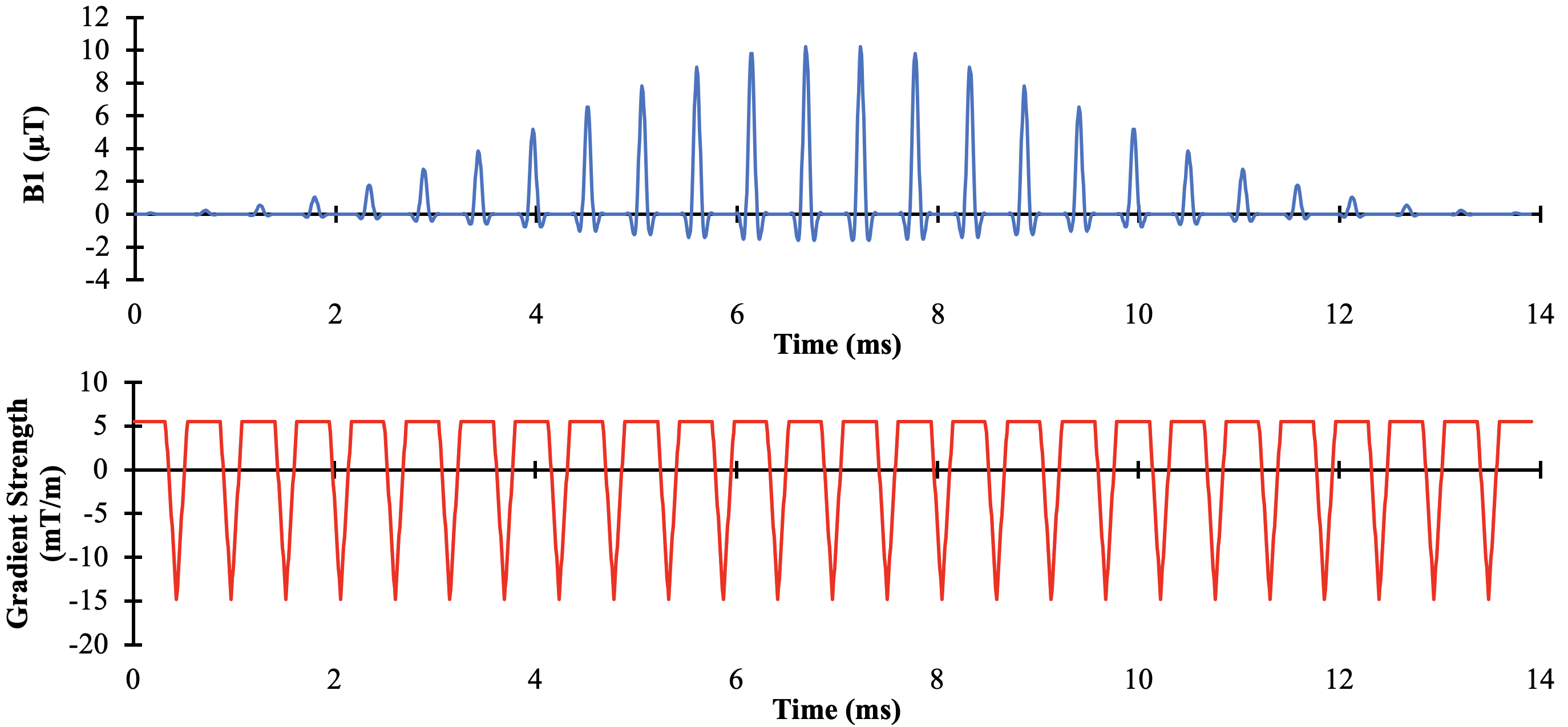
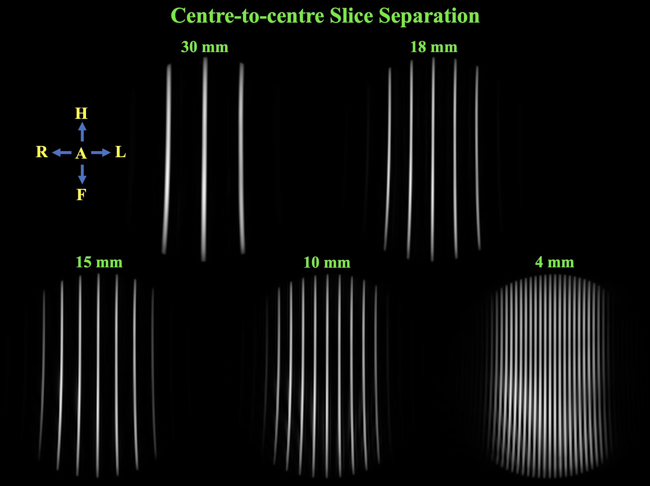
Pulse design and simulation was conducted in MATLAB using a modified Multiband RF toolbox8 with a maximum B1 of 10.2 µT, maximum gradient strength of 45 mT/m, maximum slew rate of 200 mT/m/ms, and dwell time of 6.4 µs. A 90º PINS pulse was designed with a time-bandwidth product (TBW) of 2.6, 3 mm slice thickness, and 30 mm centre-to-centre slice separation. A 90º SB pulse was designed with a TBW of 6.0 and 80 mm slice thickness. The PIFINS pulse was designed by replacing each subpulse with a down-sampled SB pulse (50 dwell point duration). The PIFINS gradient is a constant gradient superimposed with triangular gradient blips to maintain the slice thickness and centre-to-centre slice separation.The PIFINS pulse was integrated into a gradient echo sequence with a cartesian acquisition and TR 250 ms on a 3T Philips Ingenia Elition X. Sample phantom images were acquired: slice profiles from Braino with varying slice separations and three slices in an ACR phantom.
Results
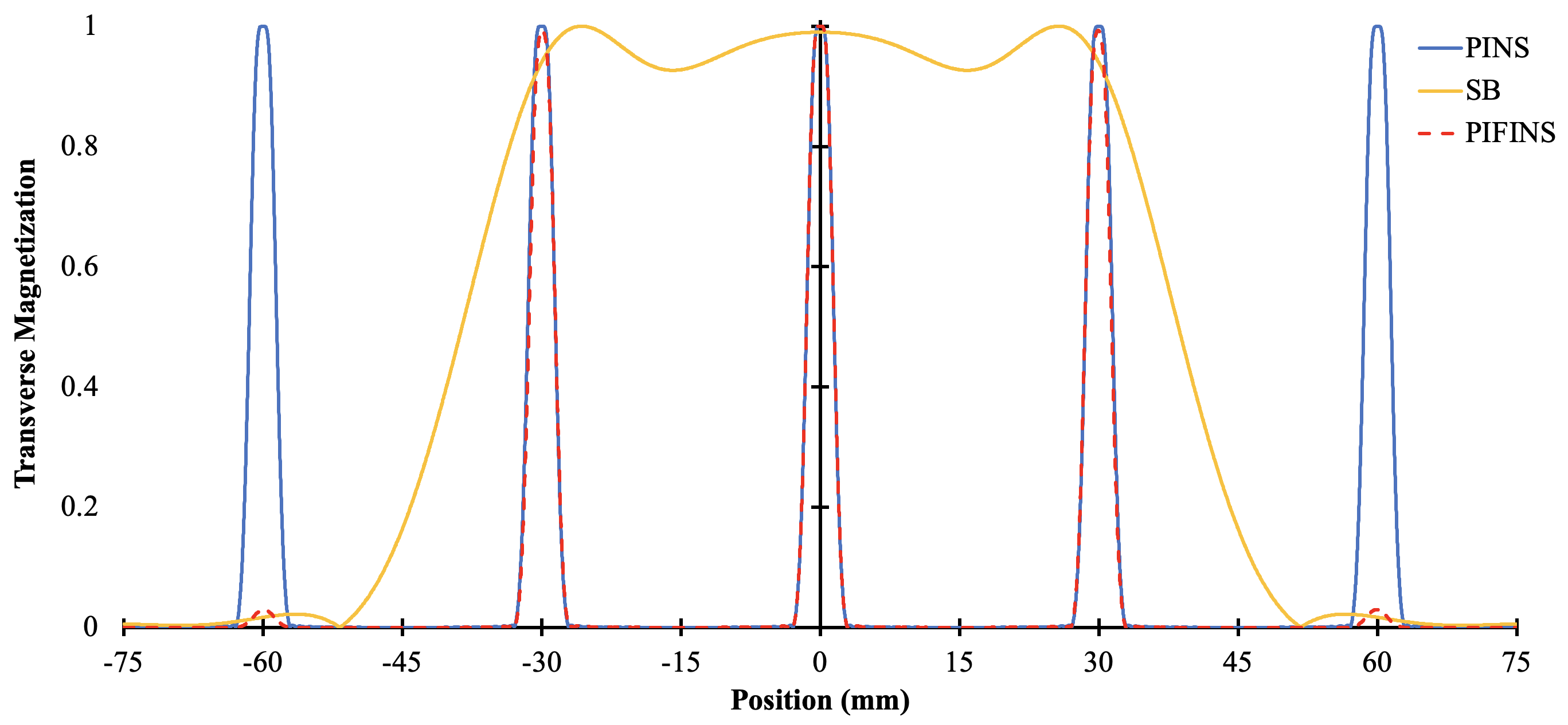
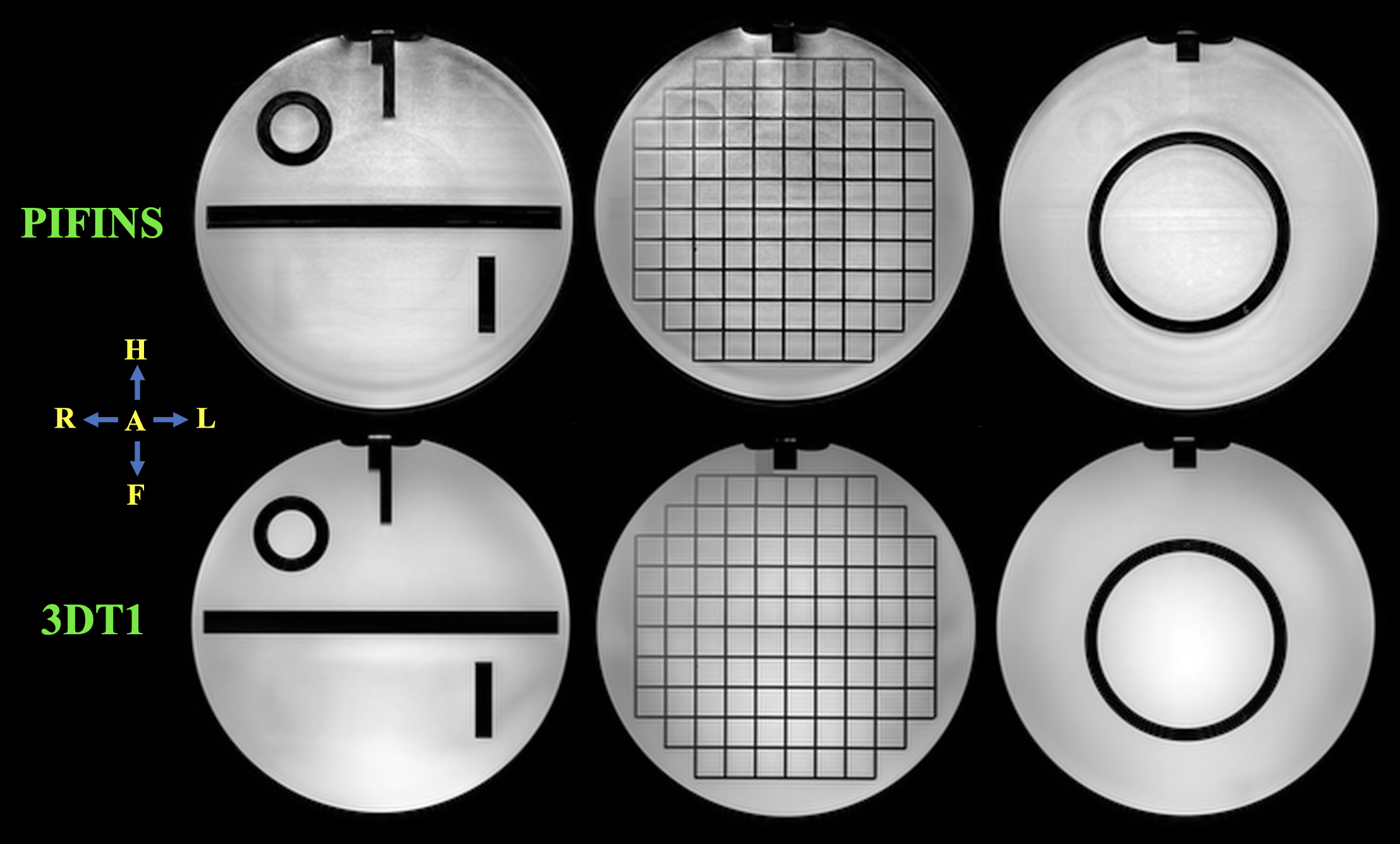
The RF and gradient waveforms of the PINS and SB pulses are shown in Figure 1. The RF and gradient waveforms of the PIFINS pulse are shown in Figure 2. The simulated transverse magnetization profiles of the PINS, SB, and PIFINS pulses are shown in Figure 3. The slice profiles excited in a spherical Braino phantom for centre-to-centre slice separations of 30, 18, 15, 10 and 4 mm are shown in Figure 4. Three slices acquired in an ACR phantom with the PIFINS pulse are shown in Figure 5 along with 3DT1 images.Discussion
The PIFINS pulse was able to excite any number of slices over an 80 mm FOV with power deposition that is independent of the number of excited slices. The PIFINS pulse is more flexible for SMS excitation and may therefore be integrated into gradient echo sequences to reduce power deposition and scan times. Applying the PIFINS pulse in thoracic9 and cardiac10 imaging could enable broader application of SMS techniques, especially given that previous research has demonstrated how accelerated scan times with SMS imaging lead to reduced motion artifacts.The PIFINS pulse has a power deposition that is the same as a SB pulse that excites the same slice profile and an 11.0 times longer duration. Compared to the PINS pulse shown in Figure 1, the PIFINS pulse has a power deposition that is reduced by a factor of 0.81 and a 3.3 times longer duration.
To optimize the window shape for specific applications, the SB subpulse TBW can be increased to increase window sharpness and pulse duration, or to increase window width and power deposition. To optimize the profiles of the excited slices, the PINS pulse TBW can be increased to increase the slice sharpness and number of subpulses, or to increase slice thickness and power deposition. Future work will focus on broadening the range of applications of the PIFINS pulse by optimizing the trade-offs to reduce duration and power deposition.
Conclusion
Flexible SMS thoracic or cardiac imaging could be enabled by the new PIFINS pulse presented here. The PIFINS pulse excites a any number of slices over the defined FOV, with a power deposition that is independent of the number of slices.Acknowledgements
We acknowledge the support of the Natural Sciences and Engineering Research Council of Canada, Canadian Foundation for Innovation, University of British Columbia, and Walter C. Sumner Memorial Foundation. Special thanks to the Technologists at UBC MRI Research. Rebecca Feldman and Erin MacMillan contributed equally to supervising this research.References
- Barth M, Breuer F, Koopmans PJ, et al. Simultaneous mutlislice (SMS) imaging techniques. Magn Reson Med. 2015;75(1):63-81.
- Norris DG, Koopmans PJ, Boyacioglu R, et al. Power independent of number of slices (PINS) radiofrequency pulses for low-power simultaneous multislice excitation. Magn Reson Med. 2011;66(5):1234-1240.
- Koopmans PJ, Boyacioglu R, Barth M, et al. Whole brain, high resolution spin-echo resting state fMRI using PINS multiplexing at 7T. Nueroimage. 2012;62(3):1939-1946.
- Feldman RE, Islam HM, Ku J, et al. A SEmi-Adiabatic matched-phase spin echo (SEAMS) PINS pulse-pair for B1-insensitive simultaneous multislice imaging. Magn Reson Med. 2016;75(2):709-717.
- Eichner C, Setsompop K, Koopmans PJ, et al. Slice accelerated diffusion-weighted imaging at ultrahigh field strength. Magn Reson Med. 2014;71(4):1518-1525.
- Schulz J, Marques JP, Ter Telgte A et al. Clinical application of Half Fourier Acquisition Single Shot Turbo Spin Echo (HASTE) imaging accelerated by simultaneous multi-slice acquisition. Eur J Radiol. 2018;98:200-206.
- Hilbert T, Schulz J, Marques JP, et al. Fast model-based T2 mapping using SAR-reduced simultaneous multislice excitation Magn Reson Med. 2019;82(6):2090-2103.
- Abo Seada S, Price AN, Schneider T, et al. Multiband RF pulse design for realistic gradient performance. Magn Reson Med. 2019;81(1):367-376.
- Riffle P, Attenberger UI, Kannengiesser S, et al. Highly accelerated T1-weighted abdominal imaging using 2-dimensional controlled aliasing in parallel imaging results in higher acceleration: a comparison with generalized autocalibrating partially parallel acquisitions parallel imaging. Invest Radiol. 2013;48(7):554-561.
- Lau AZ, Tunnicliffe EM, Frost R, et al. Accelerated human cardiac diffusion tensor imaging using simultaneous multislice imaging. Magn Reson Med. 2014;73(3):995-1004.
Figures