3242
A novel sequence for Simultaneous imaging of cerebral Arteries and VEins (SAVE) based on velocity-selective arterial spin labeling1Institute of Science and Technology for Brain-inspired Intelligence, Fudan University, Shanghai, China, 2MR Research Collaboration Team, Siemens Healthineers Ltd., Shanghai, China, 3The Department of Neurosurgery, The Affiliated Changzhou Second People's Hospital of Nanjing Medical University, Changzhou Medical Center, Nanjing Medical University, Changzhou, China, 4Key Laboratory of Computational Neuroscience and Brain-Inspired Intelligence (Fudan University), Ministry of Education, Shanghai, China, 5Human Phenome Institute, Fudan University, Shanghai, China
Synopsis
Keywords: Data Acquisition, Vessels
Motivation: The conventional velocity-selective ASL-based (VSASL) MRA couldn’t separate arteries and veins unless additional module is used, which would cause long scan time. In addition, its acquisition efficiency is low since a long waiting time for blood refreshing.
Goal(s): To proposed a VSASL-based sequence for Simultaneous imaging of cerebral Arteries and VEins (SAVE).
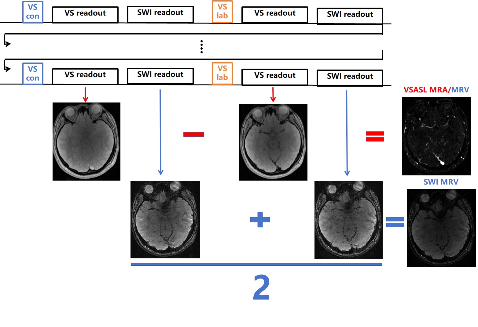
Approach: SAVE uses the waiting period between consecutive shots of VSASL MRA to acquire SWI data. VSASL MRA/MRV were obtained by subtracting Control images from Label images, while SWI images were averaged to increase SNR.
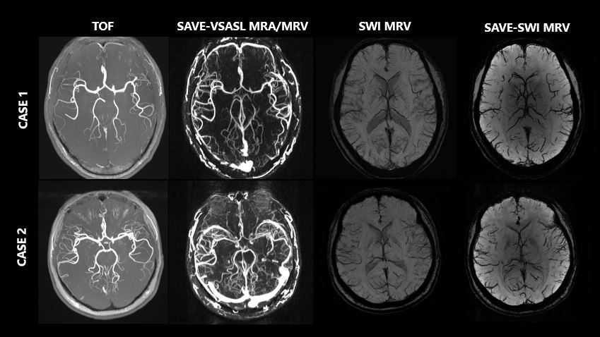
Results: SAVE-VSASL MRA/MRV can well depict arteries and veins, and SAVE-SWI MRV could depict veins well.
Impact: The novel sequence can obtain decent VSASL MRA/MRV and SWI MRV images. The combination of the VSASL MRA/MRV and SWI MRV in SAVE should allows separation of arteries and veins by post-processing, that may be useful in clinical settings.
Introduction
Imaging of cerebral arteries and veins are important for diagnosis and treatment of cerebrovascular diseases. Many methods exist for imaging either arteries or veins, such as time-of-flight (TOF) MRA, phase contrast (PC) MRA and susceptibility weighted imaging (SWI). Simultaneous imaging of cerebral arteries and veins in a single scan offers several potential advantages, such as increased acquisition efficiency and inherent co-registration of the images, which would enable more indepth disease diagnosis and easier post-processing1. Compared with the TOF sequence, velocity selective arterial spin labeling-based (VSASL) angiogram is capable of visualizing more distal branches of cerebral vessels2. However, the technique could not separate the arteries and veins unless additional module is used to suppress signal of either type of vessel, which would cause a long scan time. In addition, its acquisition efficiency is low since a long waiting time is used for blood refreshing. In this study, we proposed a VSASL-based sequence for Simultaneous imaging of cerebral Arteries and VEins (SAVE).Methods
Sequence design: As shown in Figure 1, SAVE sequence is based on the VSASL angiogram sequence, which is made up of VS labeling modules and multi-shot high resolution turbo gradient echo readouts with a centric-out trajectory. SAVE uses the waiting period between the consecutive shots, which is originally for blood refresing, to acquire SWI data. At the moment, we used the same imaging resolutions and k-space sampling trajectory for the VS data and SWI data in SAVE, and two SWI datasets were acquired, one following the VS Control readout, the other following the VS Label readout. Eight segment B1-insensitive rotation pulse (BIR-8) was used for the VS module.In vivo experiment: Ethical approval was obtained for the study. Two healthy volunteers were recruited. The experiments were performed on a Siemens Prisma 3T MRI scanner with a 32-channel head coil. SAVE imaging parameters were: FOV = 200×200×72 mm3; TRVS/TEVS = 7.62/4.34 ms; TRSWI/TESWI = 32.62/29.34 ms; FA = 8°; voxel size = 0.65×0.68×1.0 mm3; lines per shot = 100; VS cutoff velocity = 3.11 cm/s; total scan time = 14 min 26 s. SWI imaging parameters were: FOV = 200×200×72 mm3; TR/TE = 28/20 ms; FA = 15°; voxel size = 0.52×0.52×1.2 mm3; total scan time = 5 min 34 s. TOF imaging parameters were: FOV = 200×181×100 mm3; TR/TE = 21/3.42 ms; FA = 18°; voxel size = 0.78×0.82×0.6 mm3; total scan time = 4 min 54 s.
Image analysis: As shown in Figure 1, VSASL MRA/MRV were obtained by subtracting Control images from Label images, while the two SWI images were averaged to increase SNR followed by a conventional SWI image post-processing. The ability of SAVE to visualize arteries and veins was assessed by comparison with conventional TOF and SWI.
Results
As shown in Figure 2, SAVE-VSASL MRA/MRV can well depict the cerebral vessels, including both arteries and veins, and more distal vessels are visible compared to TOF MRA. In addition, decent SAVE-SWI images were obtained. The SAVE-SWI MRV could depict the veins well.Discussion and Conclusion
Both decent VSASL MRA/MRV and SWI MRV images were obtained from SAVE. This indicates the great potential of the SAVE sequence. The combination of the VSASL MRA/MRV and SWI MRV in SAVE should allows separation of the arteries and veins, either visually or by using some post-processing methods. It should be also noted that there is still large room of optimization for the SAVE sequence. For example, centric-in trajectory can be used for the SWI readout to reduce the potential influence of the VS preparation on SWI vascular signals. The current scan time is unnecessarily long because the current implementation of SAVE overestimates the SAR. In the near future, we will design studies to perform a more rigorous, more comprehensive and fairer comparison between SAVE sequence and the TOF sequence, SWI sequence as well as the conventional VSASL angiogram sequence.Acknowledgements
This work was supported by Natural Science Foundation of Shanghai (22ZR1403900) and the Clinical Project of Changzhou Medical Center (CMCC202210).References
1. Chen Y, et al. An interleaved sequence for simultaneous magnetic resonance angiography (MRA), susceptibility weighted imaging (SWI) and quantitative susceptibility mapping (QSM). Magn Reson Imaging. 2018 Apr;47:1-6.
2. Qin Q, et al. Velocity-selective magnetization-prepared non-contrast-enhanced cerebral MR angiography at 3 Tesla: Improved immunity to B0/B1 inhomogeneity. Magn Reson Med. 2016 Mar;75(3):1232-41.
Figures