3238
Spiral Time Resolved Imaging (SPTI)1Radiology & Biomedical Imaging, University of California San Francisco, San Francisco, CA, United States
Synopsis
Keywords: Data Acquisition, Quantitative Imaging
Motivation: Spiral is the most efficient k-space coverage strategies in current MR gradient systems with robustness to motion and flow, but the implementation of long spiral trajectories keeps challenging due to accumulated off resonance effect and local susceptibility.
Goal(s): SPTI is proposed for artifact- and distortion-free spiral imaging and quantification.
Approach: Modified segmented spiral readout and subspace reconstruction were performed in SPTI technique.
Results: SPTI sequence was validated in a phantom study and feasibility was evaluated on a brain volunteer. In human study, SPTI method showed consistent results with MRF-EPTI on quantitative mapping, including PD, T1, T2 and T2*.
Impact: SPTI method can simultaneously quantify PD, T1, T2 and T2* mapping and is potential to expand to other body regions, especially in the liver and cardiac MRI.
Introduction
Spiral is one of the most efficient k-space coverage strategies in current MR gradient systems with robustness to motion and flow. Therefore, spiral readout was widely used in ultra-fast quantitative MR techniques such as magnetic resonance fingerprinting1(MRF) and quantitative transient-state imaging2. However, the implementation of long spiral trajectories remains challenging due to accumulated off resonance effect and local susceptibility. In Cartesian imaging, echo planar time-resolved imaging3 (EPTI) was recently developed to eliminate these effects to acquire artifact-free images and quantifications. In addition, our previous study has proposed an MRF-EPTI technique4 to simultaneously quantify multiple parameters efficiently. In this study, a spiral time-resolved imaging (SPTI) technique is proposed for artifact- and distortion-free spiral imaging with long spiral trajectories, and enables simultaneous quantification of T1, T2 and T2* relaxations with motion and flow insensitivity.Methods
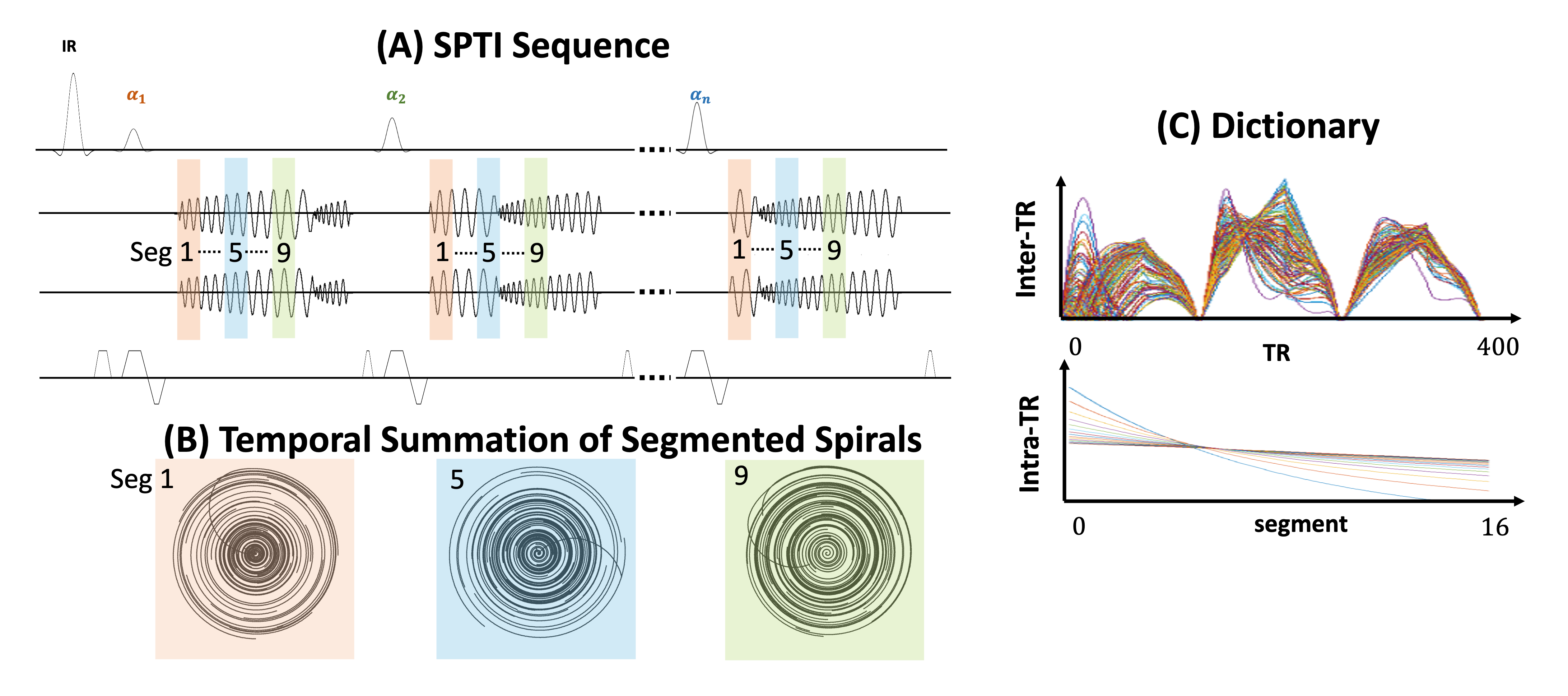
The SPTI pulse sequence is based on an inversion recovery unbalanced steady state free precession (SSFP) sequence with a modified spiral readout as shown in Figure 1. The trajectory is a spiral-out-in periodic trajectory, and in each TR, a random period of this trajectory was selected as readout. The readout can be divided into numbers of segments, and each segment across different TRs is constructed as a segmented spiral5,6 sampling pattern. The acquired temporal signal can be represented by two parts: Intra-TR signals and Inter-TR signals due to the fact that Intra-TR T2* decay is independent to the global signal change across TRs, which enables the simultaneous quantification of T2 and T2*. Two dictionaries were accordingly built with extended phase graph (EPG) for both subspace reconstruction and parametric matching. The time-resolved subspace reconstruction was performed with:$$\mathop{\text{argmin}}_{\alpha} \frac{1}{2}||U_{1,k_1}FS\Phi U_{2,k_2}\alpha - y||_2^2 + \lambda\sum_r||R_r(\alpha)||_*$$where $$$y$$$ is the kspace data, $$$\alpha$$$ is the subspace images, $$$ \Phi $$$ is the off resonance and eddy current induced phase calculated from each segment echo, and $$$\lambda\sum_r||R_r(\alpha)||_* $$$ is the locally low rank regularization term. $$$U_1$$$ and $$$U_2$$$ are subspace transform operators for Inter-TR and Intra-TR signals, respectively. After subspace reconstruction, T1, T2 and proton density (PD) are then obtained by Inter-TR dictionary matching, and T2* is obtained by Intra-TR dictionary matching.All scans were performed on a GE 3T scanner (MR750, Waukesha, WI) with a 32-channel head coil. The spectral-spatial selective (SPSP) pulse was applied to saturate the fat signal (duration = 8.7 ms, passband = 317 Hz). Other parameters included: TR = 40 ms, FOV = 224x224 mm2, resolution = 1x1 mm2, number of segments = 16, and echo spacing = 1.8 ms. The scan time is 16 s/slice.
Results and Discussion
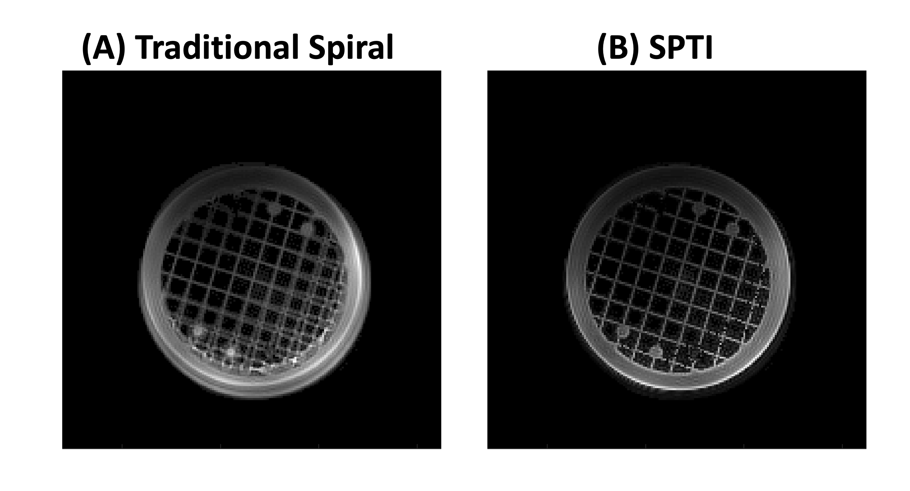
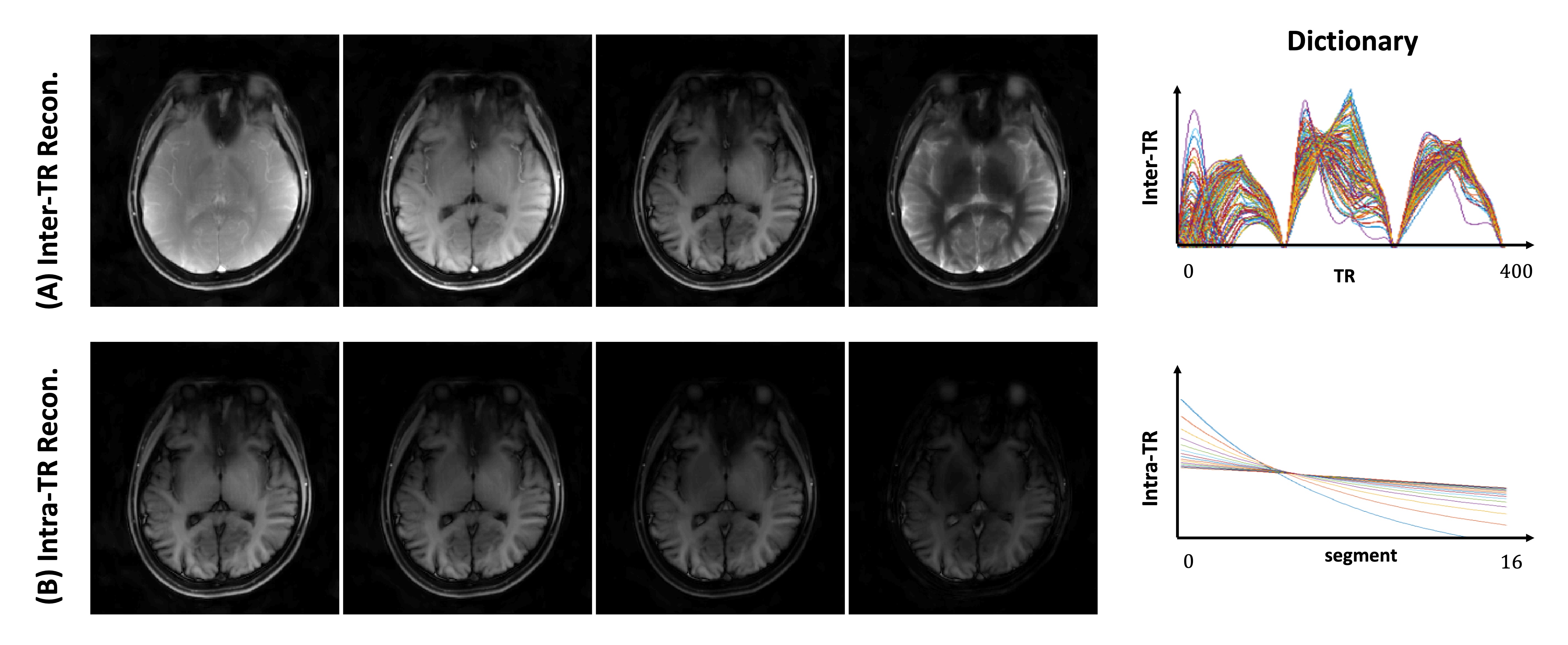
The imaging performance of SPTI sequence was validated by a phantom study, shown in Figure 2. In the traditional spiral imaging with long trajectories, there were significant blurring artifacts due to the long readout time. In SPTI, the blurring artifacts could be almost removed.Figure 3 shows the Inter-TR and Intra-TR reconstructed images and the Inter-TR and Intra-TR dictionaries, respectively. The different T1/T2 weighted images were reconstructed in the Inter-TR reconstruction, and multi-echo images were reconstructed in the Intra-TR reconstruction. The subspace Inter-TR and Intra-TR reconstructed images were fitted by the Inter-TR and Intra-TR dictionaries to estimate the following quantitative parameters.
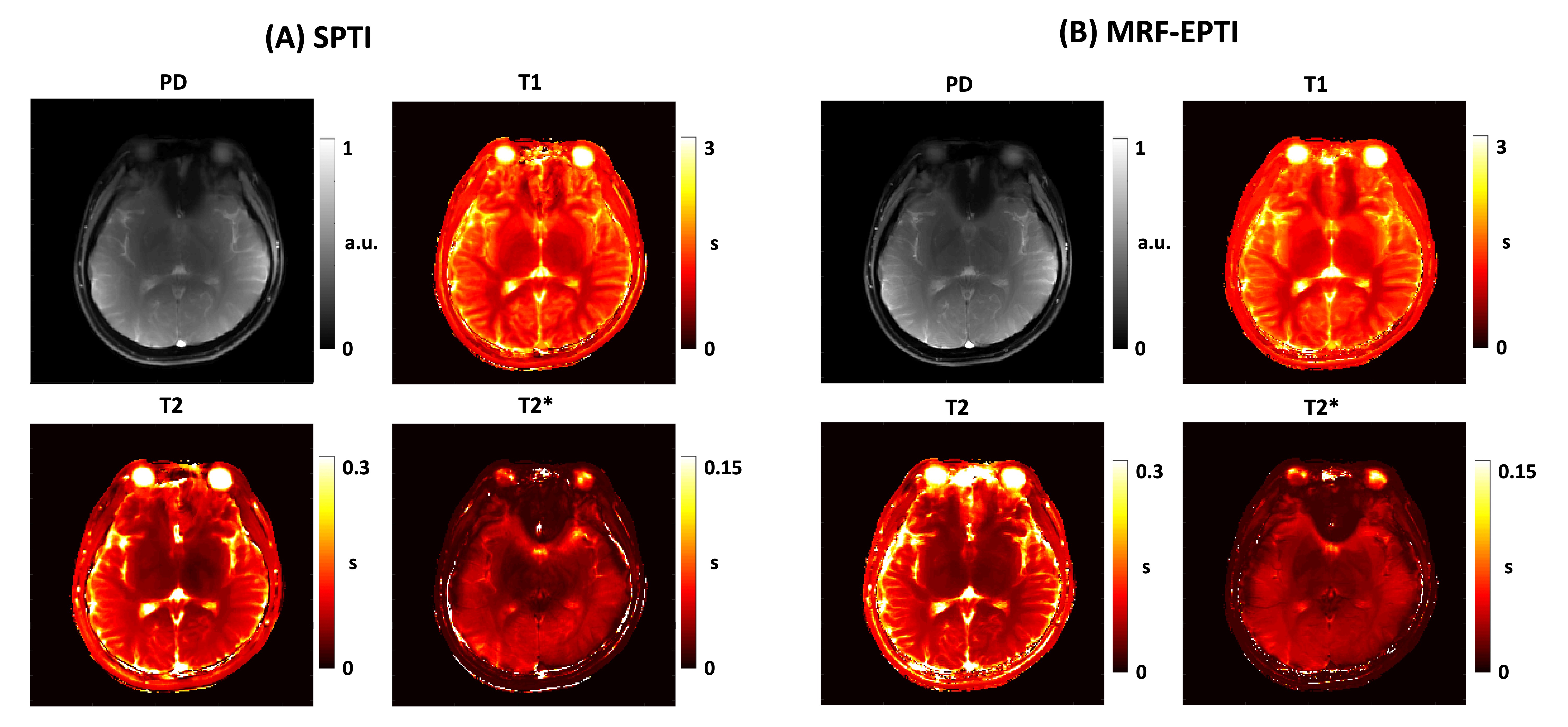
Figure 4 shows the capability for simultaneous multiparametric quantification using SPTI. T1, T2, T2* and proton density (PD) maps are shown with comparison to MRF-EPTI. Due to the long echo time caused by the SPSP pulse, the front lobe of the brain showed lower proton density in both SPTI acquisition and MRF-EPTI acquisition. Compared to MRF-EPTI results, SPTI showed similar fitted quantitative values.
Conclusion
In this study, we presented an efficient and high-quality SPTI technique for spiral imaging and parameter quantifications without off resonance and susceptibility induced artifacts. We validated the SPTI sequence in a phantom study and evaluated the feasibility of SPTI method on a healthy brain volunteer. SPTI method showed consistent results with MRF-EPTI on quantitative mapping, including PD, T1, T2 and T2*. Due to the motion and flow insensitivity of spiral trajectory, our SPTI method has the potential to expand to other body regions, especially in the liver and cardiac MRI.Acknowledgements
This work was supported by UCSF Department of Radiology & Biomedical Imaging Seed Grant #24-01.References
1. Ma D, Gulani V, Seiberlich N, et al. Magnetic resonance fingerprinting. Nature. 2013;495(7440):187-192. doi:10.1038/nature11971
2. Gómez PA, Molina-Romero M, Buonincontri G, Menzel MI, Menze BH. Designing contrasts for rapid, simultaneous parameter quantification and flow visualization with quantitative transient-state imaging. Scientific reports. 2019 Jun 11;9(1):8468.
3. Wang F, Dong Z, Reese TG, et al. Echo planar time-resolved imaging (EPTI). Magn Reson Med. 2019;81(6):3599-3615. doi:10.1002/mrm.27673
4. Cui D, Liu X, Larson P, Xu D. Simultaneous T1 T2 T2* quantification using 2D EPI-MRF by shuffled sampling and compressed time-resolved reconstruction with B0 B1+ correction. In: International Society for Magnetic Resonance in Medicine. ; 2023:2190.
5. Hennig J, Barghoorn A, Zhang S, Zaitsev M. Single shot spiral TSE with annulated segmentation. Magnetic Resonance in Medicine. 2022 Aug;88(2):651-62.
6. Hennig J. Multiecho imaging sequences with low refocusing Flip angles. J Magn Reson. 1988;78:397-407.
Figures