3231
Analysis of Temporal Lobe White Matter Volume in Patients With Hippocampal Sclerosis Based on Automatic Segmentation And its Application Value.1Clinical Medicine School of Ningxia Medical University, Yinchuan, China, 2Radiology, General Hospital of Ningxia Medical University, Yinchuan, China, 3GE HealthCare MR Research, Beijing, China
Synopsis
Keywords: Epilepsy, Segmentation, Automatic Temporal Subregion Segmentation
Motivation: Accurately identifying the range of white matter (WM) involved in epileptogenic lesions of temporal lobe epilepsy with hippocampal sclerosis (TLE-HS) is crucial prior to surgery. However, previous studies have been limited in their analysis of temporal lobe subregions due to technical constraints.
Goal(s): Determine the extent of WM damage in TLE temporal lobe subregions to provide more imaging basis for preoperative evaluation and clinical surgical method selection.
Approach: FreeSurfer software was used to perform whole-brain WM segmentation on 3D T1WI images.
Results: There are differences in the extent of WM damage in temporal lobe subregions between with left TLE-HS and right TLE-HS.
Impact: Automatic brain segmentation technology can be utilized to assess the degree of white matter damage in the subregions of the temporal lobe in patients with TLE-HS. Furthermore, this technology holds potential for investigating various other brain disorders.
Introduction
Temporal lobe epilepsy (TLE) is one of the most common drug-resistant epilepsy in adults [1], hippocampal sclerosis (HS) is considered to be the main pathological type and etiology of TLE [2-3]. Surgery can significantly reduce the frequency of epilepsy and alleviate or even stop epileptic seizures in patients with drug-refractory epilepsy [4-5]. Therefore, it is crucial to preoperatively identify the epileptogenic foci and understand their extent in order to select appropriate surgical methods and procedures, as the final outcome is of utmost importance [6]. In this study, we utilized Automatic brain segmentation technology (FreeSurfer) to analyze the WM volume of each subregion of the temporal lobe in patients diagnosed with temporal lobe epilepsy with hippocampal sclerosis (TLE-HS). By precisely identifying the epileptogenic focus and its associated regions, this technology offers valuable preoperative information for TLE. Additionally, it provides imaging evidence that aids in the evaluation and selection of surgical interventions.Methods
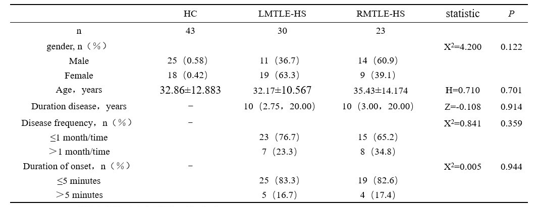
ParticipantsThis study was approved by the Ethics Review Committee of our institution. The case group consisted of 53 patients diagnosed with unilateral mesial TLE, meanwhile 43 healthy individuals were recruited as the control group (HC), There were 30 cases of left TLE-HS (LTLE-HS) and 23 cases of right TLE-HS (RTLE-HS). There were no statistically significant differences in age and gender between the three groups, The detailed inclusion and exclusion criteria were shown in Table 1.
Data acquisition
All participants were scanned after obtaining written informed consent. All MR examinations were performed on a 3.0T MR scanner (SIGNATM Architect, GE Healthcare, Milwaukee WI, USA) equipped with a 48-channel head coil. The main scan parameters were listed in Table 2.
Image post-processing
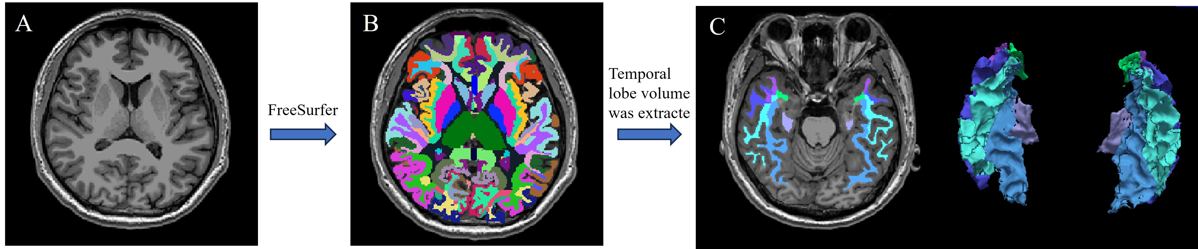
Convert 3D T1WI images to.nii/.nii.gz format, FreeSurfer(V7.3.2), developed by MIT Health Sciences & Technology and Massachusetts General Hospital, was used. http://surfer.nmr.mgh.harvard.edu/) software for whole brain segmentation [7-8], from the volume value each subregion extract temporal lobe white matter volume (figure 1).
Statistical analysis
Statistical analysis was conducted using SPSS 26.0 software. The Kruskal-Wallis test and χ2 test were employed to analyze age and gender differences among the control group, LTLE-HS group, and RTLE-HS group. The two-sample Mann-Whitney U test and χ2 test were used to compare the LTLE-HS group and RTLE-HS group in terms of course of disease, frequency of onset, and duration of onset. For normally distributed data, the independent samples t-test was used, while the two-sample Mann-Whitney U test was used for non-normally distributed data, to compare the white matter in the temporal region between the control group, LTLE-HS group, and RTLE-HS group on the affected side and contralateral side. The difference in volume was found to be statistically significant at P<0.05.
Results
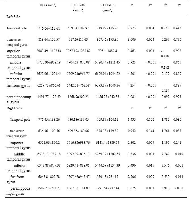
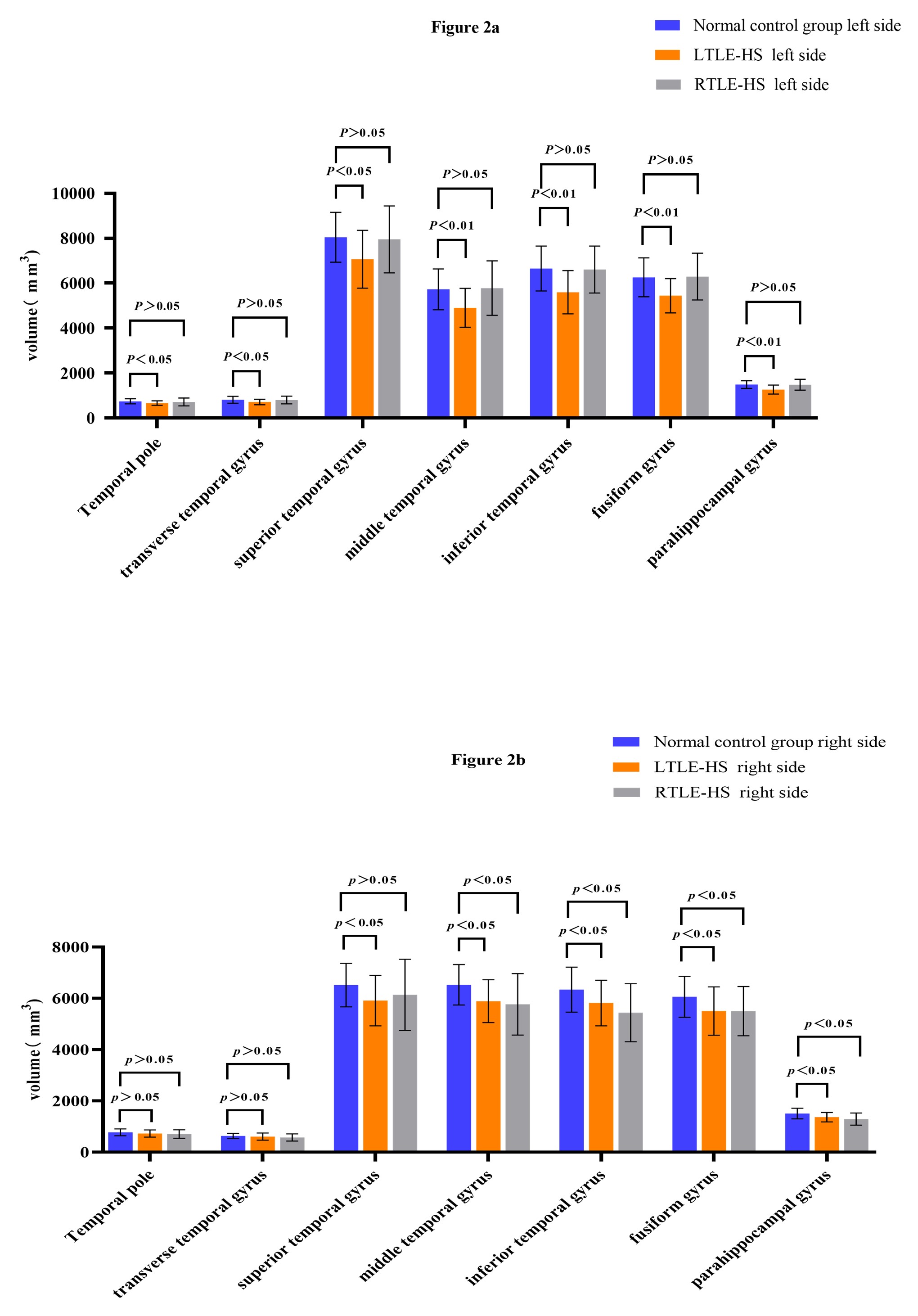
Statistically significant differences were observed in white matter volume in all subregions of the temporal lobe between the left side of the control group and the affected side of the LTLE-HS group. Similarly, significant differences were found in the right side of the control group and the contralateral superior temporal gyrus, middle temporal gyrus, inferior temporal gyrus, fusiform gyrus and parahippocampal gyrus of the LTLE-HS group. Additionally, the white matter volume of the superior temporal gyrus, middle temporal gyrus, inferior temporal gyrus, fusiform gyrus group, no and parahippocampal gyrus showed significant differences between the right side of the control group and the affected side of the RTLE-HS group. However, no statistically significant difference was found in white matter volume between the left side of the control group and the contralateral temporal region of the RTLE-HS group (Figure 2, Table 3).Discussion
The volume of WM in each subregion of the bilateral temporal lobes decreased to varying degrees in both the LTLE-HS group and the RTLE-HS group. LTLE-HS had a more widespread impact on the WM of both sides of the brain, The effect of RTLE-HS on WM was mostly confined to the affected side, and the effect on the opposite side was small.Conclusion
Conclusion This study suggests that measuring the WM volume of temporal lobe subregions using automatic brain segmentation technology is important in accurately locating the epileptogenic focus and its affected area before surgery for patients with focal mesial temporal lobe epilepsy. This measurement can also provide guidance for selecting the appropriate clinical surgical methods based on imaging findings.Acknowledgements
No acknowledgement found.References
1. Tai XY, Bernhardt B, Thom M, et al. Review: Neurodegenerative processes in temporal lobe epilepsy with hippocampal sclerosis: Clinical, pathological and neuroimaging evidence[J]. Neuropathol Appl Neurobiol. 2018;44(1):70-90.
2. Zaidan BC, Cardoso ICDS, de Campos BM, et al. Histopathological Correlations of Qualitative and Quantitative Temporopolar MRI Analyses in Patients With Hippocampal Sclerosis[J]. Front Neurol. 2021;12:801195. Published 2021 Dec 24.
3. Blumcke I, Spreafico R, Haaker G, et al. Histopathological Findings in Brain Tissue Obtained during Epilepsy Surgery[J]. N Engl J Med. 2017;377(17):1648-1656.
4. ayalakshmi S, Vasireddy S, Sireesha J, et al. Long-Term Seizure Freedom, Resolution of Epilepsy and Perceived Life Changes in Drug Resistant Temporal Lobe Epilepsy With Hippocampal Sclerosis: Comparison of Surgical Versus Medical Management.[J]. Neurosurgery. 2023;92(6):1249-1258.
5. Meng Q, Liu Y, Ren Y, et al. Multivariate analysis of seizure outcomes after resective surgery for focal epilepsy: a single-center study on 833 patients[J/OL]. Neurosurg Rev. 2023;46(1):89. Published 2023 Apr 18.
6.Johnson GW, Cai LY, Narasimhan S, et al. Temporal lobe epilepsy lateralisation and surgical outcome prediction using diffusion imaging[J]. Neurol Neurosurg Psychiatry. 2022;93(6):599-608.
7. Velasco-Annis C, Akhondi-Asl A, Stamm A, Warfield SK. Reproducibility of Brain MRI Segmentation Algorithms: Empirical Comparison of Local MAP PSTAPLE, FreeSurfer, and FSL-FIRST[J]. Neuroimaging. 2018;28(2):162-172.
8. Benjamin H, Napolon P, Esmaeil D, et al. Inter- and intra-operator variations in manual segmentation of hippocampus from MRI[J]. Informatics in Medicine Unlocked,2023,39.
Figures