3229
Machine Learning-Based Lateralization of Mesial Temporal Lobe Epilpepsy Using ASL MRI1Department of Biomedical Engineering and Medical Physics, Shahid Beheshti University of Medical Sciences, Tehran, Iran (Islamic Republic of), 2Research Center for Molecular and Cellular Imaging, Tehran University of Medical Sciences, Tehran, Iran (Islamic Republic of), 3Department of Biophysics, Faculty of Biological Sciences,, Tarbiat Modares University, Tehran, Iran (Islamic Republic of)
Synopsis
Keywords: Epilepsy, Perfusion, ASL, CBF . mTLE, Machin learning
Motivation: Utilizing perfusion features to differentiate left and right mesial temporal lobe epilepsy (mTLE) using machine learning.
Goal(s): The study aims to assess ASL MRI's perfusion analysis ability to identify abnormalities in brain regions for distinguishing between mTLE cases and normal cohorts.
Approach: Cerebral blood flow obtained features used by different machine learning classifiers to separate right and left mTLE form control cohort.
Results: The utilization of CBF features proved valuable and effective in the machine learning-based classification of right and left mTLE data from the control cohort.
Impact: This study's outcomes benefit medical professionals and drug-resistant mTLE patients by expediting surgical assessments and enhancing treatment outcomes through improved lateralization and epilepsy classification.
Mesial temporal lobe epilepsy (mTLE) is a common neurological disorder affecting millions worldwide 1. Accurate localization of seizure foci in the left or right temporal lobe is crucial for epilepsy surgery in drug-resistant cases. Advanced neuroimaging techniques, including functional MRI, DTI have shown promise in aiding lateralization 2-6. Previous research highlights the usefulness of ASL MRI in distinguishing left and right mTLE patients 4, 8. While ASL MRI has previously demonstrated its utility in Alzheimer's disease classification, its potential in the context of epilepsy categorization through machine learning presents a promising avenue for future research and clinical applications. 9, 10. This study investigates the effectiveness of features derived from cerebral blood flow (CBF) data in differentiating left and right mTLE using machine learning.
Methods:
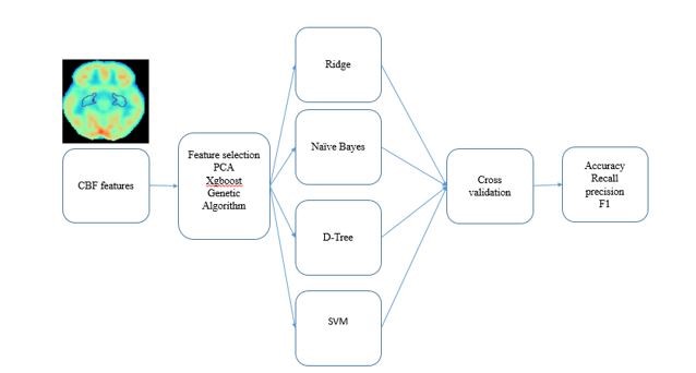
T1-weighted and Pulsed ASL MRI Data from 42 patients (22 left mTLEs and 20 right mTLEs) and 15 healthy volunteers as control at Iranian National Brain Mapping Laboratory (NBML) were gathered for this investigation. The analysis was conducted using the Bayesian Inference for Arterial Spin Labeling MRI (BASIL) toolbox, providing pre-processing and post-processing procedures for CBF mapping. CBF maps were parcellated using the automated anatomical labeling atlas 3 to 118 brain regions. A perfusion asymmetry index was obtained from the mean values of the ROIs. The study initially utilized three feature selection techniques: XGBoost, PCA, and GA, where PCA and GA were prominent methods for feature selection. Following feature selection, four distinct classifiers (ridge, naïve bayes, decision tree, and SVM) were employed for further analysis. To ensure robustness, 50 iterations of a repeated 5-fold cross-validation test were conducted, mitigating the impact of chance on model performance evaluation. Various performance metrics, including accuracy, f1-score, precision, and recall, were reported to assess the models' effectiveness (Figure 1).
Results:
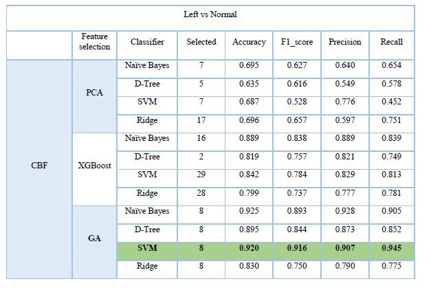
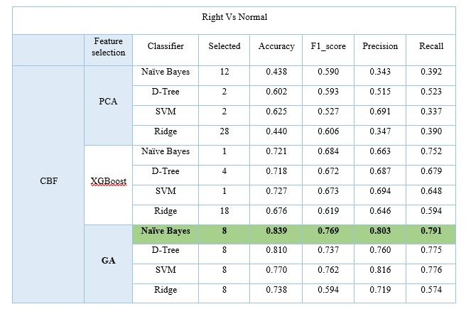
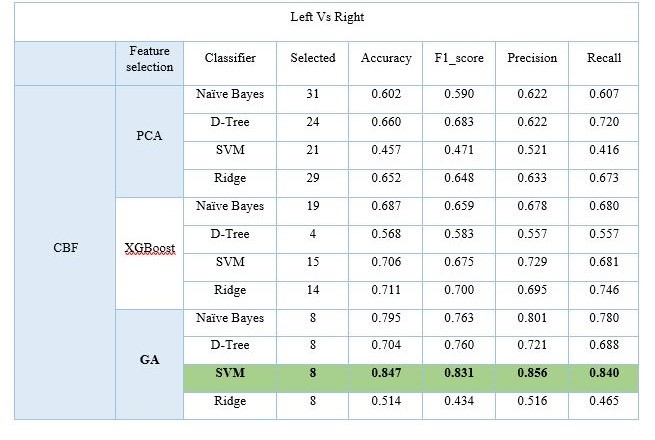
Based on the data presented in Tables 1, 2, and 3, it is evident that the genetic algorithm feature selection approach, utilizing eight features, consistently outperformed both PCA and XGBoost on all six datasets. Notably, when assessing the distinction between left epilepsy and normal cohorts in terms of perfusion, the SVM classifier exhibited exceptional performance with an accuracy of 0.92 and an f1_score of 0.91. In the comparison of the right mTLE and normal cohorts, Naïve Bayes emerged as the top-performing classifier, achieving an accuracy of 0.83 and an F1 score of 0.76 in the context of perfusion. When discerning between the left and right mTLE cohorts, SVM demonstrated its superiority within the CBF dataset, attaining an accuracy of 0.84 and an F1 score of 0.83.
Discussion:
Based on the results, perfusion MRI showed promising results in classifying right and left from control cohort. It is apparent from the result that the comparison of left TLE vs. normal are higher than those for the comparison of right TLE vs. normal. In the study that has been carried out 4, it has been shown that the perfusion pattern is different for the right and left mTLE data, so that the intra-group analysis for the left mTLE perfusion data did not show any significant difference. Meanwhile, the use of machine learning found that the left CBF features had better results. This shows the power of machine learning in improving lateralization. The superior performance of Support Vector Machine (SVM) and Naïve Bayes classifiers in our study can be attributed to their adaptability to different data distributions, robustness, and simplicity10. SVM's generalization ability and minimal need for parameter tuning make it well-suited for complex datasets like cerebral blood flow (CBF) data. Naïve Bayes' assumption of feature independence and its simplicity are advantageous, especially in cases with limited data, such as our study. The strengths of SVM and Naïve Bayes have proven to be valuable in accurately distinguishing between right mTLE patients and normal cohorts based on perfusion data, demonstrating their potential for clinical applications in epilepsy lateralization.
Conclusion:
This study explored the use of ASL MRI with machine learning to distinguish left and right temporal lobe epilepsy (mTLE) from normal controls. CBF data from ASL MRI proved to be valuable in this differentiation.
Acknowledgements
No acknowledgement found.References
References:1. Sheikh SR, Nair D, Gross RE, Gonzalez‐Martinez J. Tracking a changing paradigm and the modern face of epilepsy surgery: a comprehensive and critical review on the hunt for the optimal extent of resection in mesial temporal lobe epilepsy. Epilepsia. 2019;60(9):1768-93.
2. Fallahi A, Pooyan M, Lotfi N, Baniasad F, Tapak L, Mohammadi-Mobarakeh N, et al. Dynamic functional connectivity in temporal lobe epilepsy: a graph theoretical and machine learning approach. Neurological Sciences. 2021;42:2379-90.
3. Fallahi A, Pooyan M, Mehvari-Habibabadi J, Tabatabaei NH, Ay M, Nazem-Zadeh M-R. Application of Machine Learning in Comparison between Multimodal Neuroimaging Markers of Laterality in Temporal Lobe Epilepsy.
4. Rahimzadeh H, Kamkar H, Hoseini-Tabatabaei N, Mobarakeh NM, Habibabadi JM, Hashemi-Fesharaki SS, et al. Alteration of intracranial blood perfusion in temporal lobe epilepsy, an arterial spin labeling study. Heliyon. 2023;9(4):e14854.
5. Yoganathan K, Malek N, Torzillo E, Paranathala M, Greene J. Neurological update: structural and functional imaging in epilepsy surgery. Journal of Neurology. 2023;270(5):2798-808.
6. Hosseini M-P, Nazem-Zadeh MR, Mahmoudi F, Ying H, Soltanian-Zadeh H, editors. Support vector machine with nonlinear-kernel optimization for lateralization of epileptogenic hippocampus in mr images. 2014 36th Annual International Conference of the IEEE Engineering in Medicine and Biology Society; 2014: IEEE.
7. Mahmoudi F, Elisevich K, Bagher-Ebadian H, Nazem-Zadeh M-R, Davoodi-Bojd E, Schwalb JM, et al. Data mining MR image features of select structures for lateralization of mesial temporal lobe epilepsy. Plos one. 2018;13(8):e0199137.
8. Sanjari Moghaddam H, Rahmani F, Aarabi MH, Nazem-Zadeh M-R, Davoodi-Bojd E, Soltanian-Zadeh H. White matter microstructural differences between right and left mesial temporal lobe epilepsy. Acta Neurologica Belgica. 2020;120(6):1323-31.
9. Collij LE, Heeman F, Kuijer JP, Ossenkoppele R, Benedictus MR, Möller C, et al. Application of machine learning to arterial spin labeling in mild cognitive impairment and Alzheimer disease. Radiology. 2016;281(3):865-75.
10. Miranda Â, Lavrador R, Júlio F, Januário C, Castelo-Branco M, Caetano G. Classification of Huntington’s disease stage with support vector machines: a study on oculomotor performance. Behavior research methods. 2016;48:1667-77.
Figures