3228
Abnormal apparent diffusion coefficient of ultra-high b-values in the bilateral thalamus and striatum in MRI-negative refractory epilepsy1First Affiliated Hospital of Jinan University, Guangzhou, China, 2MR Research, GE Healthcare, Beijing, China, Guangzhou, China, 3Affiliated Brain Hospital of Jinan University, Guangzhou, China
Synopsis
Keywords: Epilepsy, Diffusion/other diffusion imaging techniques
Motivation: Subcortical nuclei such as the thalamus and striatum have been shown to be related to seizure modulation and termination, especially in refractory epilepsy.
Goal(s): This study aimed to assess AQP4 function reflected by the apparent diffusion coefficient (ADC) from ultra-high b-values (ADCuh) in MRI-negative refractory epilepsy.
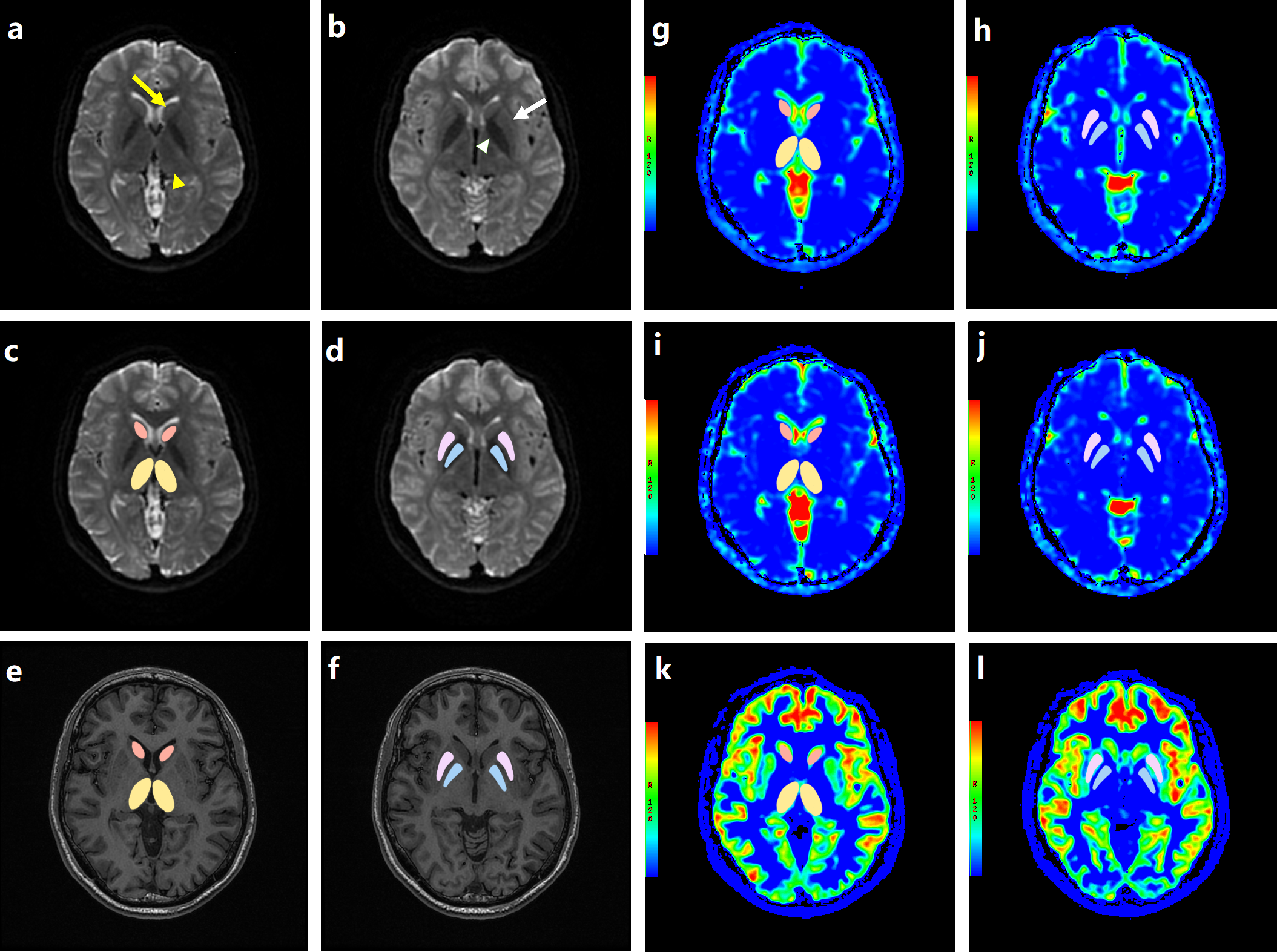
Approach: The eDWI parameters such as standard ADC (ADCst), pure water diffusion (D) and ADCuh were calculated from the 15 b-values.
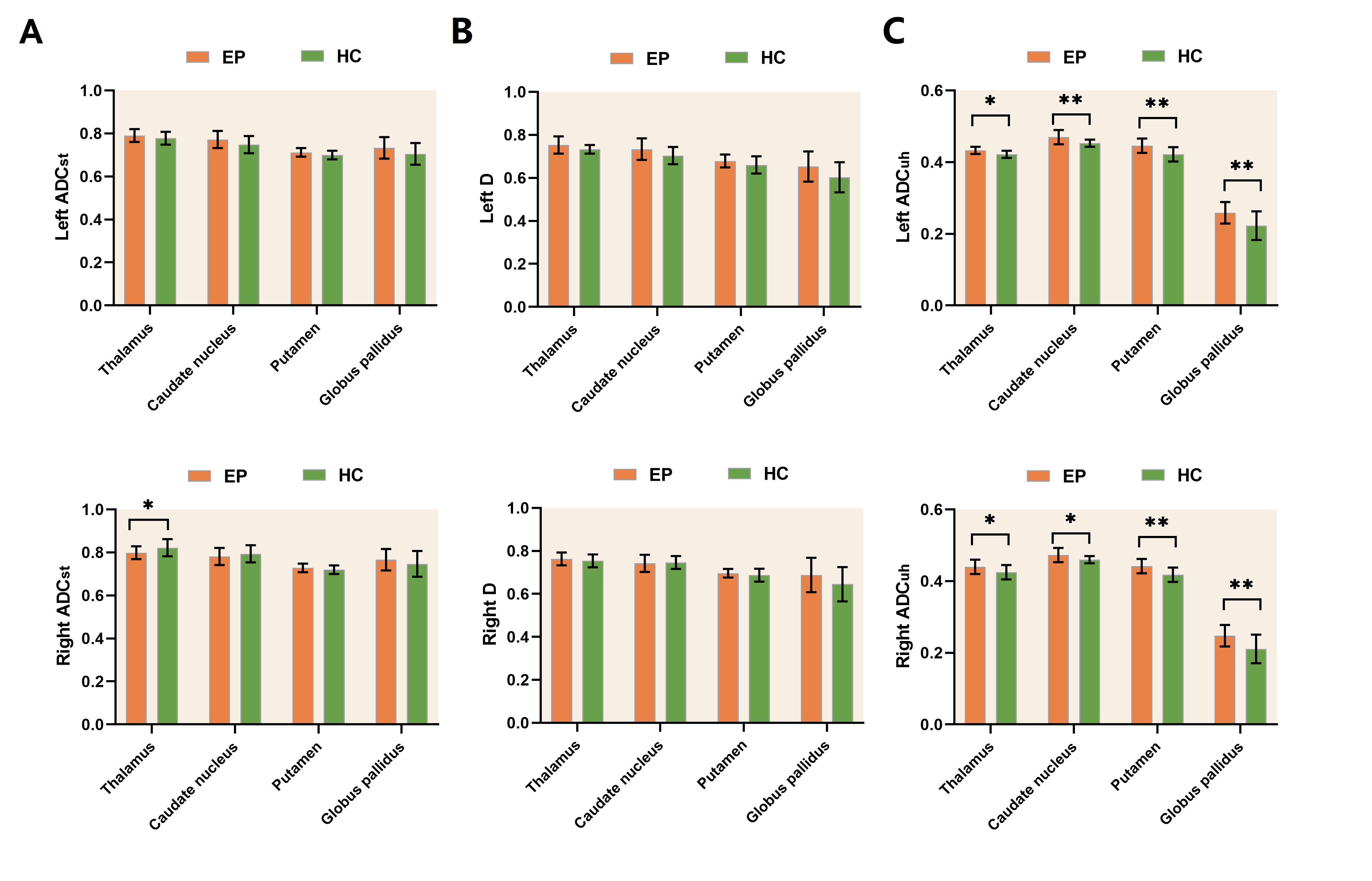
Results: ADCuh values in the bilateral thalamus, caudate nucleus, putamen and globus pallidus in MRI-negative refractory epilepsy were significantly higher than those in the healthy control subjects.
Impact: The alterations of the ADCuh values in the bilateral thalamus and striatum may reflect abnormal AQP4 function in MRI-negative refractory epilepsy. ADCuh might be a useful measurement for evaluating subcortical nuclei related brain damage in epilepsy patients.
Objective
Epilepsy is a common and severe neurological disorder characterized by an enduring predisposition to generate epileptic seizures [1], which affects over 70 million people worldwide [2]. Patients with refractory epilepsy have increased risks of premature death, injuries, psychosocial dysfunction, and a reduced quality of life [3]. However, its underlying neuropathological mechanism remains largely unknow. In the past, epilepsy has been considered, for the most part, to be a cortical disease. However, cumulative findings have demonstrated that epileptic seizures involve widespread network interactions between cortical and subcortical structures [4, 5]. Subcortical structures play a crucial role in behavioral manifestations, propagation, and, in some cases, initiation of epileptic seizures [5], thus emerging as a critical area to help understanding the pathological mechanism of epilepsy. Aquaporins (AQP) are a family of at least ten homologous water transporting proteins in mammals that are expressed in many epithelial, endothelial and other tissues [6]. Aquaporin 4 (AQP4) is the predominant water channel in the mammalian brain [7]. It is densely expressed in astrocyte end-feet and is an important factor in water and potassium homeostasis in the central nervous system (CNS) [8]. AQP4 regulates multiple biological processes in astrocytes, such as maintaining CNS water balance, spatial buffering of extracellular potassium, regulation of neurotransmission, synaptic plasticity [9], etc. The potential roles of the AQP4 in modulating brain excitability and in epilepsy have been mentioned in previous studies [10, 11]. Dysregulation of AQP-4 expression has been demonstrated in studies using surgical samples from patients with refractory epilepsy related to hippocampal sclerosis [12] and focal cortical dysplasia [13]. However, no study has investigated the alteration of AQP4 alterations in the subcortical structures in vivo in epilepsy patients so far. Diffusion-weighted magnetic resonance imaging (DWI) is a widely used non-invasive technique that is sensitive to the random motion of water molecules in biological tissues, and offers information about tissue architecture and pathological changes on a cellular level [14]. Recently, several studies using ultra-high b-value-DWI suggested that ADC calculated using ultra-high b-values (ADCuh) may reflect AQP expression [15-17]. To date, researchers have used enhance diffusion-weighted imaging (eDWI) technique and tri-component model to calculate standard ADC (ADCst), ADCuh, D* and D in diseases such as parkinson's disease [15], prostate cancer [18] and bipolar disorder [16]. However, no eDWI studies have been published to report the abnormal diffusion parameters in epilepsy. Therefore, in the current study, we aimed to investigate the possible alterations of ADCuh in the thalamus and the striatum in MRI-negative refractory epilepsy by using eDWI. To the best of our knowledge, this is the first study to reveal the changes of ADCuh in the thalamus and the striatum in MRI-negative refractory epilepsy, which might reflect the AQP4 function of the brain and help to elucidate the neuropathophysiological mechanism of the disease.Methods
Twenty-nine patients with MRI-negative refractory epilepsy and 18 healthy controls underwent enhance diffusion weighted imaging (eDWI) with 15 b-values (0-5,000 s/mm2). The eDWI parameters such as standard ADC (ADCst), pure water diffusion (D) and ADCuh were calculated from the 15 b-values. Regions-of-interest analyses were conducted in the bilateral thalamus, caudate nucleus, putamen and globus pallidus and ADCst, D and ADCuh values were compared between the epilepsy patients and controls.Results
ADCuh values in the bilateral thalamus, caudate nucleus, putamen and globus pallidus in MRI-negative refractory epilepsy were significantly higher than those in the healthy control subjects (all P < 0.05), ADCst value in the right thalamus was significantly lower than those in the healthy control subjects (P < 0.05).Conclusion
The alterations of the ADCuh values in the bilateral thalamus and striatum may reflect abnormal AQP4 function in MRI-negative refractory epilepsy. ADCuh might be a useful measurement for evaluating subcortical nuclei related brain damage in epilepsy patients.Acknowledgements
This work was financially supported by the National Natural Science Foundation of China (No. 81871383), Basic and Applied Basic Research Foundation of Guangdong (No.2020A1515011192), Foundation of Guangzhou Science Brain (No.2023A03J0610), and Frontier Technology Program of the Affiliated of Jinan University, China (No. JNU1AF-CFTP-2022-n1214).References
1. Fisher, R.S., et al., ILAE official report: a practical clinical definition of epilepsy. Epilepsia, 2014. 55(4): p. 475-82.
2. Thijs, R.D., et al., Epilepsy in adults. Lancet, 2019. 393(10172): p. 689-701.
3. Löscher, W., et al., Drug Resistance in Epilepsy: Clinical Impact, Potential Mechanisms, and New Innovative Treatment Options. Pharmacol Rev, 2020. 72(3): p. 606-638.
4. Badawy, R.A., et al., Subcortical epilepsy? Neurology, 2013. 80(20): p. 1901-7.
5. Norden, A.D. and H. Blumenfeld, The role of subcortical structures in human epilepsy. Epilepsy Behav, 2002. 3(3): p. 219-231.
6. Verkman, A.S., Physiological importance of aquaporin water channels. Ann Med, 2002. 34(3): p. 192-200.
7. Assentoft, M., B.R. Larsen, and N. MacAulay, Regulation and Function of AQP4 in the Central Nervous System. Neurochem Res, 2015. 40(12): p. 2615-27.
8. Vandebroek, A. and M. Yasui, Regulation of AQP4 in the Central Nervous System. Int J Mol Sci, 2020. 21(5).
9. Xiao, M. and G. Hu, Involvement of aquaporin 4 in astrocyte function and neuropsychiatric disorders. CNS Neurosci Ther, 2014. 20(5): p. 385-90.
10. Binder, D.K., E.A. Nagelhus, and O.P. Ottersen, Aquaporin-4 and epilepsy. Glia, 2012. 60(8): p. 1203-14.
11. Hsu, M.S., D.J. Lee, and D.K. Binder, Potential role of the glial water channel aquaporin-4 in epilepsy. Neuron Glia Biol, 2007. 3(4): p. 287-97.
12. Eid, T., et al., Loss of perivascular aquaporin 4 may underlie deficient water and K+ homeostasis in the human epileptogenic hippocampus. Proc Natl Acad Sci U S A, 2005. 102(4): p. 1193-8.
13. Medici, V., et al., Aquaporin 4 expression in control and epileptic human cerebral cortex. Brain Res, 2011. 1367: p. 330-9.
14. Lenz, C., et al., Assessing extracranial tumors using diffusion-weighted whole-body MRI. Z Med Phys, 2011. 21(2): p. 79-90.
15. Xueying, L., et al., Investigation of Apparent Diffusion Coefficient from Ultra-high b-Values in Parkinson's Disease. Eur Radiol, 2015. 25(9): p. 2593-600.
16. Zhao, L., et al., Abnormalities of aquaporin-4 in the cerebellum in bipolar II disorder: An ultra-high b-values diffusion weighted imaging study. J Affect Disord, 2020. 274: p. 136-143.
17. Wang, Y., et al., Investigation of aquaporins and apparent diffusion coefficient from ultra-high b-values in a rat model of diabetic nephropathy. Eur Radiol Exp, 2017. 1(1): p. 13.
18. Kwak, J.T., et al., Automated prostate cancer detection using T2-weighted and high-b-value diffusion-weighted magnetic resonance imaging. Med Phys, 2015. 42(5): p. 2368-78.
Figures