3225
The altered brain activity in children with electrical status epilepticus during sleep: a preliminary study of resting-state fMRI1Department of Radiology, Shenzhen Children’s Hospital, Shenzhen, China, 2Philips Healthcare, Guangzhou, China, 3Philips Healthcare, Shanghai, China
Synopsis
Keywords: Epilepsy, fMRI (resting state)
Motivation: The micro-structure alteration related to cognitive function impairment, and the mechanism of functional impairment in children withESES remains unclear.
Goal(s): To investigate the brain activity in children with ESES, and the associations of the brain activity with cognitive performance.
Approach: We observed the ReHo difference between the ESES group and the HCs. And the correlation analysis was used to explore the relationship between Reho values and cognitive test.
Results: Compared with HCs, patients with ESES showed ReHo alteration in some brain regions. The correlation analysis revealed that increased ReHo in the right frontal gyrus was correlated with the decreased PRI.
Impact: The distinct changes of spontaneous brain activity were measured by ReHo, which may be further suggest that the potential of RS-fMRI as a novel biomarker tool for identifying cognitive impairments on neural aspect.
Introduction
Electrical Status Epilepticus During Sleep (ESES) is a special EEG phenomenon that occurs in the process of sleep, especially in the Non-Rapid Eye Movement (NREM). It was characterized by the presence of focal or extensive persistent spike slow waves [1]. Previous research result drew the conclusion that children with ESES get cognitive function impairments and poor prognosis. However, the micro-structure alteration related to cognitive function impairment, and the mechanism of functional impairment remains unclear. In this study, we aimed to investigate the brain activity in children with ESES, and the associations of the brain activity with cognitive performance.Methods
Twenty-five children with ESES, including 13 boys and 12 girls, aging from 6.75 to 12.33 years old (mean: 8.571.99 years), and 16 gender and age matched healthy controls (HCs) were enrolled in this study. Resting state fMRI (rs-fMRI) data were acquired on a 3.0T MRI system (Ingenia, Philips Healthcare, Best, the Netherlands). Then, the rs-fMRI data preprocessing was carried out using the toolbox Data Processing & Analysis for Brain Imaging (DPABI) [2]. The regional homogeneity (ReHo) was calculated to characterize the local synchronization of spontaneous brain activity. Wechsler Intelligence scale for children-Fourth Edition (WISC-Ⅳ) was used for cognitive test. The difference between the ESES group and the HCs were detected using the two-sample t test. And the correlation analysis was used to explore the relationship between Reho values and cognitive test.Results
In cognitive test, patients with ESES exhibited significant lower scores in Full-Scale Intelligence Quotient (FSIQ), Comprehension Index (VCI) and Perceptual Reasoning Index (PRI) (P values were 0.007, 0.034 and 0.008, respectively) compared with HCs (Tabel 1). Moreover, compared with HCs, patients with ESES also showed decreased ReHo in left postcentral gyrus, right superior temporal gyrus, right supplementary motor area, right cerebellar lobule Ⅵ, right precentral gyrus and increased ReHo in bilateral middle occipital gyrus, left middle frontal gyrus, left lingual gyrus, cerebellar hemisphere Crus1 area and Crus2 area (Figure 1). The further correlation analysis revealed that increased ReHo in the right frontal gyrus was positively correlated with the decreased PRI (r = 0.761; P = 0.028).Discussion
In this study, the cognitive test showed that patients with ESES have cognitive impairment in language, perceptual and reasoning abilities. In addition, we identified synchronization of neural activities alterations compared with the HC group. The cortex-cerebellar system has been well documented in humans and primates, with separate and closed circuits between the cerebellum and cortex. Middleton, Kelly and Strick et al. [3, 4] discovered two cortical-cerebellar circuits: the motor circuit and the frontal circuit. In the motor circuit, the primary motor cortex projects signals to the V, VI, HVIIB and HVIII parts of the cerebellar cortex, and then returns to the primary motor cortex through the dorsal cerebellar dentate nucleus and thalamus. In the prefrontal loop, area 46 of the prefrontal cortex projects information through the pontine nucleus to the HVIIA part of the cerebellar cortex lobules (mainly Crus1 and Crus2 areas), and then returns to the prefrontal cortex through the ventral dentate nucleus and thalamus of the cerebellum [5-7]. Therefore, cerebellum Cru1 and Crus2 regions are closely related to the functions of the prefrontal cortex. Language function of ESES group was impaired compared with the normal control group, while Crus1 and Crus2 the ReHo values had increased. We might get the conclusion that the language centers of the cerebral hemisphere and the language activation areas of the cerebellum have a synergistic effect. When the function of the language centers of the brain declines, the language function of Cru1 and Crus2 regions may increase compensatively, or some of the language function of the brain may shift to the cerebellum. This is consistent with the findings of Lin et al. [8] that the ReHo value in the cerebral hemisphere decreased and the ReHo value in the cerebellum increased compensatively in patients with cirrhosis. These compensatory changes may be related to the redistribution of blood flow and metabolism [9, 10].Conclusion
Cognitive test showed cognitive impairments in patients with ESES. The distinct changes of spontaneous brain activity were measured by ReHo, which may be further suggest that the potential of RS-fMRI as a novel biomarker tool for identifying cognitive impairments on neural aspect.Acknowledgements
We would like to acknowledge the generous support and contribution of all our trial participants.References
[1] Patry G, Lyagoubi S, Tassinari CA. Subclinical "electrical status epilepticus" induced by sleep in children. A clinical and electroencephalographic study of six cases. Arch Neurol. 1971. 24(3): 242-52.
[2] Yan, CG., Wang, XD., Zuo, XN. et al. DPABI: Data Processing & Analysis for (Resting-State) Brain Imaging. Neuroinform 14, 339–351 (2016).
[3] Middleton FA, Strick PL. Basal ganglia and cerebellar loops: motor and cognitive circuits. Brain Res Brain Res Rev. 2000. 31(2-3): 236-50.
[4] Kelly RM, Strick PL. Cerebellar loops with motor cortex and prefrontal cortex of a nonhuman primate. J Neurosci. 2003. 23(23): 8432-44.
[5] Middleton FA, Strick PL. Cerebellar projections to the prefrontal cortex of the primate. J Neurosci. 2001. 21(2): 700-12.
[6] Barbas H, Henion TH, Dermon CR. Diverse thalamic projections to the prefrontal cortex in the rhesus monkey. J Comp Neurol. 1991. 313(1): 65-94.
[7] Goldman-Rakic PS, Porrino LJ. The primate mediodorsal (MD) nucleus and its projection to the frontal lobe. J Comp Neurol. 1985. 242(4): 535-60.
[8] Lin WC, Hsu TW, Chen CL, Lu CH, Chen HL, Cheng YF. Resting State-fMRI with ReHo Analysis as a Non-Invasive Modality for the Prognosis of Cirrhotic Patients with Overt Hepatic Encephalopathy. PLoS One. 2015. 10(5): e0126834.
[9] Lockwood AH, Yap EW, Wong WH. Cerebral ammonia metabolism in patients with severe liver disease and minimal hepatic encephalopathy. J Cereb Blood Flow Metab. 1991. 11(2): 337-41.
[10] Ahl B, Weissenborn K, van den Hoff J, et al. Regional differences in cerebral blood flow and cerebral ammonia metabolism in patients with cirrhosis. Hepatology. 2004. 40(1): 73-9.
Figures
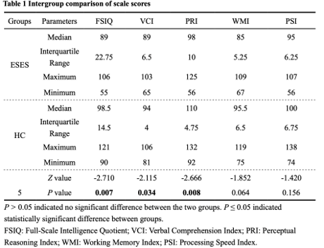
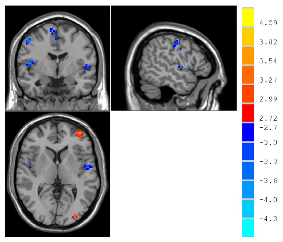
Figure 1. Differences in regional homogeneity (ReHo) values between patients with ESES and controls (P < 0.01, minimum cluster > 15, with AlphaSim correction). T score bars are shown on the right. Hot (red) and cold (blue) colors indicate ESES group ReHo increases and decreases, respectively.