3222
Unsupervised MR2PET Synthesis Provides Pseudo-Normal Reference to Improve Epileptic Lesion Detection1School of Biomedical Engineering, Shanghai Jiao Tong University, Shanghai, China, 2University of Michigan-Shanghai Jiao Tong University Joint Institute, Shanghai Jiao Tong University, Shanghai, China, 3Global Institute of Future Technology, Shanghai Jiao Tong University, Shanghai, China, 4Department of Nuclear Medicine, Ruijin Hospital, Shanghai Jiao Tong University School of Medicine, Shanghai, China
Synopsis
Keywords: Epilepsy, PET/MR, Imaging Translation
Motivation: It is challenging to obtain demographically matched controls for every patient who underwent FDG PET examinations.
Goal(s): We aim to generate pseudo-normal PET for each epilepsy patient leveraging recent progress in cross modality image translation.
Approach: We employed diffusion model to learn the translation between T1w-MRI and FDG PET of healthy subjects, then generated pseudo-normal PET for a cohort of 104 patients with focal epilepsy, who underwent PET/MR scanning for presurgical evaluation.
Results: Unsupervised SynDiff achieved comparable performance as supervised Pix2pixGAN in PET synthesis. Improved DICE coefficient and lesion detection were achieved using synthesized reference compared with traditional group reference.
Impact: Imaging translation provides a personalized pseudo-normal reference for each epilepsy patient. Pseudo-normal PET is poised for potential adoption as an auxiliary tool to enhance the capability of PET imaging in detecting epileptic lesions within clinical settings.
Introduction
Positron Emission Tomography (PET) labeled with [18F] Fluorodeoxyglucose (FDG) is a valuable tool for epileptic lesion detection, especially when routine MRIs are inconclusive1,2. Previous studies indicated that demographically matched control groups improved lesion detection of FDG PET3. However, for every clinical site to obtain such control images can be challenging.Recent progress in generative model allowed cross modality image translation4. For example MRI-to-PET translation has been attempted to generate PET images without using radioactive tracer5. Yaakub et al. proposed the use of conditional GAN (cGAN) for synthesizing pseudo-normal PET images from T1w-MRI scans to aid epilepsy lesion detection6. However, it still requires pre-existing high quality paired FDG PET and T1w-MRI training data.
In this study, we aim to investigate the capability of unsupervised diffusion model SynDiff7 in synthesizing FDG PET from T1w-MRI, and to evaluate potential improvements using pseudo-normal PET images in patients lesion detection.
Methods
Participants and Data AcquisitionIn this IRB approved study, we recruited 36 healthy control without any history of neurological or psychiatric disease and 104 patients diagnosed with refractory focal epilepsy. Lesion masks were either obtained by comparing pre- and post- surgical T1w images (N =56), or manually drawn (N =48) based on routine MRI, FDG PET, and clinical diagnosis under supervision of experienced radiologists. Images were collected on the Siemens PET/MR Synchronous integrated scanner (Biograph mMR), including T1-weighted structural images and 18F-FDG PET images. We acquired T1 MPRAGE images (TR/TE = 1900/2.44 ms, resolution=1.0 × 1.0 × 1.0 mm3, FOV = 256 × 256 mm2, 192 slices). They were also administered [18F] FDG intravenously using a mean dose of 184.8 ± 29.0 MBq (range 133.2–247.9 MBq), and scanned 30-50 min after the injection. Static PET data were acquired in a sinogram mode for 15 min, matrix size 344 × 344, and post-filtered with an isotropic full-width half-maximum (FWHM) Gaussian kernel of 2 mm.
Preprocessing
FreeSurfer v7.0 package (https://surfer.nmr.mgh.harvard.edu) was used to obtain processed T1-MPRAGE and then processed by skull-stripping using SynthStrip tool8. [18F] FDG PET were rigidly aligned to its corresponding MR images.
Training and Generation of Pseudo-Normal PET
For model training, we used 4,503 2D slices of preprocessed MRI and PET images from 36 healthy subjects. Among these, 3,484 (80%) were for training, and 1,019 (20%) for testing. Computation was done on a single NVIDIA GeForce RTX 3090 Ti GPU, with an initial learning rate of 2e-4 and a batch size of 1 over 2000 iterations. We employed minibatch SGD with the Adam solver (learning rate 0.0002, momentum parameters β1 = 0.5, β2 = 0.999). Three models were used: supervised Pix2pixGAN9, unsupervised CycleGAN10, and SynDiff7 (Fig.1A).
Evaluation of Epileptic Lesion Detection Performance
Gold standard of epileptic lesion segmentation was defined by manually drawn lesion mask.
To extract hypometabolic regions in epilepsy patients, we calculated the z-score value between the real PET and the pseudo-normal PET (Fig.1B). We chose the z-score value and cluster size threshold, Z = 1.5 and K = 1500 to restrict the prediction result. We calculated dice coefficient between predict lesion and actual lesion mask. If Dice>0, the lesion was thought to be detected successfully11. All the calculation was limited in gray matter regions.
We also compared those results to the statistical parametric mapping (SPM) framework implemented in the SPM12 software (fil.ion.ucl.ac.uk/spm). Decreased metabolism was regarded as statistically significant if the uncorrected P value was under 0.005, with a cluster level above 50 voxels (selection of threshold mainly considered the balance between detection rate and false positive rate).
Results
The unsupervised diffusion model SynDiff reached comparable performance in terms of structural similarity (SSIM), peak signal to noise ratio (PSNR), fréchet inception distance (FID) as Pix2pixGAN, out-performing CycleGAN (Table 1).Representative cases of pseudo-normal PET aided detection were shown in Figure 2. Anatomical structure was better preserved in pseudo-normal PET images generated by SynDiff and Pix2pixGAN, as compared to those generated by CycleGAN. Lesion segmentation of each method was overlayed with manually drawn lesion masks. Further, synthesized personalized reference help achieve higher lesion detection rate (Pix2pixGAN 77%, CycleGAN 67%, SynDiff 79%) compared to traditional statistical parametric mapping using group reference (53%); while distribution of Dice coefficients plotted in Figure 3.
Conclusions
The unsupervised diffusion model had comparable performance in imaging synthesis compared to supervised Pix2pixGAN. Our findings also confirmed that using pseudo-normal PET as personalized reference can bolster epileptic lesion detection for FDG PET.Acknowledgements
N/AReferences
1.Duncan, J. S.; Winston, G. P.; Koepp, M. J.; Ourselin, S. Brain Imaging in the Assessment for Epilepsy Surgery. Lancet Neurol. 2016, 15 (4), 420–433.
2. Niu, N.; Xing, H.; Wu, M.; Ma, Y.; Liu, Y.; Ba, J.; Zhu, S.; Li, F.; Huo, L. Performance of PET Imaging for the Localization of Epileptogenic Zone in Patients with Epilepsy: A Meta-Analysis. Eur. Radiol. 2021, 31 (8), 6353–6366.
3. De Blasi, B.; Barnes, A.; Galazzo, I. B.; Hua, C.-H.; Shulkin, B.; Koepp, M.; Tisdall, M. Age-Specific 18F-FDG Image Processing Pipelines and Analysis Are Essential for Individual Mapping of Seizure Foci in Pediatric Patients with Intractable Epilepsy. J. Nucl. Med. Off. Publ. Soc. Nucl. Med. 2018, 59 (10), 1590–1596.
4. Yi, X.; Walia, E.; Babyn, P. Generative Adversarial Network in Medical Imaging: A Review. Med. Image Anal. 2019, 58, 101552.
5. Sikka, A.; Peri, S. V.; Bathula, D. R. MRI to FDG-PET: Cross-Modal Synthesis Using 3D U-Net for Multi-Modal Alzheimer’s Classification. In Simulation and Synthesis in Medical Imaging; Gooya, A., Goksel, O., Oguz, I., Burgos, N., Eds.; Lecture Notes in Computer Science; Springer International Publishing: Cham, 2018; pp 80–89.
6. Yaakub, S. N.; McGinnity, C. J.; Clough, J. R.; Kerfoot, E.; Girard, N.; Guedj, E.; Hammers, A. Pseudo-normal PET Synthesis with Generative Adversarial Networks for Localising Hypometabolism in Epilepsies. In: Burgos, N., Gooya, A., Svoboda, D. (eds) Simulation and Synthesis in Medical Imaging. SASHIMI 2019.
7. Ozbey M, Dalmaz O, Dar SU, Bedel HA, Ozturk S, Gungor A, Cukur T. Unsupervised Medical Image Translation with Adversarial Diffusion Models. IEEE Trans Med Imaging. 2023 Jun 28;PP.
8. Hoopes, A.; Mora, J. S.; Dalca, A. V.; Fischl, B.; Hoffmann, M. SynthStrip: Skull-Stripping for Any Brain Image. NeuroImage 2022, 260, 119474.
9. Isola, P.; Zhu, J.-Y.; Zhou, T.; Efros, A. A. Image-to-Image Translation with Conditional Adversarial Networks. In 2017 IEEE Conference on Computer Vision and Pattern Recognition (CVPR); 2017; pp 5967–5976.
10. Zhu, J.-Y.; Park, T.; Isola, P.; Efros, A. A. Unpaired Image-to-Image Translation Using Cycle-Consistent Adversarial Networks. In 2017 IEEE International Conference on Computer Vision (ICCV); 2017; pp 2242–2251.
11. Walger, L.; Adler, S.; Wagstyl, K.; Henschel, L.; David, B.; Borger, V.; Hattingen, E.; Vatter, H.; Elger, C. E.; Baldeweg, T.; Rosenow, F.; Urbach, H.; Becker, A.; Radbruch, A.; Surges, R.; Reuter, M.; Cendes, F.; Wang, Z. I.; Huppertz, H.-J.; Rüber, T. Artificial Intelligence for the Detection of Focal Cortical Dysplasia: Challenges in Translating Algorithms into Clinical Practice. Epilepsia 2023, 64 (5), 1093–1112.
Figures
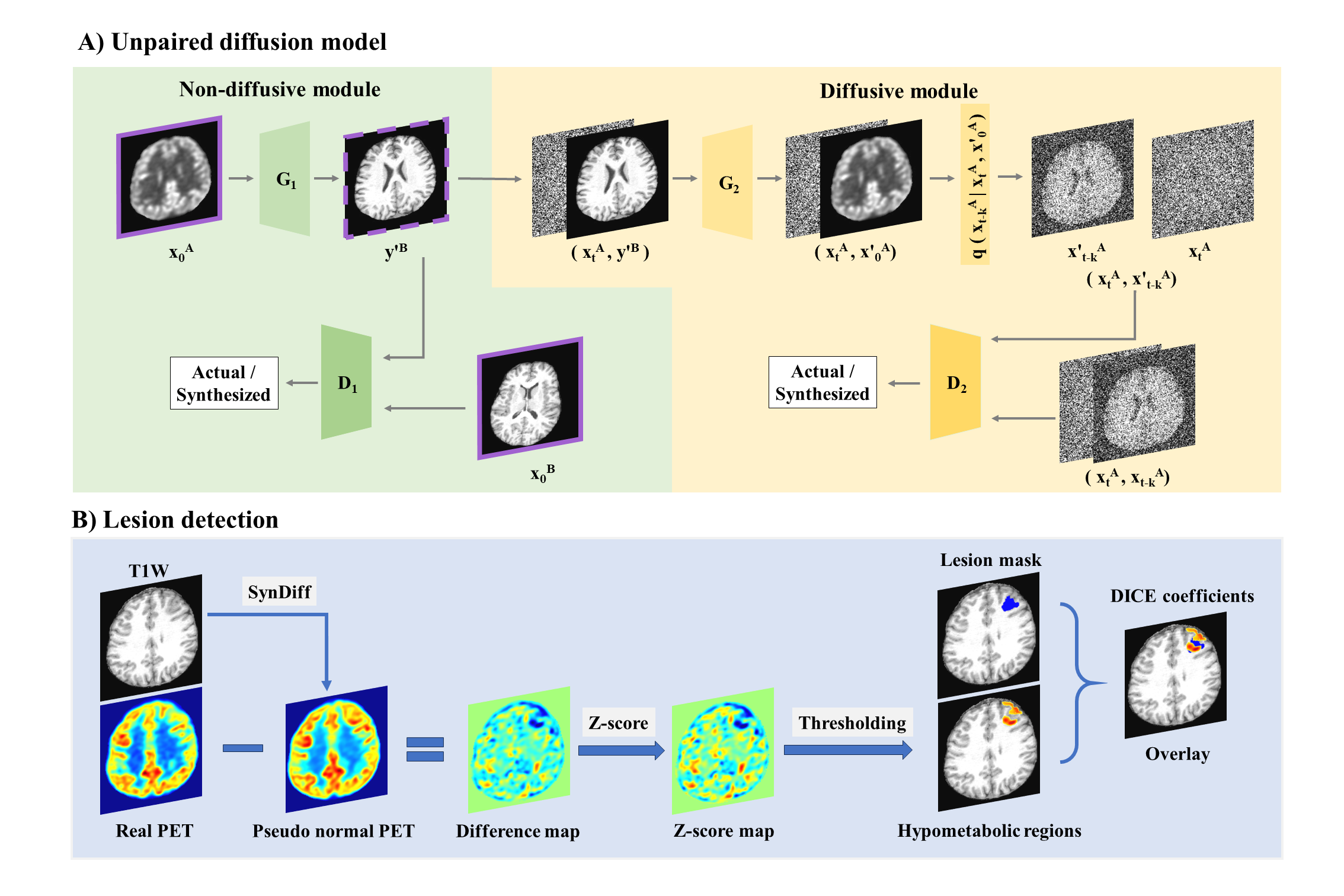
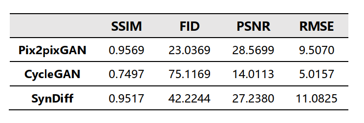
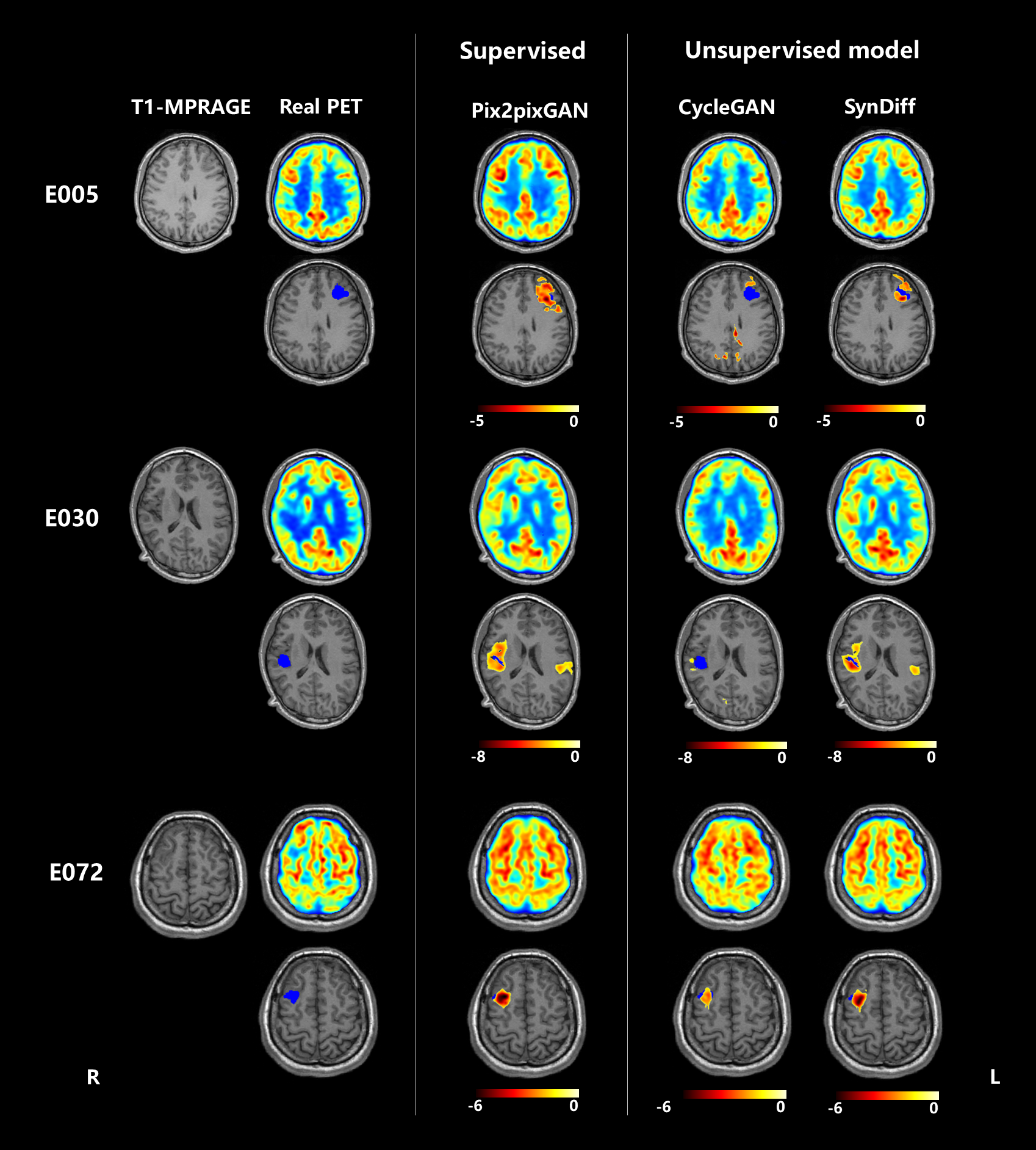
Figure 2. Representative cases of epileptic patients. The left column showed anatomical MRI and corresponding FDG PET images. The middle column and the right column showed pseudo-normal PET generated by supervised model Pix2pixGAN, unsupervised model CycleGAN and SynDiff, as well as the predicted epileptic lesion overlaying on the anatomical MRI. Blue region represents lesion mask. Red and yellow region represents predicted lesion.
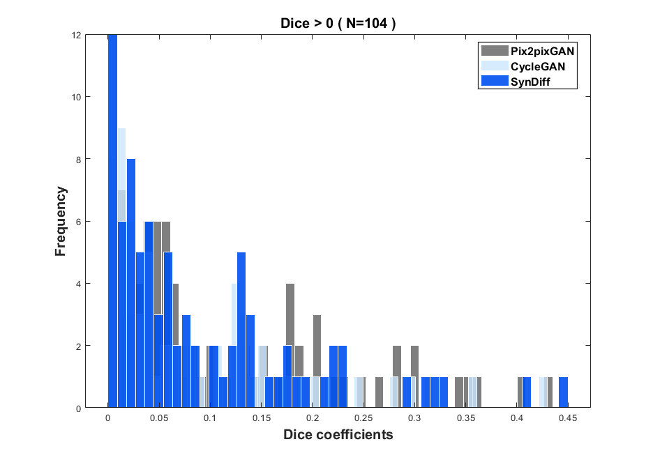
Figure 3. Histogram of Dice coefficients. All the value were greater than 0. The Dice calculated from Pix2pixGAN, CycleGAN and SynDiff were found to be 0.060 (0.432), 0.046 (0.427), and 0.055 (0.449), respectively, with the median (range) values represented.