3218
Comparison of Spontaneous Brain Activity between Hippocampal Sclerosis and MRI-Negative Temporal Lobe Epilepsy1MRI, the first affiliated hospital of zhengzhou university, zhengzhou, China, 2MR Research Collaboration, Siemens Healthineers, Beijing, China
Synopsis
Keywords: Epilepsy, Epilepsy, fMRI
Motivation: There has been controversy regarding the pathophysiological basis of MRI-negative temporal lobe epilepsy (TLE-N) and its similarities and differences relative to TLE with hippocampal sclerosis (TLE-HS).
Goal(s): Static and dynamic abnormalities of spontaneous brain activity (SBA) in TLE-HS and TLE-N were examined.
Approach: Six static SBA indicators and corresponding temporal dynamic indicators were calculated using a sliding window approach, then compared.
Results: Patterns of change in SBA abnormalities were generally similar between TLE-HS and TLE-N groups; they were more pronounced in the TLE-HS group. VMHC in the hippocampus showed promise for differential diagnosis. Many indicators were correlated with cognition.
Impact: The use of static and dynamic metrics can convey a more detailed and reliable description of abnormal neuronal activity, facilitating exploration of epileptic activity and cognitive impairment mechanism in MRI-negative temporal lobe epilepsy (TLE) and TLE with hippocampal sclerosis.
INTRODUCTION
Hippocampal sclerosis (HS) is the most common etiology of temporal lobe epilepsy (TLE)1. However, ~30% of individuals with TLE exhibit negative magnetic resonance imaging (MRI) findings2. The characterization of similarities and differences in brain activity between distinct TLE subtypes will facilitate treatment and future research. Here, we comprehensively examined static and dynamic abnormalities of spontaneous brain activity (SBA) in TLE with hippocampal sclerosis (TLE-HS) and MRI-negative TLE (TLE-N). We also explored whether these alterations were correlated with epilepsy duration and cognition, then sought to find out a clinically useful differential diagnostic index.METHODS
This study included 38 patients with TLE-HS, 51 patients with TLE-N, and 53 healthy control (HC) volunteers. All participants underwent MRI scans on a 3T system (MAGNETOM Prisma, Siemens Healthcare, Erlangen, Germany), including resting-state functional imaging with BOLD sequence and three-dimensional T1-weighted structural imaging with MPRAGE sequence. We recorded epilepsy duration and scores on the Mini-Mental State Examination, Montreal Cognitive Assessment-Basic, Auditory Verbal Learning Test, and Shape Trail Making Test-A/B. Using DPABI3 software, we calculated 6 static SBA indicators (amplitude of low-frequency fluctuation [ALFF], fractional ALFF [fALFF], regional homogeneity [ReHo), degree centrality [DC], voxel-mirrored homotopic connectivity [VMHC], and global signal correlation [GSCorr]) and corresponding temporal dynamic indicators (dynamic ALFF [dALFF], dynamic fALFF [dfALFF], dynamic ReHo [dReHo], dynamic DC [dDC], dynamic VMHC [dVMHC], and dynamic GSCorr [dGSCorr]) using a sliding window approach with a window length of 50 repetition times (TR) and a shifting step size of 10 TRs. Differences in SBA metrics among TLE-HS, TLE-N, and HC groups were examined using one-way analysis of variance (ANOVA), with age, sex, education, and mean frame-wise displacement as covariates. Then, the mask produced by ANOVA analyses was utilized in post hoc comparisons via two-sample t-tests. Corrections for multiple comparisons were performed based on Gaussian random field theory (pvoxelwise < 0.005, pcluster-wise < 0.05). Relationships between SBA metrics in regions with between-group differences and the epilepsy duration and cognitive scores were analyzed by Spearman correlation (p < 0.01). Receiver operating characteristic curve analyses of abnormalities in TLE-HS and TLE-N were conducted to investigate the discriminatory power.RESULTS
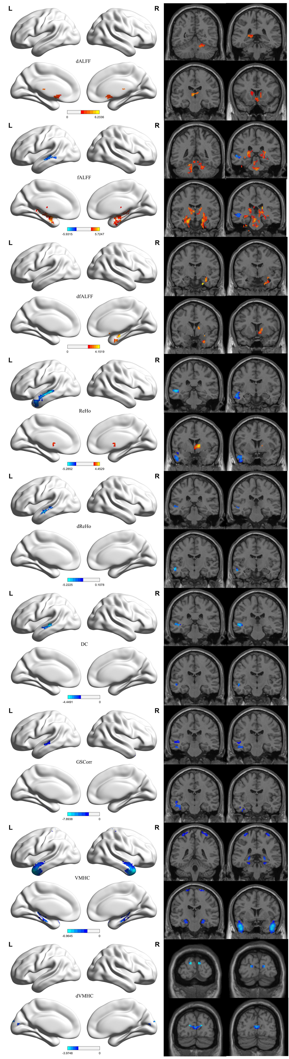
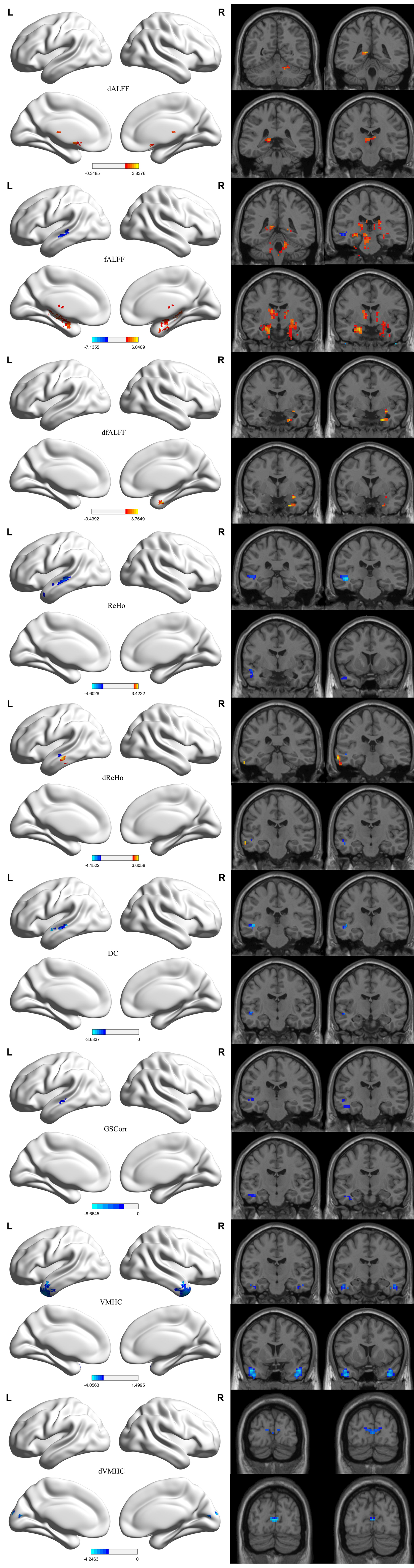
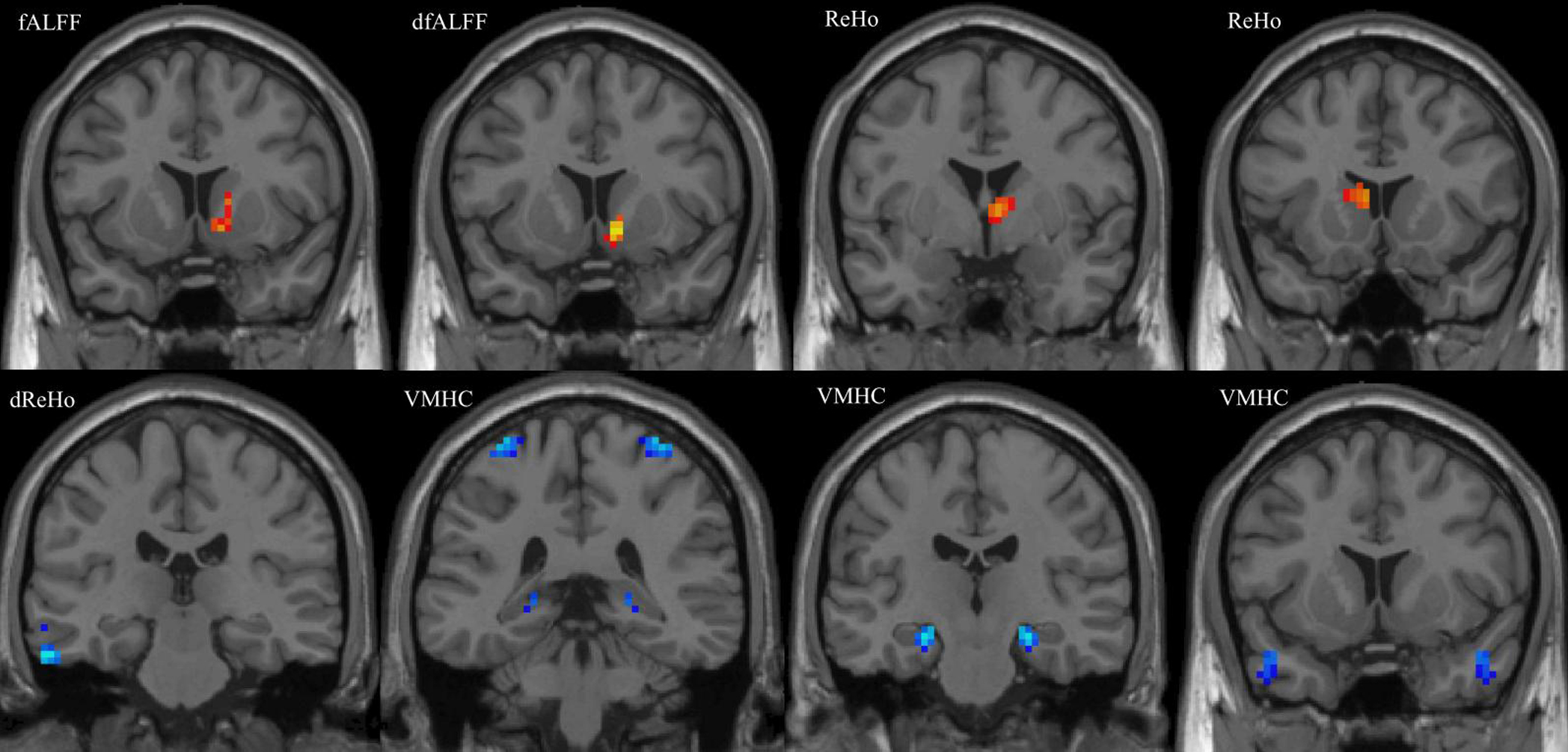
Cognitive decline was observed in the TLE-HS and TLE-N groups; it was more pronounced in the TLE-HS group. Compared with the HC group, the TLE-HS and TLE-N groups both demonstrated abnormal changes in SBA with similar patterns: (1) elevated fALFF, dALFF, and dfALFF values in the mesial temporal lobe, thalamus, basal ganglia, pons, and cerebellum; (2) reduced fALFF, ReHo, dReHo, DC, GSCorr, and VMHC values in the lateral temporal lobe ipsilateral to the epileptogenic focus; and (3) reduced dVMHC in the bilateral calcarine cortex. However, in the TLE-HS group, the cluster sizes were larger; additional reductions in VMHC were detected in the bilateral hippocampus, as well as the precentral and postcentral gyri. Compared with the TLE-N group, the TLE-HS group exhibited increased fALFF, dfALFF, and ReHo values in the caudate; reduced VMHC values in the bilateral postcentral gyrus, mesial, and lateral temporal lobes; and decreased dReHo in the ipsilateral temporal neocortex. VMHC in the hippocampus displayed the best discriminatory power, with an area under the curve of 0.759. Correlation analysis demonstrated that many SBA indicators in regions with abnormalities were significantly correlated with epilepsy duration or cognitive scores (P < 0.01).DISCUSSION
This comprehensive study of SBA, ranging from local to global and from static to dynamic, provides important insights concerning the effects of epileptic activity on brain function. Previously, most studies in patients with TLE only focused on ALFF, fALFF, and ReHo; a few studies focused on VMHC, DC, GSCorr, and novel dynamic SBA indicators4-6. Additionally, SBA alterations have rarely been investigated in patients with TLE-N. The increased dALFF, fALFF, and dfALFF values in TLE network-related brain regions suggest that epileptic activity induces hyperactivity and a particularly unstable state. Functional abnormalities in the temporal neocortex constituted a decrease in SBA intensity (fALFF), as well as lower connectivity to adjacent areas (ReHo, dReHo), bilateral hemispheres (VMHC), and other regions of the brain (DC, GSCorr).CONCLUSIONS
The simultaneous use of static and dynamic SBA metrics can convey a more detailed and reliable description of abnormal neuronal activity, facilitating exploration of epileptic activity and cognitive impairment mechanism in TLE. Overall, patterns of change in SBA abnormalities were generally similar between TLE-HS and TLE-N groups; they were more pronounced in the TLE-HS group. VMHC showed promise for differential diagnosis, indicating progress in defining a new family of biomarkers to distinguish TLE-HS and TLE-N.Acknowledgements
No acknowledgement found.References
[1] Engel J Jr; International League Against Epilepsy (ILAE). A proposed diagnostic scheme for people with epileptic seizures and with epilepsy: report of the ILAE Task Force on Classification and Terminology. Epilepsia. 2001;42(6):796-803.
[2] Muhlhofer W, Tan YL, Mueller SG, et al. MRI-negative temporal lobe epilepsy-What do we know?. Epilepsia. 2017;58(5):727-742.
[3] Yan CG, Wang XD, Zuo XN, et al. DPABI: Data Processing & Analysis for (Resting-State) Brain Imaging. Neuroinformatics. 2016;14(3):339-351.
[4] Zhao B, Yang B, Tan Z, et al. Intrinsic brain activity changes in temporal lobe epilepsy patients revealed by regional homogeneity analysis. Seizure. 2020;81:117-122.
[5] Zhang Z, Lu G, Zhong Y, et al. fMRI study of mesial temporal lobe epilepsy using amplitude of low-frequency fluctuation analysis. Hum Brain Mapp. 2010;31(12):1851-1861.
[6] Song C, Zhang X, Han S, et al. More than just statics: Static and temporal dynamic changes in intrinsic brain activity in unilateral temporal lobe epilepsy. Front Hum Neurosci. 2022;16:971062.
Figures