3217

Noninvasive Visualization of Molecular Signatures of Liver Fibrosis Progression by Collagen Targeted Protein MRI Contrast Agent
Zongxiang Gui1, Dongjun Li1, Jingjuan Qiao1, Sophia Bamishaye1, and Jenny Yang2
1Georgia State University, Atlanta, GA, United States, 2Chemistry, Georgia State University, Atlanta, GA, United States

Synopsis

Keywords: Contrast Agents, Liver, chronic diseases, fibrosis, precision MRI

Motivation: Hepatic fibrosis can develop in patients with any type of chronic liver disease which increase the risk of death for an estimated 1–2% of the world’s population.

Goal(s): Our goal is to develop novel MRI contrast agents for liver diseases which enable us to detect the liver diseases at early stages.

Approach: We developed the novel MRI contrast agent for precision MRI. We achieved the molecular imaging on different animal models with liver diseases.

Results: Our results shows that our protein based MRI contrast agent can target the small lesions and fibrotic areas in the liver.

Impact: The Protein based contrast agent with specific targeting capabilities is able to stage the liver diseases accurately.

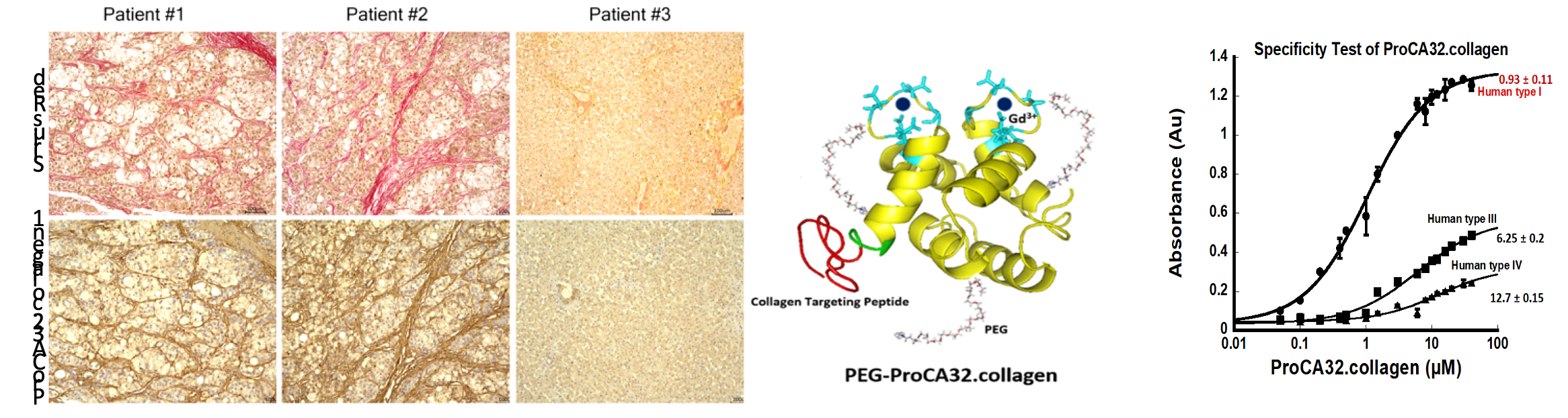
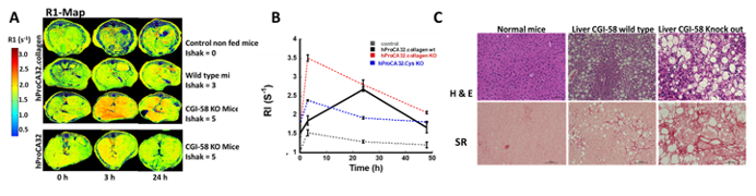
Introduction: The largest and fastest-growing population at risk for developing hepatic fibrosis are individuals with nonalcoholic fatty liver disease (NAFLD) and Alcoholic steatohepatitis (ASH). There is a pressing unmet medical need to noninvasively detect and stage various types of NAFLD/NASH, and define determinants for heterogeneity among different types of liver fibrosis. Tempo-spatial overexpression of Collagen I is used to stage liver fibrosis regardless of etiology, and to decide the risk of disease progression. Our team has pioneered a new class of protein-based gadolinium MRI contrast agents, including a non-targeting blood pool agent hProCA32, and biomarker targeting hProCA32.Collagen1, which specifically targets collagen I with strong affinity and specificity (Fig. 1). In this work, we report our validation of quantitative early detection and staging of in vivo non-alcoholic steatohepatitis (NASH) and liver fibrosis in several mouse models using a new model of Precision MRI (pMRI) enabled by hProCA32.collagen1. Methods:MRI contrast agent preparation: ProCA32.Collagen1 was expressed, purified, and characterized, according to established protocol1.Mice models: TET2 knockout mice were fed a NASH diet to study the progression of NASH, and early-stage fibrosis was induced in HDAC1 knockout mice (Jackson Laboratories, Bar Harbor, ME) by daily feeding of a high-fat diet containing 4.2% fructose in the drinking water. To create a more aggressive model of NASH and liver fibrosis, CGI-58 gene knockout mice were used and fed the same high-fat diet. Normal mice fed the NASH diet were designated as the fatty liver model, while normal mice fed a chow diet were designated as the wild-type (WT) control model.In-Vivo MRI: All animal experiments were approved by the institutional animal care and use committee (IACUCs) of Georgia State University and Emory University. MRI images of mice were acquired with a 7.0 T Bruker MRI scanner, generating MRI-proton density fat fraction (MRI-PDFF) data files. T1- and T2-weighted images, and T1- and T2-mapping files, were collected before and after a single bolus injection of hProCA32.Collagen1/hProCA32 (0.025 mmol/kg) at 3, 24, and 48 h.Results and Discussion: We first developed an imaging methodology by optimization of pulse sequences and other parameters to achieve the highest signal-to-noise ratio (SNR) and sensitivity for in vivo and ex vivo imaging, taking advantage of both r1 and r2 of ProCA32.collagen1. MRI enhancements in both R1 and R2 enabled by ProCA32.collagen1 resulted in noninvasive detection of early-stage fibrosis induced by DEN, ASH, and NASH (Fig. 2). In contrast, non-targeting ProCA32 did not produce any significant enhancement in R1 maps at these same time points (Fig. 2). Diversified heterogeneity and its change were captured by pMRI and dynamic molecular contrast enhancement (DMCE) as it evolved during disease progression (Fig. 3). High fat results in the formation of steatosis with a cooperative change during NAFLD progression due to DNA methylation modification in a Ten-Eleven-Translocation 2 knock out (TET2 KO) mouse model by PDFF (Fig. 4). For the first time, we demonstrate that pMRI has a unique capability to map liver changes associated with steatosis and inflammation in the pre-fibrosis stage (Fig. 5). Molecular radiomics using AI-assistant analysis was found to be an effective approach to detecting the tempo-spatial distribution of fibrosis pattern related to angiogenesis, steatosis, and sinusoid distribution with immunofluorescence imaging.Conclusion: The development of hProCA32.Collagen1 and pMRI imaging methodology has enabled the detection and staging of early stage liver fibrosis, and subtyping of fibrosis heterogeneity. hProCA32.Collagen1 is expected to have strong application in monitoring of liver fibrosis progression and response to treatment, and to facilitate drug discovery.

Acknowledgements

No acknowledgement found.

References

1) Salarian, Mani, et al. "Early detection and staging of chronic liver diseases with a protein MRI contrast agent." Nature communications 10.1 (2019): 1-14. 2) Yang, Jenny J., et al. "Rational design of protein-based MRI contrast agents." Journal of the American Chemical Society 130.29 (2008): 9260-9267.
Figures

Fig. 1. Specific Binding to Overexpressed Collagen in Patient Samples compared with Sirius Red staining Liver sample. hProCA32.Collagen1 shows high binding affinity to Collagen type I with ELISA measurement.

Fig. 2. (A) R1 Maps with (B) change of control (Ishak stage 0 of 6), wild type early-stage (Ishak stage 3 of 6), and late-stage (Ishak stage 5 of 6) of CGI58 mouse model liver fibrosis before and 3 h after injection of hProCA32.collagen1and hProCA32. (C) Sirius red and H&E staining of liver tissue.

Proc. Intl. Soc. Mag. Reson. Med. 32 (2024)
3217
DOI: https://doi.org/10.58530/2024/3217