3216
Resotran - A positive direction for negative contrast: clinically approved Ferucarbotran reintroduced1Experimental Physics 5 (Biophysics), Julius-Maximilians University, Würzburg, Germany, 2Department of Diagnostic and Interventional Radiology, University Hospital Würzburg, Würzburg, Germany, 3Department of Diagnostic and Interventional Neuroradiology, University Hospital Würzburg, Würzburg, Germany, 4Radiologie Augsburg Friedberg, Augsburg, Germany, 5Pure Devices GmbH, Rimpar, Germany
Synopsis
Keywords: Hybrid & Novel Systems Technology, Magnetization transfer, Magnetic particle spectroscopy, MPS, COMPASS, contrast agents, iron oxide based
Motivation: Contrast agents allow a significant improvement of MRI performance. Resotran is a new iron oxide-based and biocompatible MRI contrast agent that is now commercially available in Europe, which still needs to be characterized in more detail.
Goal(s): Classification of the performance of Resotran compared to Resovist.
Approach: Analyzation and characterization with various spectroscopic and imaging methods.
Results: Resotran shows a comparable performance to the former liver contrast agent Resovist. Furthermore, it was shown that Resotran is feasible as a tracer for the new imaging modality magnetic particle imaging (MPI). This is an important step towards paving the way for MPI in clinical routine.
Impact: In this work it is shown that the new clinically approved contrast agent Resotran shows a comparable performance as the well-known contrast agent Resovist by a detailed comparison applying different spectroscopic characterization methods.
Introduction
At least 40% of all clinical MR examinations are performed using contrast agents, which significantly improve diagnocstic quality or certain pathologies. Most contrast agents in clinical use are based on gadolinium. Despite the significant enhancement of contrast, gadolinium is suspected to be nephrotoxic1.Alternatively, iron-oxide-based contrast agents can be used for negative contrast imaging as known from liver contrast agent Resovist. These contrast agents consist of superparamagnetic iron oxide nanoparticles (SPIONs) promising high biocompatibility1.
Additionally, the potential of SPIONs is not limited to the application in MRI. They gained increasing relevance for the emerging imaging modality magnetic particle imaging (MPI), that shows promising results for radiation-free real-time imaging of SPIONs as a tracer2.
However, in the recent years clinically approved iron oxide-based contrast agent were not commercially available. This changed recently on the european market with the commercially available Ferucarbotran based contrast agent Resotran (b.e.imaging, Germany).
Due to its novelty, there is a lack of data concerning the characterization of Resotran. To fill this gap, we provide first results of characterization-measurements with Resotran, which are performed using an established method, namely magnetic particle spectroscopy (MPS)3, and a recently proposed method named critical offset magnetic particle spectroscopy (COMPASS) that shows high potential in the field of SPION characterization4.
Additionally, it is tested if Resotran is also applicable as a tracer for MPI, which is an important aspect for the development of MPI towards clinical approvement. The characterization of Resotran is compared to the well characterized SPION system Resovist.
Methods
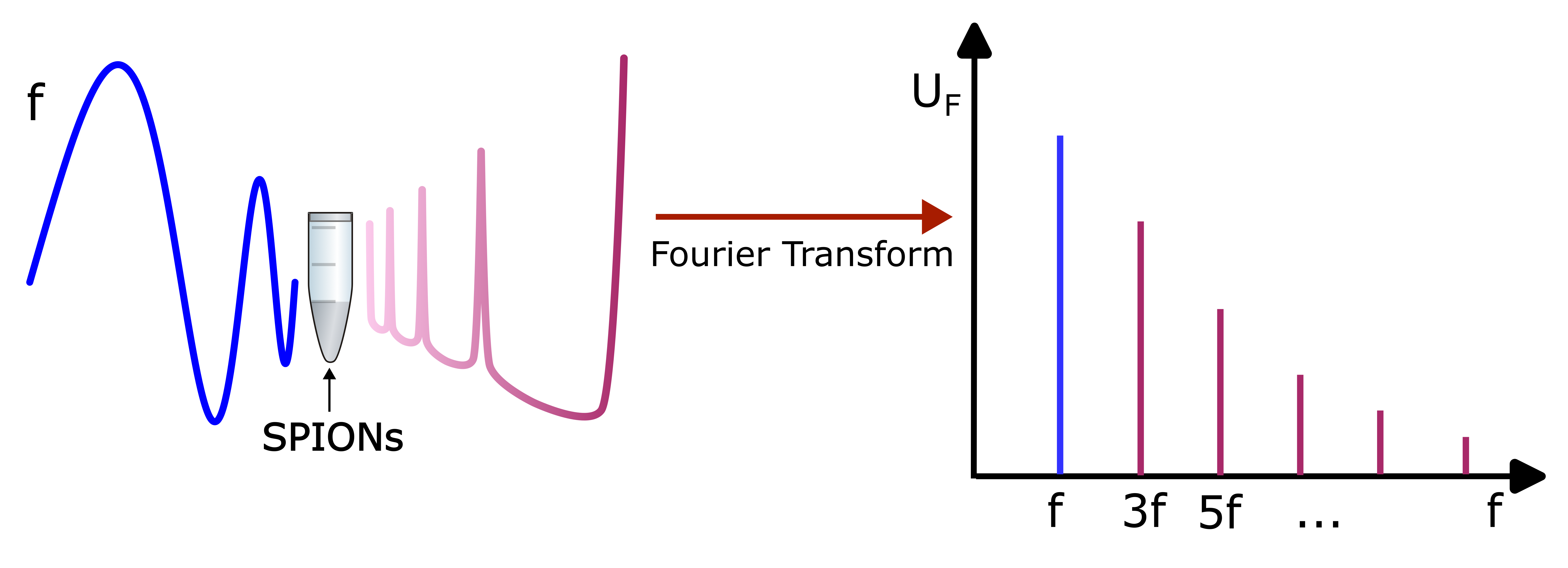
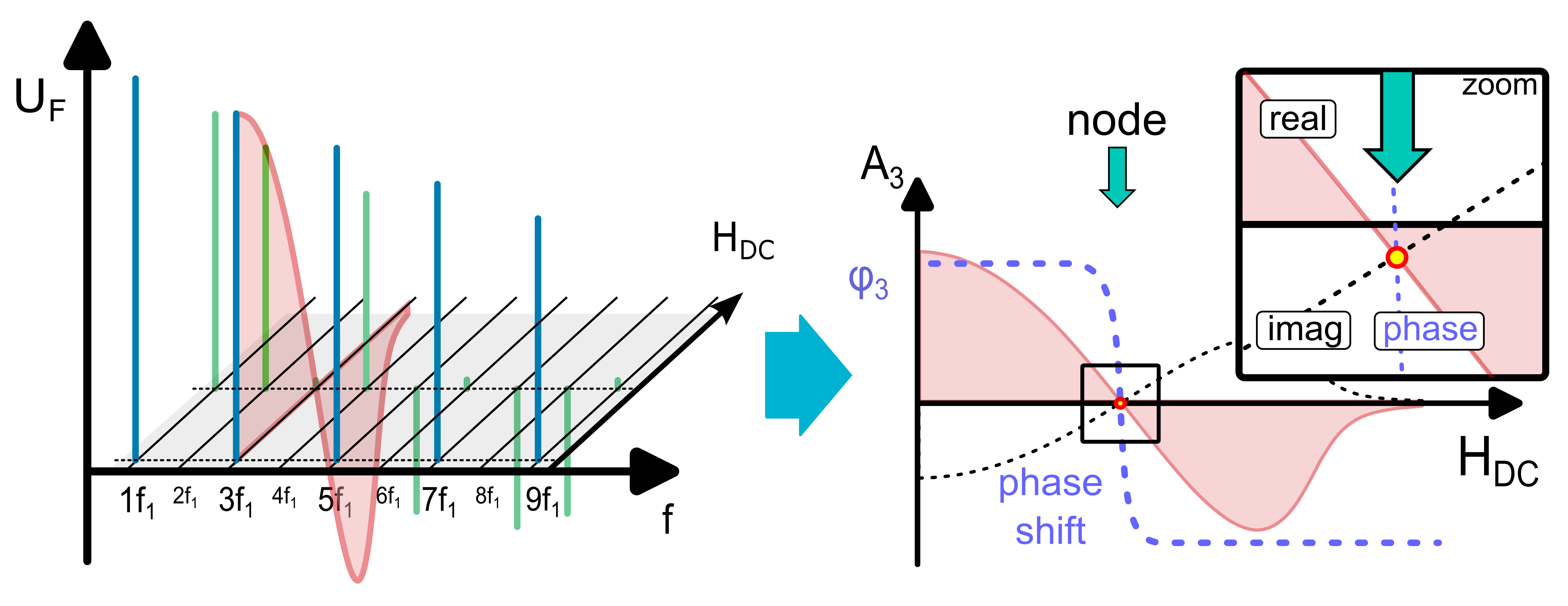
For reliable comparison both SPION types were analyzed with MPS and COMPASS. Both modalities are highly sensitive and easy-to-use systems providing rapidly specific information about the particle core, particle-particle interaction, relaxation parameters as well as the s urrounding medium3,4. MPS is a spectroscopy method using a sinusoidal magnetic field with a sufficient magnitude to saturate the SPIONs’ magnetization . The inductively detected SPION-signal is Fourier transformed resulting in a frequency spectrum with higher harmonics of the excitation frequency. This provides a characteristic fingerprint for the measured SPION (see figure 1).The concept of MPS can be extended by adding a static offset field HDC to the sinusoidal excitation field , which is referred to as COMPASS. Following the course of the phase of a single harmonic during varying the offset field strength as shown in figure 2, multiple phase jumps (critical points - CPs) occur at specific HAC/HDC ratios. For this type of measurement, the location of each CP is characteristic for the SPION-system.
Additionally, the imaging performance of the SPIONS are compared using a human-sized MPI-scanner developed for assisting in percutaneous transluminal angioplasty5.
Results and Discussion
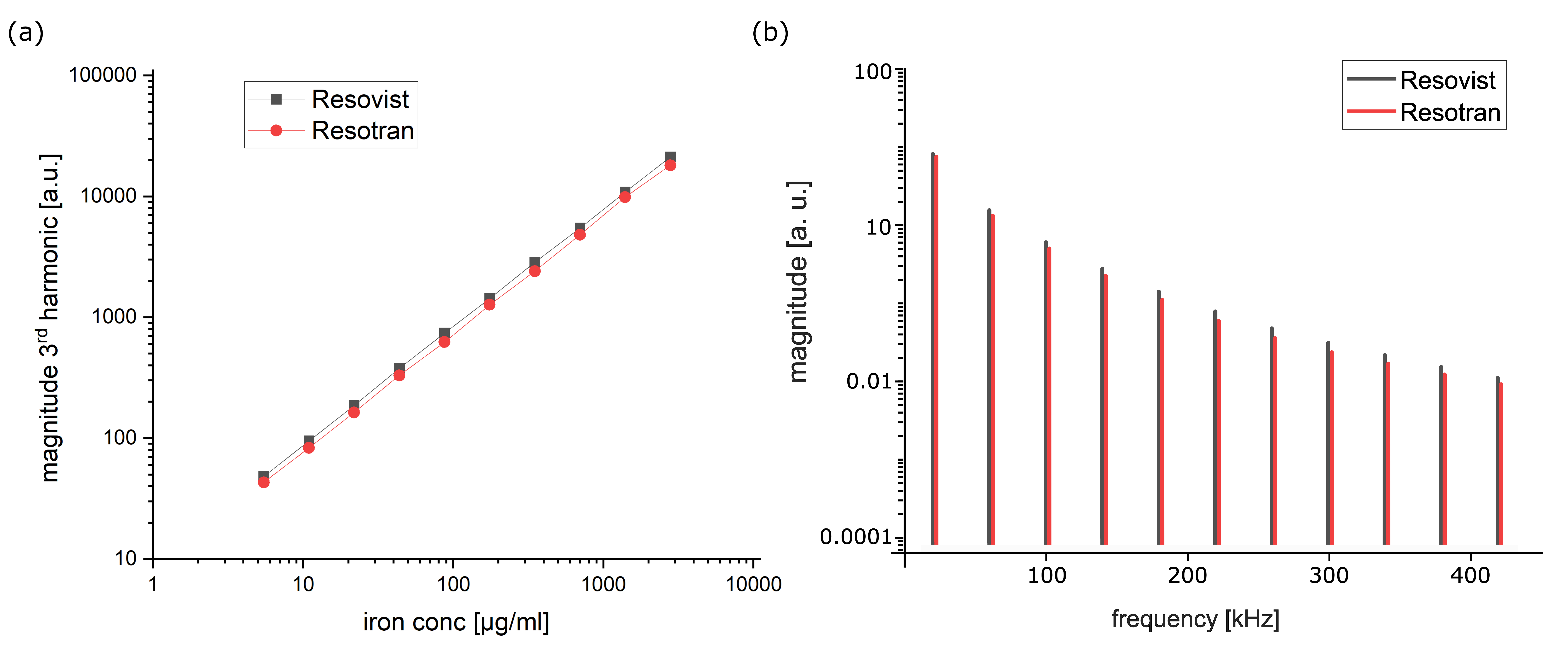
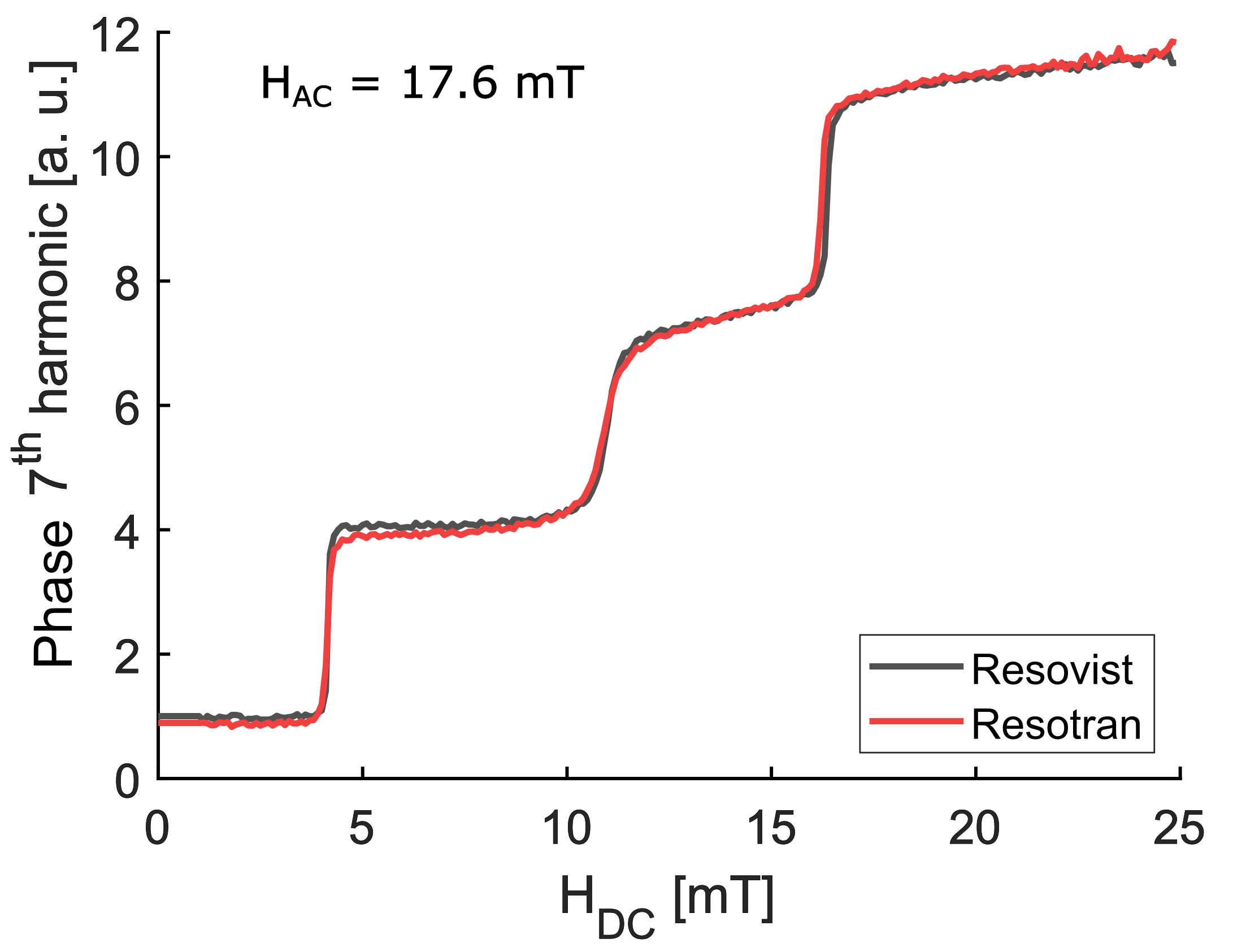
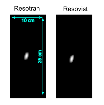
The results of MPS measurements of Resovist and Resotran are compared in figure 3. It depicts the behavior of the signal amplitude of the 3rd harmonic for samples of different iron contents and the complete spectra of undiluted Resovist and Resotran. It is evident that both SPION-types show the expected linear behavior of the amplitude. However, the amplitudes of Resotran show a 10% smaller signal than Resovist. The COMPASS measurement shown in figure 4 aims for creating a unique fingerprint of the measured SPION ensemble by the characteristic position of the CP. Comparing the complex phase of the 7th harmonic the two curves show a high agreement indicating a high similarity of the principal structure of Resotran and Resovist. As a third comparison, both undiluted samples have been imaged with a human sized portable MPI scanner. The reconstructed images of a 50µl sample are shown in figure 5. The graphic indicates that both SPIONS have sharp point spread functions and thus provide a good imaging quality.Conclusion
Here, we presented a first characterization of Resotran, a new clinically approved Ferucarbotran-based contrast agent for MRI using MPS and COMPASS for a spectroscopic analysis. The characterization is accompanied with a systematic comparison to the well-known former contrast agent Resovist. The results show that the signal amplitude of Resotran is reduced by 10% compared to Resovist. Still, the COMPASS fingerprints indicate a relatively similar structure of the two SPION-types. Additionally, both Resotran and Resovist show a similar imaging performance for MPI.Thus, Resotran promises a performance similar to the quality of Resovist for MRI as well as MPI, which opens up new possibilities for the clinical applicability of iron oxide-based contrast agents in MRI and MPI in general.
Acknowledgements
Research funding: The work was supported by theGerman Research Council (DFG), grant numbers: VO-2288/1-1, VO-2288/3-1, and BE 5293/1-2.References
- Shen Z, Wu A, Chen X. Iron Oxide Nanoparticle Based Contrast Agents for Magnetic Resonance Imaging. Mol. Pharmaceutics 2017;14(5):1352–1364.
- Gleich B, Weizenecker J. Tomographic imaging using the nonlinear response of magnetic particles. Nature 2005;435:1214–1217.
- Wu K, Su D, Saha R, et al. Magnetic Particle Spectroscopy: A Short Review of Applications Using Magnetic Nanoparticles. ACS Appl. Nano Mater. 2020;3(6):4972–4989.
- Vogel P, Rückert MA, Friedrich B, et al. Critical Offset Magnetic PArticle SpectroScopy for rapid and highly sensitive medical point-of-care diagnostics, Nat. Commun 2022;13(1):7230.
- Vogel P, Rückert MA, Greiner C, et al. iMPI: portable human-sized magnetic particle imaging scanner for real-time endovascular interventions. Sci. Rep. 2023;13:10472.
Figures