3214
Detection of low calcium channel activity in pig hearts with manganese-enhanced MRI1Department of Physics and Computational Radiology, Oslo University Hospital, OSLO, Norway, 2Department of Physics and Computational Radiology, Oslo University Hospital, Oslo, Norway, 3IC Targets AS, Oslo, Norway
Synopsis
Keywords: Contrast Agents, Myocardium, MEMRI, Manganese, Mangafodipir, pig study, T tubuli, Ca2+ channel activity
Motivation: Evaluate manganese-enhanced MRI (MEMRI) for heart failure assessment.
Goal(s): Quantify the manganese uptake rate in the myocardium as an indicator of L-type calcium channel activity and T-tubule density.
Approach: A high temporally-resolved dynamic SatRec sequence was used to track the Mangafodipir bolus instead of using the less precise, but higher spatially-resolved, conventional T1 mapping method that was used before and after the manganese administration.
Results: Pigs have less dense T-tubule than humans leading to fewer L-type calcium channels and low Mn uptake in cardiomyocytes. Therefore, we believe that MEMRI can become a promising tool for assessing heart failure diseases in humans.
Impact: Since pigs are often used in preclinical research, there are important differences to be aware of. The low Mn uptake proved their less dense T-tubule, showing that MEMRI may be a useful tool for studying heart failure in humans.
Introduction
This study aims to test a new MRI method to quantify the uptake rate of manganese (Mn) in the myocardium as an indicator of L-type calcium channel (LTCC) activity and T-tubule density. T-tubuli ensure rapid propagation of the action potential to the LTCCs which are located in the T-tubule membranes. Reduced LTCC activity may be caused by loss of cardiomyocytes (e.g. fibrosis) or loss of T-tubuli in heart failure [1]. Mn$$$^{2+}$$$-ions are taken up by cardiomyocytes through the LTCCs in proportion to calcium influx and increases intracellular longitudinal relaxation rate (R1=1/T1) in proportion to the Mn concentration. The study also measured manganese in blood and tissue samples to calculate the relaxivities at 3T in pigs. Previous studies have used well-established T1 mapping (MOLLI and shMOLLI) methods to measure the manganese uptake rate [2], requiring relatively long periods of breath-hold which may be difficult to apply repeatedly in patients with heart failure. Since T1 mapping is recorded over approx. 12 seconds, it may also have an inadequate temporal resolution to follow the kinetics of manganese uptake particularly during and after rapid infusion.Methods
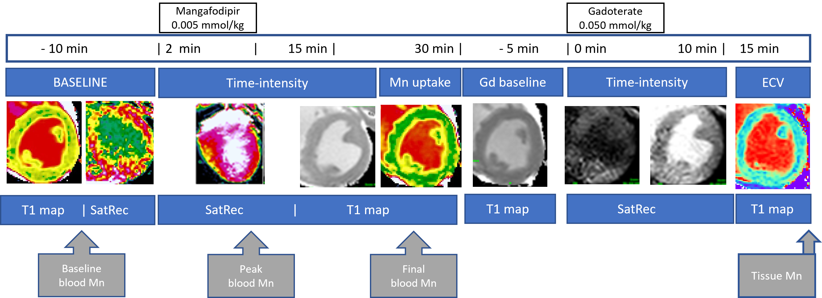
The study was performed in six normal domestic pigs with body weights of 50-60 kg. 0.005 mmol/kg Mangafodipir (MnDPDP) was infused over 2 minutes. Septum and blood relaxation rates at baseline (1/T1(0)) were measured with a native MOLLI 5s(3s)3s scheme. The SatRec signal intensities, SI(t), were converted into R1(t) values using the baseline signal intensity SI(0) and T1(0) values: $$R_{1}(t)=-\frac{1}{TD}\times{ln}[1-(1-e^{-\frac{TD}{T_{1}(0)}})\times\frac{SI(0)}{SI(t)}]$$ , where TD is the saturation time delay. Hematocrit (hct) was used to calculate plasma relaxation rates: $$$R_1^{plasma}=R_1^{blood}/(1 – hct)$$$ The integral of changes in blood relaxation rates were calculated with the trapezoid formula: $$\int_{a}^{b}ydx≈\sum_{i=1}^{n}(t_{i}-t_{i-1})\times0.5\times(y_{i}+y_{i-1})$$ Gadoterate meglumine was administered after the mangafodipir experiment to measure the extracellular volume (ECV) using an enhanced MOLLI 4s(1s)3s(1s)2s scheme (Figure 1): $$ECV=\frac{\triangle{R_1^{myo}}}{\triangle{R_1^{blood}}}\times(1-hct)$$ Mn$$$^{2+}$$$ enters cardiomyocytes through the LTCCs and is irreversibly trapped for several hours [3]. The rate constant $$$k_{Mn}^{myo}$$$ for irreversible uptake in tissue can be measured using the Patlak Plot method [4]. $$\frac{\triangle R_1^{myo}(t)}{ΔR_1^{blood}(t)}\times(1-hct)=V_{c}+k_{Mn}^{myo}\times\frac{\int_{0}^{t} \triangle R_1^{blood}(t)dt}{\triangle R_1^{blood}(t)}$$ The intercept V$$$_{c}$$$ represents the reversible tissue compartment in the model. Assuming that the cardiomyocyte volume fraction is 1 – ECV, the rate constant for cellular uptake of manganese is: $$k_{Mn}^{cell} \approx k_{Mn}^{myo} / (1 - ECV)$$ The relaxivity of manganese (r$$$_{1}$$$) was calculated from the change in relaxation rate, change in concentration of manganese [ppm] and the molecular weight (MW) of manganese (54.94 g/mol): $$r_{1} = \frac{\triangle R_1\times MW \times 1000}{\triangle C_{Mn}}$$Results
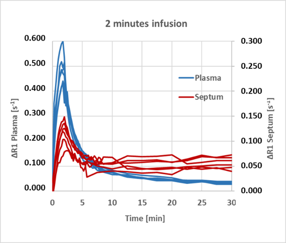
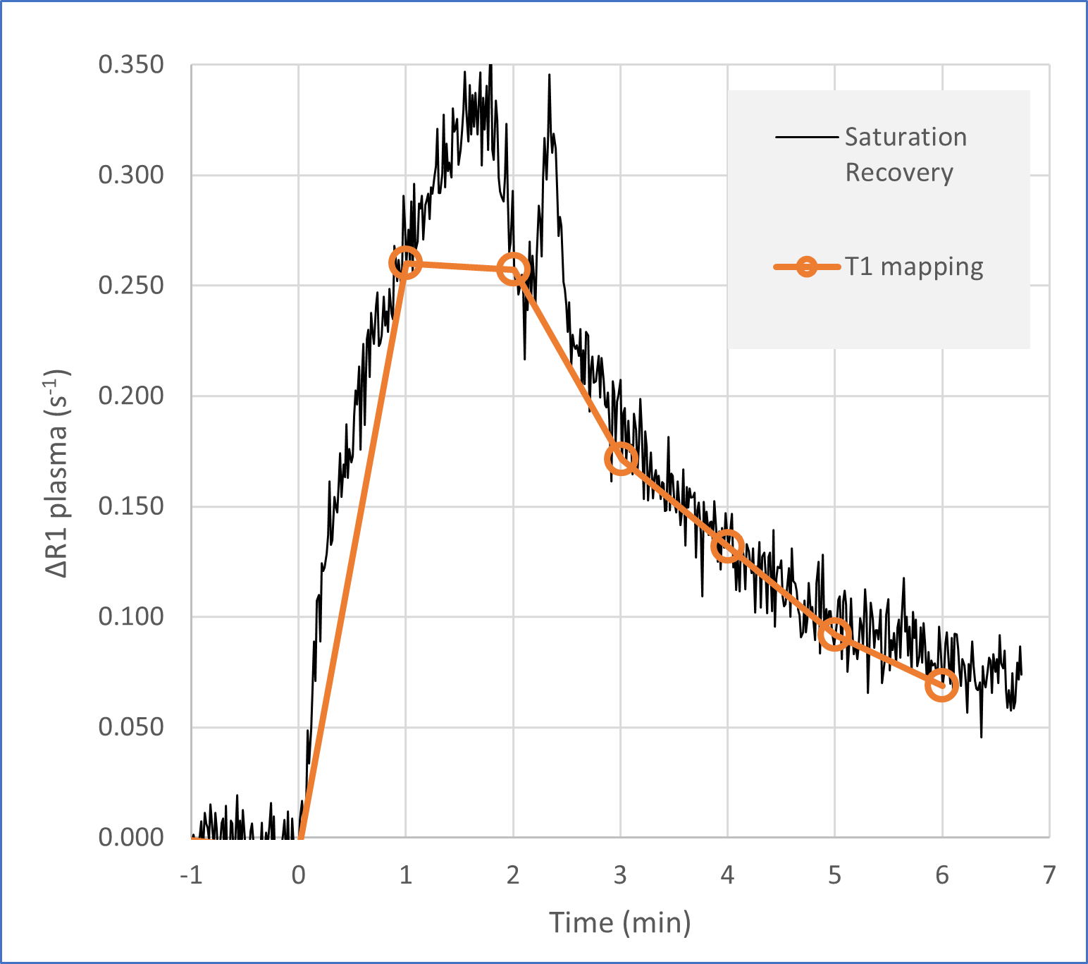
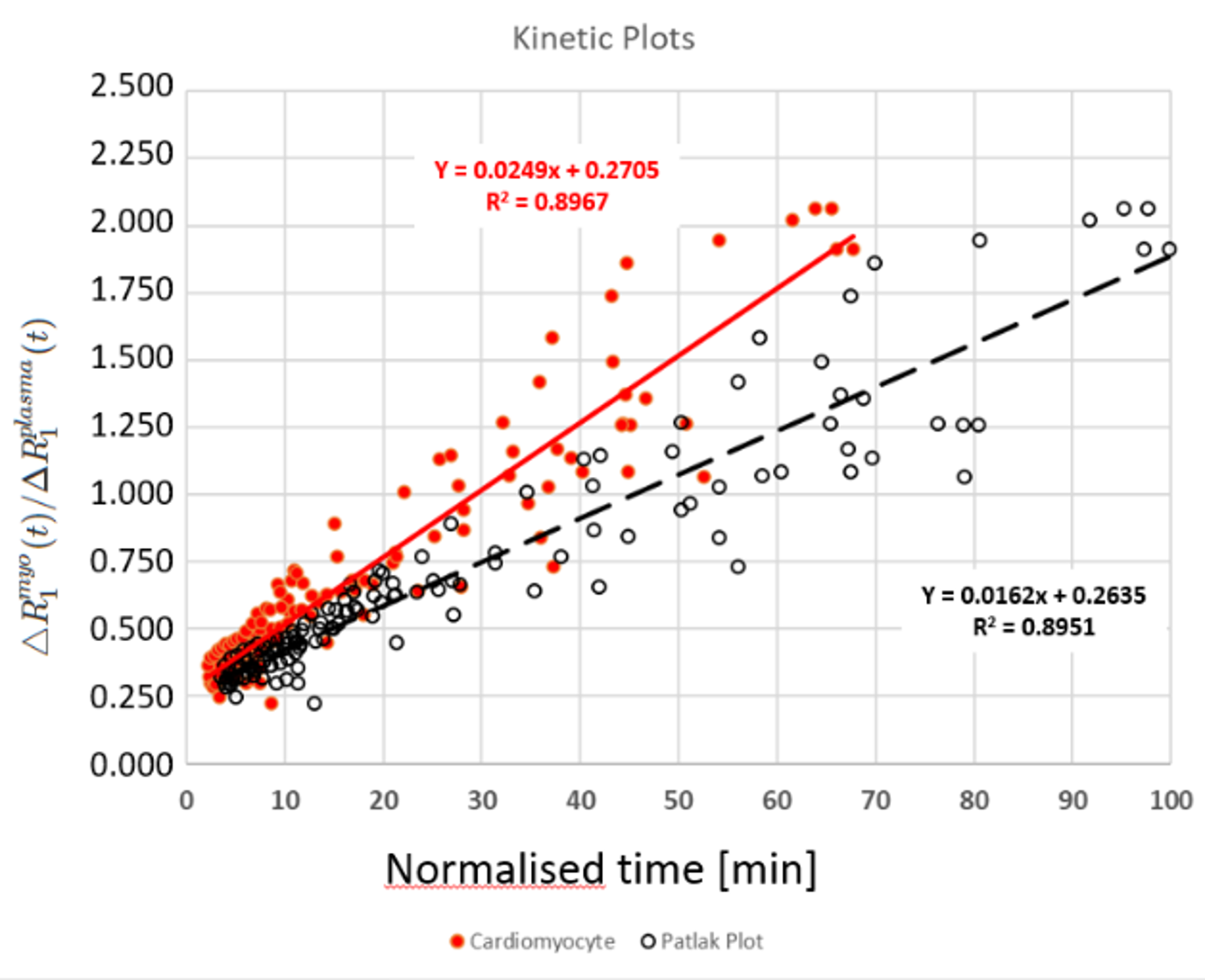
Baseline T1 in septum and blood was 1177±35 ms and 1666±64 ms, respectively. The relaxation effect ΔR1 in septum and plasma reached maxima at the end of infusion of 0.122±0.025 s$$$^{-1}$$$ and 0.495±0.069 s$$$^{-1}$$$. Immediately after the infusion there was a rapid reduction of ΔR1 in septum to less than half the peak value (Figure 2). After 30 minutes, blood T1 had returned to baseline, whereas septum T1 remained at a plateau of 1102±22 ms corresponding to an irreversible increase in myocardial R1 of 0.058±0.011 s$$$^{-1}$$$. Maximum plasma manganese concentration was 1793±260 ppm with a r$$$_{1}$$$ of 11.4±0.9 s$$$^{-1}$$$mM$$$^{-1}$$$. At the end, tissue manganese was raised by 324±64 ppm with an average tissue r$$$_{1}$$$ of 10.6±4.4 s$$$^{-1}$$$ mM$$$^{-1}$$$. The ECV was 0.337±0.017.Rapid infusion of MnDPDP leads to rapid changes in the plasma R1 (Figure 3) during conventional T1 mapping (12 seconds), which might not have adequate temporal resolution for integration of the time intensity curve with an upward convex shape, leading to a significant underestimation of the integral. This problem was solved by using SatRec instead of T1 mapping. The ΔR1 rate constant was 0.0162±0.0056 min$$$^{-1}$$$ for total tissue (Patlak method) and 0.0249±0.0085 min$$$^{-1}$$$ for the cardiomyocyte cell fraction (Figure 4).
Discussion
SatRec provided superior temporal resolution for measurement of the integral of ΔR1 in blood, which in combination with T1 mapping provided better data for the kinetic analysis than T1 maps alone. The tissue uptake rate constant 0.0162 min$$$^{-1}$$$ in these normal pigs was dramatically lower than values reported in humans, and for whom tissue R1 continues to increase after the end of infusion [2,4]. Our results support the finding that normal pig heart has fewer T-tubuli than humans [5] and indicate that Mn(DPDP)-enhanced-MRI can assess T-tubule density in human heart diseases.Conclusion
The imaging protocol tested in this study provides a better basis for kinetic analysis of MEMRI data and does not require multiple breath-hold scans. The low uptake rate in pigs show that mangafodipir has the potential to measure T-tubule density in human heart disease.Acknowledgements
We would like to give a special thanks to Grethe Løvland and Itai Chalit and co-workers for their professionality and dedication in this project in the operation theatre and MR lab at the Intervention Centre, Oslo University Hospital, Oslo, Norway. The project was funded by The Research Council of Norway (327815).References
- Louch WE, Sejersted OM, and Swift F. There Goes the Neighborhood: Pathological Alterations in T-Tubule Morphology and Consequences for Cardiomyocyte Ca$$$^{2+}$$$ Handling. J Biomed Biotech. 2010:1-17.
- Spath NB, Singh T, Papanastasiou G, et al. Assessment of stunned and viable myocardium using manganese-enhanced MRI. 2021;8:e001646. doi:10.1136/openhrt-2021-001646.
- Hunter DR, Hawort RA, and Berkoff HA. Cellular Manganese Uptake by the Isolated Perfused Rat Heart: a Probe for the Sarcolemma Calcium Channel. J. Mol. Cell. Cardiology. 1981;13:823-832.
- Skjold A, Kristoffersen A, Vangberg TR, et al. An Apparent Unidirectional Influx Constant for Manganese as a Measure of Myocardial Calcium Channel Activity. J Magn Res Imaging. 2006;24:1047-1055.
- Heinzel FR, Bito V, Volders PGA, et al. Spatial and
Temporal Inhomogeneities During Ca$$$^{2+}$$$ Release From the Sarcoplasmic Reticulum
in Pig Ventricular Myocytes Circ Res. 2002;91:1023-1030.
Figures