3209
Measurement of tumor extracellular pH (pHe) by a PET/MRI co-inject reagent1Department of Cancer Systems Imaging, UT MD Anderson Cancer Center, Houston, TX, United States, 2University of Turin, Turin, Italy, 3Stony Brook University, Stony Brook, NY, United States
Synopsis
Keywords: Contrast Agents, Cancer, Simultaneous PET/MRI, 68Ga contrast agent, extracellular pH measurement, 4T1
Motivation: The measurement of extracellular pH (pHe) is very important for studying the cancer development, progression, and therapeutic response.
Goal(s): Accurately measure the pHe for 4T1 tumor and 0.2 pH change in vitro independent on the concentration of MRI contrast reagent.
Approach: To co-inject the synthesized pH sensitive MRI contrast reagent and the PET reagent with the same pharmacokinetic functions and DCE MRI and dynamic PET scan were obtained simultaneously.
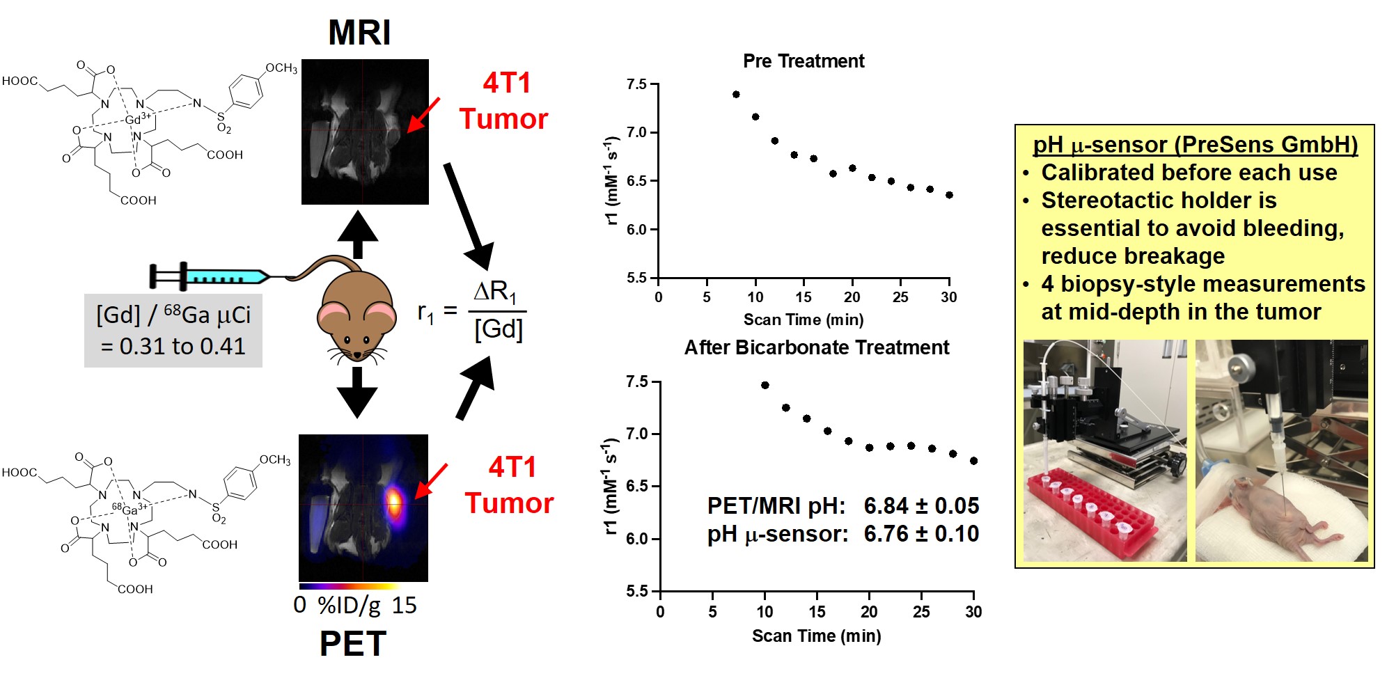
Results: The pHe change of the bicarbonate treatment for the acidosis tumor type can be measured by our PET/MRI co-inject reagent.
Impact: PET/MRI co-inject reagent makes it possible for the pHe measurement independent of the concentration of MRI contrast reagent.
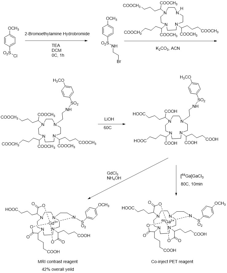
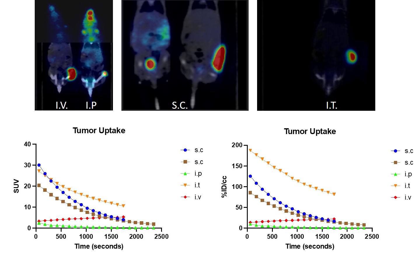
For the synthesis of both reagents, the amide group was introduced by substitution of 4-methoxybenzenesulfonyl chloride with 2-bromoethylamine hydrobromide. The amide arm was added onto the unprotected amide of hexamethyl 2,2',2''-(1,4,7,10-tetraazacyclododecane-1,4,7-triyl) triadipate followed by the deprotection of the carboxyl methyl ester with lithium hydroxide (Figure 1). The resulting free acid was used to chelate gadolinium (MRI) and 68Ga (PET) separately to obtain the MRI/PET co-agent pair. The overall yield is around 42%. We performed the kinetic PET studies in a 4T1 breast cancer model (Figure 4). The PET reagent was stable for one hour, enough for our PET/MRI co-agent imaging protocol.
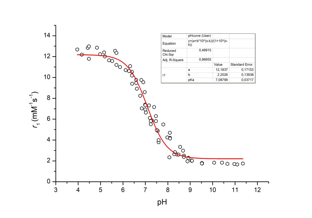
Our MRI agent can detect a pH difference of 0.2 pH units in the phantom with relatively high sensitivity (Figure 2). The pHe of 4T1 breast cancer with and without bicarbonate treatment was measured by our PET/MRI contrast reagent. The r1 relaxivity of the MRI co-agent was determined by the ratio of the measurement of PET radioactivity and MRI ∆R1 relaxation rate. The dynamic changes in ∆R1 relaxation rate and PET-derived MRI co-agent concentration were monitored at 7.6 sec and 2-min temporal resolution, respectively as averaged for the tumor region in the image. The r1 relaxivity was determined from ∆R1 and concentration from each two-minute time interval and converted to pHe based on the pH-relaxivity calibration (Figure 3). This pHe was found to be consistent for five measurements, made 20 minutes after s.c. injection, presumably when the MRI co-agent had accumulated in the tumor to a sufficient concentration for areliable measurement. The average of these five pHe measurements after 20 minutes post injection was 6.84 ± 0.05 pH units which was similar to the gold standard pH microsensor 6.76 ± 0.10 pH units.
These preliminary results showed that tumor acidosis can be evaluated with simultaneous 68Ga-PET/ Gd-MRI co-agents.
We are currently synthesizing other PET reagents with different isotopes for radiolabeling, such as 86Y and 18F. The best candidate showing the best pharmacokinetic stability and highest accuracy in pH measurement will be tested with other cancer treatment. Our PET/MRI co-inject reagent will become the indicator for tumor development, progression and treatment response.
Acknowledgements
We would like to thank the Small Animal Imaging Facility of the MD Anderson Cancer Center. This work was funded by NIH/NCI R21EB027197 and NIH/NCI P30 CA016672.References
1. Ji, K., et al., Acidosis and proteolysis in the tumor microenvironment. Cancer Metastasis Rev, 2019. 38(1-2): p. 103-112.
2. Yang, L.V., Tumor Microenvironment and Metabolism. Int J Mol Sci, 2017. 18(12).
3. Glunde, K. and Z.M. Bhujwalla, Metabolic tumor imaging using magnetic resonance spectroscopy. Semin Oncol, 2011. 38(1): p. 26-41.
4. Dou, W., et al., Chemical exchange saturation transfer magnetic resonance imaging and its main and potential applications in pre-clinical and clinical studies. Quant Imaging Med Surg, 2019. 9(10): p. 1747-1766.
5. Gao, T., et al., A Brief History and Future Prospects of CEST MRI in Clinical Non-Brain Tumor Imaging. Int J Mol Sci, 2021. 22(21).
6. Pollard, A.C., et al., Radiometal-Based PET/MRI Contrast Agents for Sensing Tumor Extracellular pH. Biosensors, 2022. 12(2): p. 134.
Figures