3205

Noninvasive Early Detection and Stage of Chronic Lung Diseases with Precision MRI (pMRI)
Dongjun Li1, Zongxiang Gui1, Jingjuan Qiao1, Sophia Bamishaye1, and Jenny Yang2
1Georgia State University, Atlanta, GA, United States, 2Chemistry, Georgia State University, Atlanta, GA, United States

Synopsis

Keywords: Contrast Agents, Lung, MRI, collagen, Protein-based contrast agent, lung fibrosis, IPF, COPD, lung cancer

Motivation: MRI diagnosis of lung has a limitation due to the air in the lungs which prevents the water signals. We use precision MRI to image the small lesions or fibrosis in the lung.

Goal(s): Our goal is to quantitatively stage the lung diseases by MRI contrast agents.

Approach: Our novel MRI contrast agent is biomarker specific which enables it target to the lung lesions or fibrosis.

Results: We are able to image the lung cancer and metastasis as well as lung fibrosis using our developed MRI contrast agent.

Impact: The imaging of lung tumor lesion or fibrosis is always hindered by the lung features. Our novol contrast agents enable us to image the lung diseases at early stages.

Chronic lung diseases, such as idiopathic pulmonary fibrosis (IPF), chronic obstructive pulmonary disease (COPD), and lung cancer, are major leading causes of death worldwide and are generally associated with poor prognoses. The heterogeneous distribution of collagen, mainly type I collagen associated with excessive collagen deposition, plays a pivotal role in the progressive remodeling of the lung parenchyma to chronic exertional dyspnea for IPF, COPD and lung cancer. To address the pressing need for noninvasive early diagnosis and drug treatment monitoring of pulmonary fibrosis and lung cancer, we report the development of human collagen-targeted protein MRI contrast agent (hProCA32.collagen1) to specifically bind to collagen I overexpressed in multiple lung diseases. When compared to clinically-approved Gd3+ contrast agents, hProCA32.collagen1 exhibits significantly better r1 and r2 relaxivity values, strong metal binding affinity and selectivity, and transmetallation resistance. Here we report the robust detection of early and late-stage lung fibrosis with stage-dependent MRI signal-to-noise ratio (SNR) increase, and with good sensitivity and specificity, using a progressive bleomycin-induced IPF mouse model. Spatial heterogeneous mapping of usual interstitial pneumonia (UIP) patterns with key features closely mimicking human IPF, including cystic clustering, honeycombing, and traction bronchiectasis, were noninvasively detected by multiple MR imaging techniques and verified by histological correlation. We further report the detection of fibrosis in the lung airway of an electronic cigarette-induced COPD mouse model, using hProCA32.collagen1-enabled precision MRI (pMRI), and validated by histological analysis. We have also achieved the first detection of subclinical invasive lung nodules of LKB1 with heterogenous distribution of collagen in our mouse model with a 3-fold increase of Contrast-Noise Ratio (CNR) compared with CT imaging. The developed hProCA32.collagen1 is expected to have strong translational potential for the noninvasive detection and staging of lung diseases, and facilitating effective treatment to halt further chronic lung disease progression.
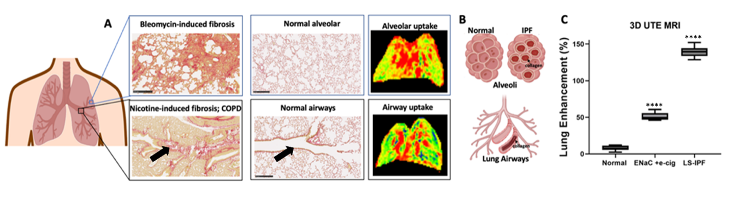
Figure. Comparison of collagen accumulation pattern and hProCA32.collagen1 pattern uptake in BM-induced IPF and E-cigarette-induced lung fibrosis in COPD mouse models. (A-B). Collagen accumulation and binding in IPF (top) and COPD (bottom) with overexpression of collagen 1 (red color) at alveolar and bronchial airways, respectively, compared to normal healthy mice (middle panel). Human collagen-targeted protein MRI contrast agent (hProCA32.collagen1) detected collagen overexpression in IPF and COPD at the alveolar (top) and bronchial airways (bottom) in 3D UTE MRI. (C) Comparison of lung signal enhancement (%) in normal mice, nicotine-induced lung fibrosis model for COPD, and bleomycin-induced mice model (LS: Late stage, ****P < 0.001 one-way ANOVA)

Acknowledgements

No acknowledgement found.

References

1. Yang, J.J., et al., Rational design of protein-based MRI contrast agents. Journal of the American Chemical Society, 2008. 130(29): p. 9260-9267.2. Xue, S., et al., Protein MRI contrast agent with unprecedented metal selectivity and sensitivity for liver cancer imaging. Proceedings of the National Academy of Sciences, 2015. 112(21): p. 6607-6612.3. Salarian, M., et al., Early detection and staging of chronic liver diseases with a protein MRI contrast agent. Nature communications, 2019. 10(1): p. 4777.4. Ibhagui, O.Y., et al., Early Detection and Staging of Lung Fibrosis Enabled by Collagen-Targeted MRI. Protein Contrast Agent. Chemical & Biomedical Imaging, 2023.

Figures

Comparison of collagen accumulation pattern and hProCA32.collagen1 pattern uptake in BM-induced IPF and E-cigarette-induced lung fibrosis in COPD mouse models.

Proc. Intl. Soc. Mag. Reson. Med. 32 (2024)
3205
DOI: https://doi.org/10.58530/2024/3205