3204
Concurrent Dual-Contrast Enhancement Using Fe3O4 Nanoparticles to Achieve a CEST Signal Controllability1Department of Radiology, Fudan University Shanghai Cancer Center, Shanghai, China, 2Department of Ultrasound, Shanghai Municipal Hospital of Traditional Chinese Medicine, Shanghai, China, 3Department of Radiology, Children’s Hospital of Fudan University, Shanghai, China, 4GE Healthcare, Beijing, China
Synopsis
Keywords: Contrast Agents, Contrast Agent
Motivation: The unique microenvironment of tumors making detection of microenvironmental alterations a valuable asset for early diagnosis of tumors.
Goal(s): This study focuses on exploring impact of Fe3O4 NPs on changes in the tumor microenvironment's acidity.
Approach: By combining them with CEST exogenous contrast agent, we evaluated the feasibility of dual-contrast imaging.
Results: In vitro MRI results suggest that Fe3O4 NPs can act as stable reference agents under varying pH conditions, providing negative contrast for T2-weighted MRI. The CEST effect shows a strong correlation with pH, allowing for quantitative assessment of pH shifts in tumor microenvironment, and can be controlled by adjusting Fe3O4 concentration.
Impact: These preliminary findings represent an initial stride toward dual-contrast imaging involving CEST and T2 contrast agents, with the potential for further expansion in future in vivo investigations.
Introduction
Conventional imaging techniques primarily focus on detecting observable anatomical or morphological changes in cells, offering insight into the cumulative impact of molecular alterations in lesioned cells. Recent advances in molecular imaging enable the detection of anomalies at the cellular and molecular levels in early lesions, even before the emergence of anatomical or morphological changes associated with diseases. The unique microenvironment of tumors deviates significantly from that of normal tissues, making early detection of microenvironmental changes a valuable asset for early diagnosis of tumors1. Fe3O4 nanoparticles (NPs) can serve as switchable T1/T2 contrast agents in MRI by adjusting the size of NPs2-6. They possess catalytic activity akin to peroxidase, facilitating the conversion of endogenous hydrogen peroxide into highly cytotoxic hydroxyl radicals via the Fenton reaction, leading to tumor cell destruction7,8. This has given rise to a promising avenue in cancer treatment known as chemodynamic therapy based on the Fenton reaction9. Given that metabolic changes are core indicators of tumors, characterizing the tumor microenvironment through in vivo monitoring of metabolite alterations is a pressing research challenge. Therefore, this study delves into the use of Fe3O4 NPs to explore their impact on the tumor microenvironment's acidity changes. We combined them with a CEST exogenous contrast agent, ioversol, investigated the modulation of CEST signals by Fe3O4 NPs to reflect the distribution of active components in tumors, and aimed to evaluate the feasibility of dual-contrast enhancement.Materials and Methods
Fe3O4 NPs were prepared using the hydrothermal method. FeCl3·6H2O was dissolved in ethylene glycol, and CH3COONH4 was added to the solution. The mixture was subjected to a hydrothermal reaction at 200 °C for 12 hours. The resulting black magnetic precipitates were collected, washed with ethanol and deionized water, and dried to obtain Fe3O4 NPs. Fe3O4 NP powder was weighed, and its Fe3+ concentration was quantified. The NPs were diluted with PBS at different pH values and combined with agar powder. Ioversol solutions at different pH values were prepared, and different concentrations of Fe3O4 were added to ioversol solutions. In vitro MRI was conducted using a 3.0 T scanner (SIGNA Pioneer; GE Healthcare, Milwaukee, WI). T1WI and T2WI were acquired using 2D FSE sequences. CEST images were obtained with a 2D SE-EPI sequence and saturation RF powers of 1.25/2.5/3.75/5 µT. From the CEST data, the Z-spectrum was plotted and the MTRasym at 4.3 ppm was calculated. The relationship between the CEST effect and pH was evaluated using linear fitting. Relaxation mapping was acquired using a multidynamic multiecho 2D FSE (MAGiC) sequence. The R2 values under different pH conditions were obtained from the MAGiC data through single exponential fitting. Cell survival rates were calculated, and the IC50 value was determined based on the concentration where the cell survival rate reached 50%.Results
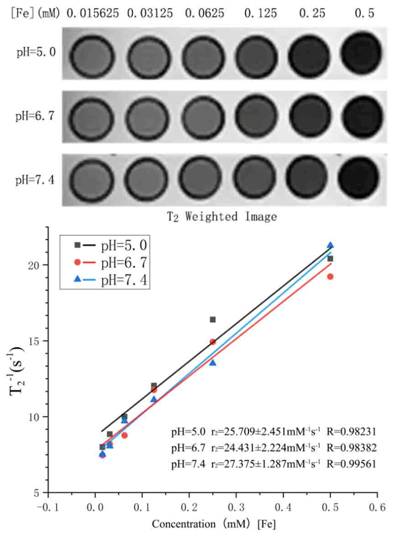
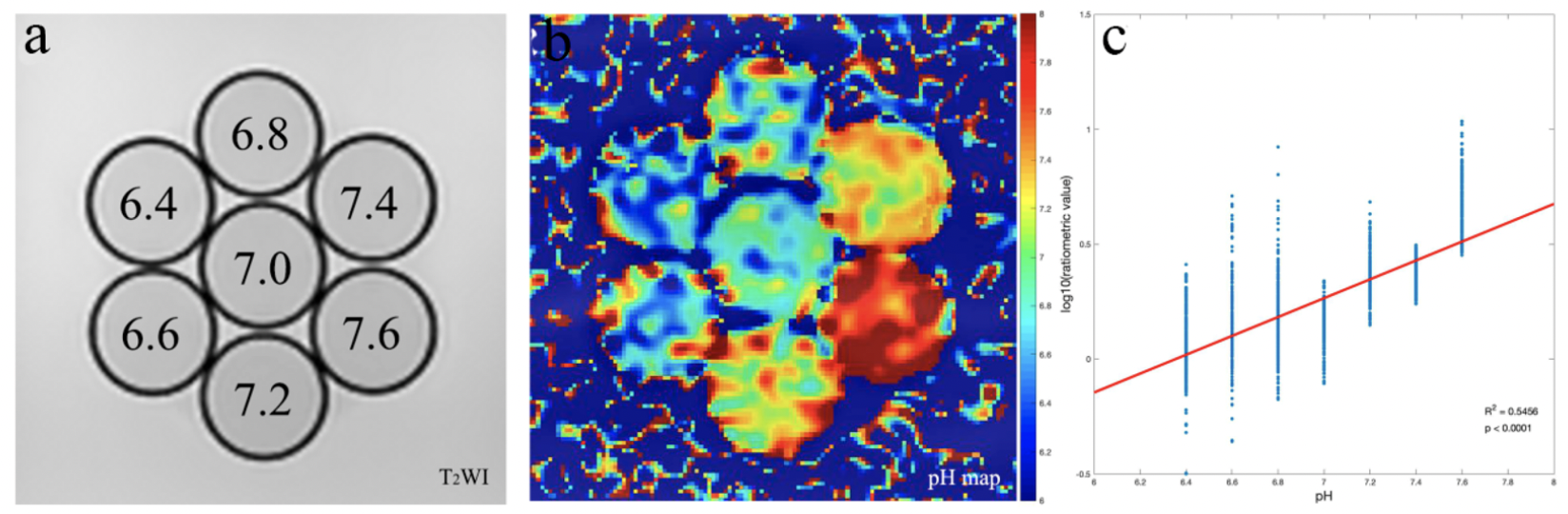
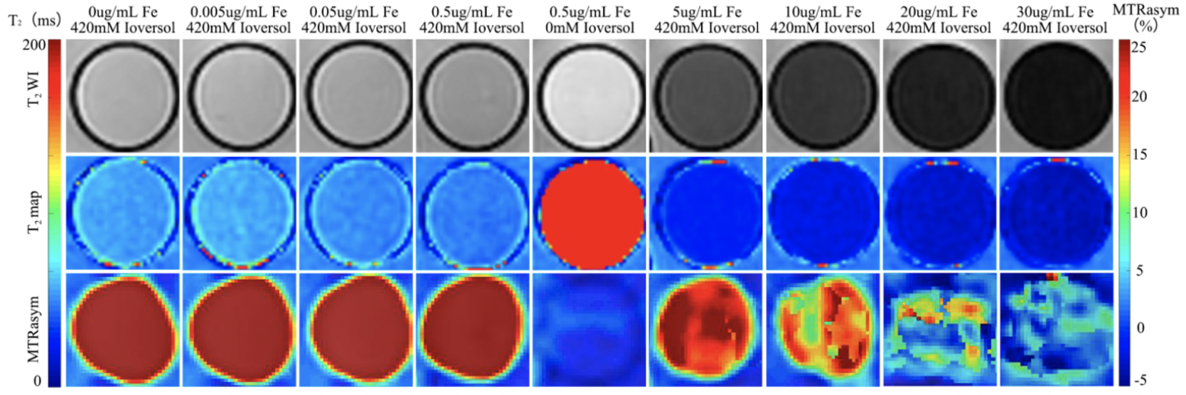
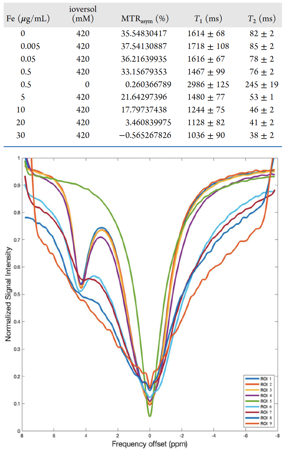
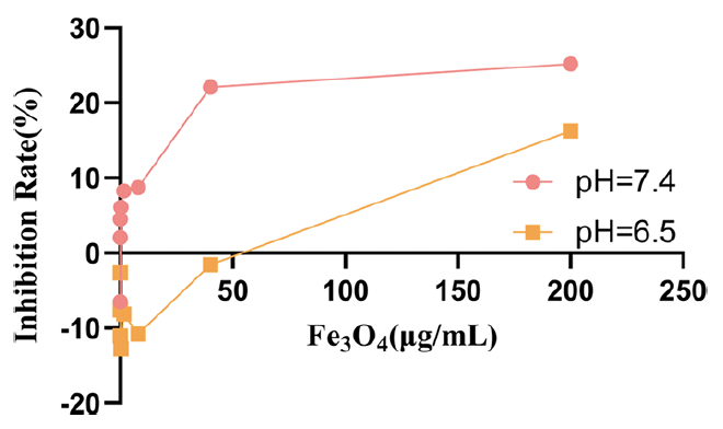
The in vitro MRI results concerning Fe3O4 NPs revealed that ratio between R2 value and Fe concentration at pH levels of 5.0, 6.7, and 7.4 were 25.709 ± 2.451, 24.431 ± 2.224, and 27.375 ± 1.287 mM-1s-1, respectively (Figure 1). These findings suggest that Fe3O4 NPs can function as stable reference agents across varying pH environments. Notably, the significant positive correlation between R2 value and Fe concentration indicated that Fe3O4 NPs have the capability to serve as negative contrast agents for T2 contrast-weighted MRI. Using the ratiometric value between RF powers of 5 and 1.25 µT10, we demonstrated that the CEST effect was influenced by the pH value and can be used to establish the quantitative relationship (Figure 2). Figure 3 shows that the CEST effect was completely inhibited at a Fe concentration of approximately 30 μg/mL, whereas at concentrations below 10 μg/mL, both contrasts could be effectively imaged. The corresponding values and Z-spectrum are presented in Figure 4. As exhibited in Figure 5, the inhibition rates were 25.168% and 16.25% at pH levels of 7.4 and 6.5, respectively, when Fe concentration was at 200 μg/mL. Notably, this concentration was lower than the IC50, indicating that the NPs prepared in this study displayed no significant cytotoxicity.Discussion
This study employed the ratiometric approach to precisely reveal the linear association between pH and the CEST effect, thereby establishing a foundational framework for quantifying shifts in the tumor microenvironment's acidity. When the concentration of Fe3O4 NPs is low, both T2 and CEST contrast can be visualized concurrently. Elevated levels of the NPs result in the suppression of the signal produced by the CEST contrast agent. These preliminary findings represent an initial stride toward dual-contrast imaging involving CEST and T2 contrast agents, with the potential for further expansion in future in vivo investigations.Acknowledgements
No acknowledgement found.References
1. Zhou, Z.; Lu, Z. R. Molecular imaging of the tumor microenvironment. Advances in Drug Delivery Reviews 2017, 113, 24−48.
2. Shen, Z.; Chen, T.; Ma, X.; Ren, W.; Zhou, Z.; Zhu, G.; Zhang, A.; Liu, Y.; Song, J.; Li, Z.; et al. Multifunctional theranostic nanoparticles based on exceedingly small magnetic iron oxide nanoparticles for t(1)-weighted magnetic resonance imaging and chemotherapy. ACS Nano2017, 11 (11), 10992−11004.
3. Deh, K.; Zaman, M.; Vedvyas, Y.; Liu, Z.; Gillen, K. M.; O’, M. P.; Bedretdinova, D.; Nguyen, T.; Lee, R.; Spincemaille, P.; et al. Validation of MRI quantitative susceptibility mapping of superparamagnetic iron oxide nanoparticles for hyperthermia applications in live subjects. Scientific Reports 2020, 10 (1), 1171.
4. Song, S.; Zhang, S.; Huang, S.; Zhang, R.; Yin, L.; Hu, Y.; Wen, T.; Zhuang, L.; Hu, B.; Wang, X. A novel multi-shelled fe3o4@mnox hollow microspheres for immobilizing u(vi) and eu(iii). Chemical Engineering Journal 2019, 355, 697−709.
5. Ghazanfari, M. R.; Kashefi, M.; Shams, S. F.; Jaafari, M. R. Perspective of fe3o4 nanoparticles role in biomedical applications. Biochemical Research International 2016, 2016, 7840161.
6. Weng, Q.; Hu, X.; Zheng, J.; Xia, F.; Wang, N.; Liao, H.; Liu, Y.; Kim, D.; Liu, J.; Li, F.; et al. Toxicological risk assessments of iron oxide nanocluster- and gadolinium-based t1mri contrast agents in renal failure rats. ACS Nano 2019, 13 (6), 6801−6812.
7. Sang, M.; Luo, R.; Bai, Y.; Dou, J.; Zhang, Z.; Liu, F.; Feng, F.; Xu, J.; Liu, W. Mitochondrial membrane anchored photosensitive nanodevice for lipid hydroperoxides burst and inducing ferroptosis to surmount therapy-resistant cancer. Theranostics 2019, 9 (21), 6209−6223.
8. Gobbo, O. L.; Sjaastad, K.; Radomski, M. W.; Volkov, Y.; Prina-Mello, A. Magnetic nanoparticles in cancer theranostics. Theranostics 2015, 5 (11), 1249−1263.
9. Torti, S. V.; Torti, F. M. Winning the war with iron. Nature Nanotechnology 2019, 14 (6), 499−500.
10. Longo, D. L.; Sun, P. Z.; Consolino, L.; Michelotti, F. C.; Uggeri, F.; Aime, S. A general MRI-CEST ratiometric approach for pH imaging: demonstration of in vivo pH mapping with iobitridol. Journal of the American Chemical Society 2014, 136 (41), 14333−14336.
Figures