3197
129Xe MRI Ventilation Texture Features and Machine Learning to Predict Response to ICS/LAMA/LABA in Moderate Asthma1Western University, London, ON, Canada
Synopsis
Keywords: Hyperpolarized MR (Gas), Hyperpolarized MR (Gas), Asthma, Machine learning, Texture analysis
Motivation: 129Xe MRI ventilation texture features provide a way to generate quantitative spatial information about ventilation heterogeneity beyond ventilation defect percent, which is important in some asthma patients with patchy (and not obviously segmental or subsegmental) ventilation abnormalities.
Goal(s): Machine-learning and 129Xe MRI ventilation texture-analysis were used to generate ventilation-imaging based models for predicting ICS/LAMA/LABA response.
Approach: Machine-learning models trained on clinical measurements were compared with those trained on ventilation texture features.
Results: MRI texture-based models outperformed clinical models for predicting 6-week response. The neighbourhood gray-tone difference matrix strength was the top-ranking texture feature, which significantly correlated with clinical measurements.
Impact: 129Xe MRI ventilation texture features provided unique information about ventilation abnormalities and ventilation patchiness; when texture features were embedded in predictive models, these features outperformed clinical models explaining response to ICS/LAMA/LABA in moderate asthma.
INTRODUCTION:
129Xe MRI provides a way to quantify ventilation abnormalities in airways disease and may be quantified as ventilation defect percent (VDP)1 MRI VDP is highly sensitive to small airways disease abnormalities and correlates with airway inflammation and remodeling in asthma.2 However, it is binary, categorizing regions of the lung as ventilated or not and disregards signal and spatial intensity differences.MRI ventilation texture analysis has been shown to provide additional information about ventilation heterogeneity, especially in asthma patients with patchy ventilation.3 In particular, the texture patterns and spatial distribution of 129Xe MRI ventilation voxel intensities, have the potential to uncover additional, information that better estimates patchy ventilation heterogeneity in asthma patients. We hypothesized that 129Xe MRI texture features and machine-learning used in combination would provide predictive models of response to inhaled combination anti-inflammatory and long-acting muscarinic antagonist/long-acting beta-agonist (ICS/LAMA/LABA) therapy in asthma patients with patchy ventilation.
METHODS:
Participants and Study DesignThirty-one patients with moderate asthma provided written informed consent (NCT04651777) to pre-/post-bronchodilator (BD) 129Xe MRI, pulmonary function tests, oscillometry and questionnaires including the asthma quality-of-life (AQLQ), asthma control (ACQ-6) and St. George's Respiratory Questionnaire (SGRQ) just prior to and after 6-weeks of daily inhaled fluticasone furoate/umeclidinium/vilanterol (FF/UMEC/VI; 200/62.5/25μg).4
Data Acquisition
Anatomic (1H) and functional (129Xe) MRI were acquired using a 3.0 Tesla Discovery MR750 (GE Healthcare). 1H MRI was acquired using a fast-spoiled gradient-recalled-echo (FGRE) sequence (partial-echo acquisition, total acquisition time=8s; TR/TE/flip-angle=4.7ms/1.2ms/30°; FOV=40x40cm2; BW=24.4kHz, matrix=128x80 zero-padded to 128x128; partial-echo percent=62.5%; 15-17x15mm slices), 129Xe MRI was acquired using a three-dimensional FGRE sequence (total acquisition time=14s; TR/TE=6.7ms/1.5ms; variable flip-angle; FOV=40x40cm2; BW=15.63kHz; 128x128 matrix; 14x15mm slices). Supine participants were instructed to inhale and hold 1.0L of gas (1.0L N2 for anatomical scan, 400mL hyperpolarized 129Xe + 600mL 4He for static ventilation scan) to ensure volume-matched images.
Image Processing and Statistics
MRI VDP was semi-automatically quantified as previously described.1 Texture features were extracted from the 3D-application of gray-level run-length, gap-length, zone-size, dependence, gray-tone difference, and co-occurrence-matrices via open-source PyRadiomics platform.5 Feature selection was performed using Boruta analysis via a random forests classifier to identify MRI features contributing to the machine-learning model’s accuracy. Classification Learner application (MATLAB R2021b) was used to test single and ensemble models. Model performance was evaluated using area under the receiver-operator-curve (AUC), sensitivity, specificity and accuracy. Participants were dichotomized on the basis of the FEV1 minimal-clinically-important-difference (MCID) (≥230 mL)6 at 6-weeks. Univariate relationships were evaluated using Spearman (ρ) correlations and differences between subgroups were evaluated using independent samples t-tests.
RESULTS:
Table 1 provides baseline measurements for 27 of 31 participants with a complete dataset (three withdrew after the first visit and one did not complete spirometry at the second visit). They were dichotomized by FEV1 change ≥MCID at 6-weeks and there were no differences between subgroups. Table 2 summarizes the models for predicting improved FEV1≥MCID where a Medium Gaussian support vector machine (SVM) model exclusively trained on MRI texture features was the top performer (AUC=0.84) and significantly (p=.03) outperformed the model trained on clinical measures (AUC=0.75). Figure 1 shows 129Xe MRI for representative participants in each group. Figure 2 shows the individual components of each model, where the top-performing feature was neighbourhood gray-tone difference matrix (NGTDM) strength (AUC=0.74). Figure 3 shows that, of the features in the MRI-model, NGTDM strength significantly correlated with ΔFEV1 (ρ=-.41, p=.04), FEV1 (ρ=-.49, p=.02), forced vital capacity (FVC; ρ=-.50, p=.01) and lung clearance index (LCI; ρ=.48, p=.02).DISCUSSION:
We used a machine-learning model to predict FEV1 response to ICS/LAMA/LABA using 129Xe MRI ventilation texture features. The models indicate the presence of non-linear, complex relationships of texture features and therapy response. The correlation between NGTDM strength with FEV1 and FVC suggests a linkage with obstruction. Furthermore, NGTDM strength also correlated with LCI, which may be complementary to MRI in identifying ventilation heterogeneity,7 air trapping8 and small airway dysfunction. MRI texture features outperformed all measurements, including MRI VDP.CONCLUSION:
Unique information about patchy ventilation patterns was provided by 129Xe MRI ventilation texture features and machine-learning that was not captured using VDP and clinical measurements. When ventilation patchiness textures were included in predictive models, these outperformed clinical models in explaining 6-week response to ICS/LAMA/LABA in moderate asthma.Acknowledgements
Study 217438 is a Supported Collaborative Study with GSKReferences
1 Kirby, M. et al. Acad Radiol (2012).
2 Svenningsen, S. et al. Thorax (2014).
3 Zha, N. et al. Acad Radiol (2016).
4 Kerwin, E. et al. Respir Res (2020).
5 Lafata, K. J. et al. Sci Rep (2019).
6 Santanello, N. C. et al. Eur Respir J (1999).
7 Svenningsen, S. et al. European Respiratory Journal (2016).
8 Thompson, B. R. et al. J Allergy Clin Immunol (2013).
Figures
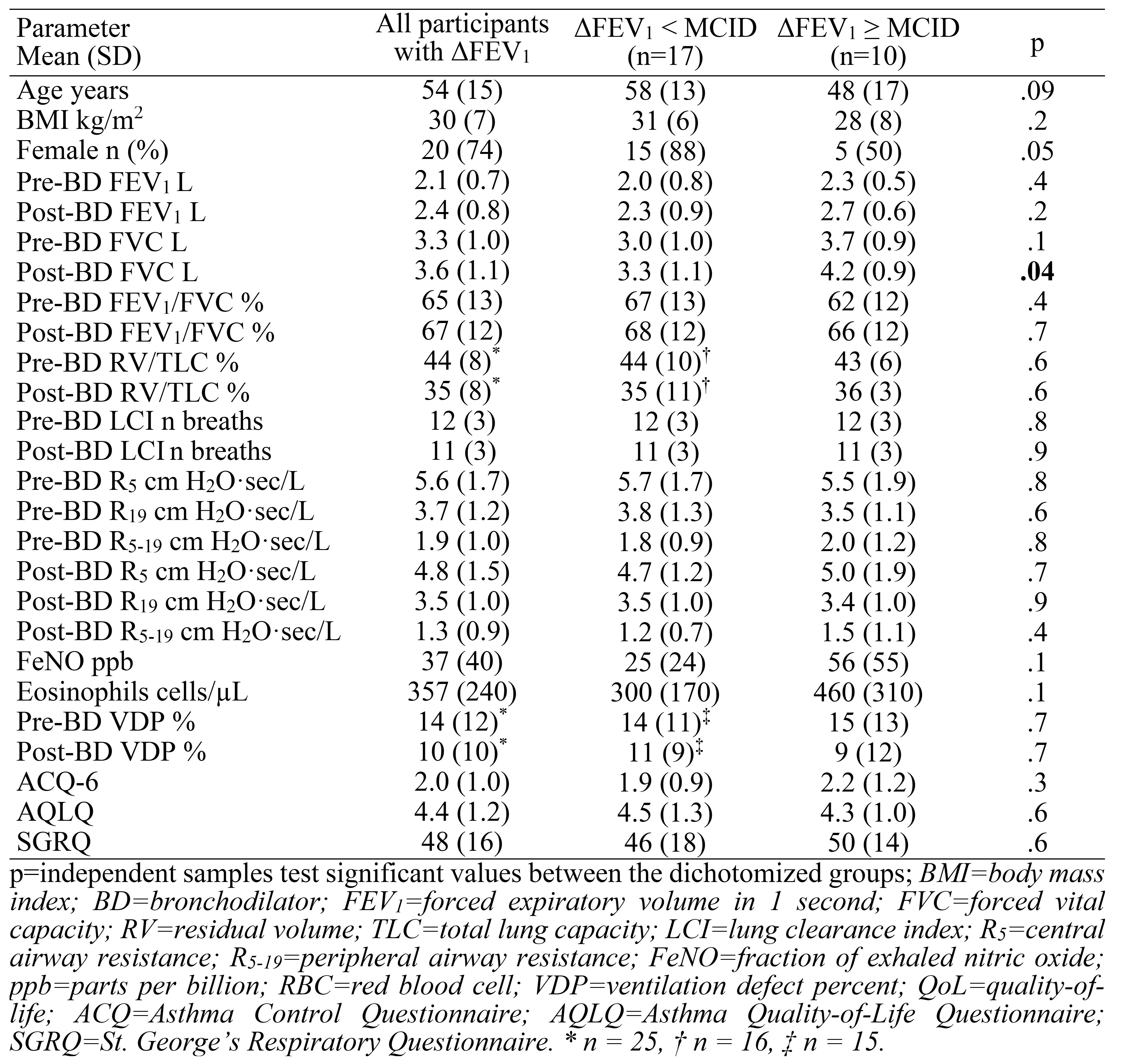
Table 1. Demographics, Pulmonary Function, Questionnaire and Imaging Measurements
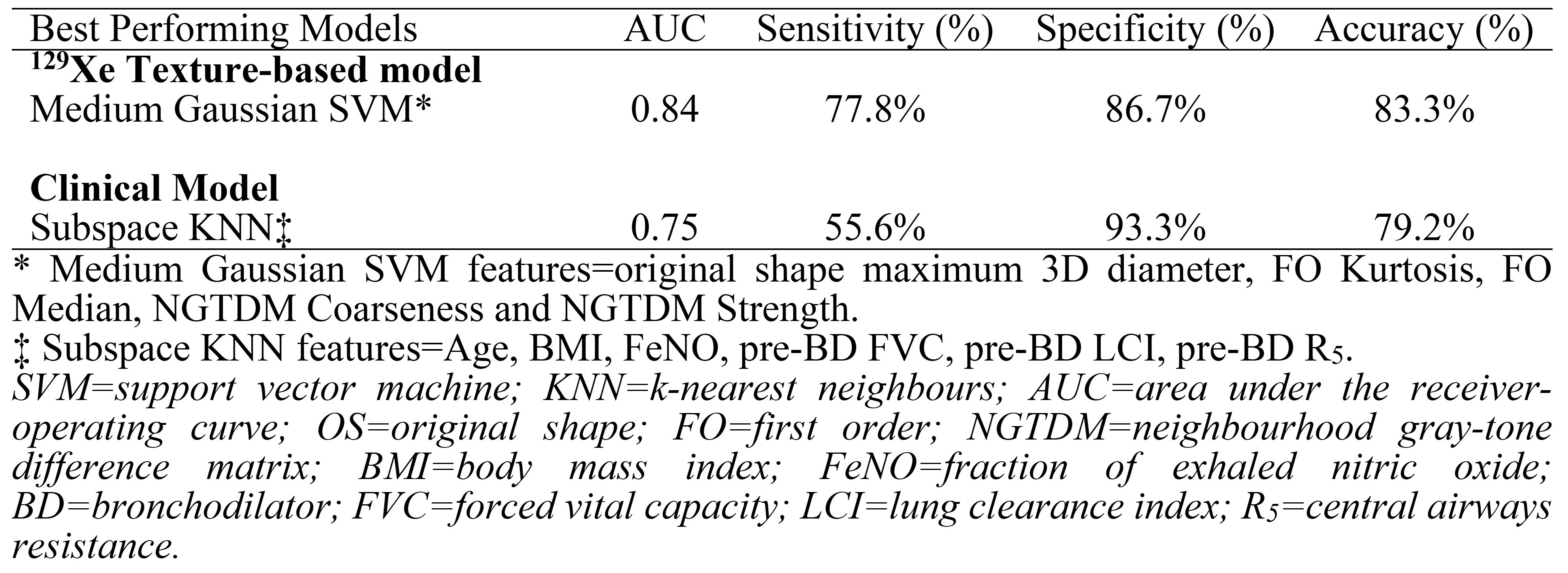
Table 2. Comparison of machine-learning models exclusively trained on MRI ventilation textures and exclusively trained on clinical measurements
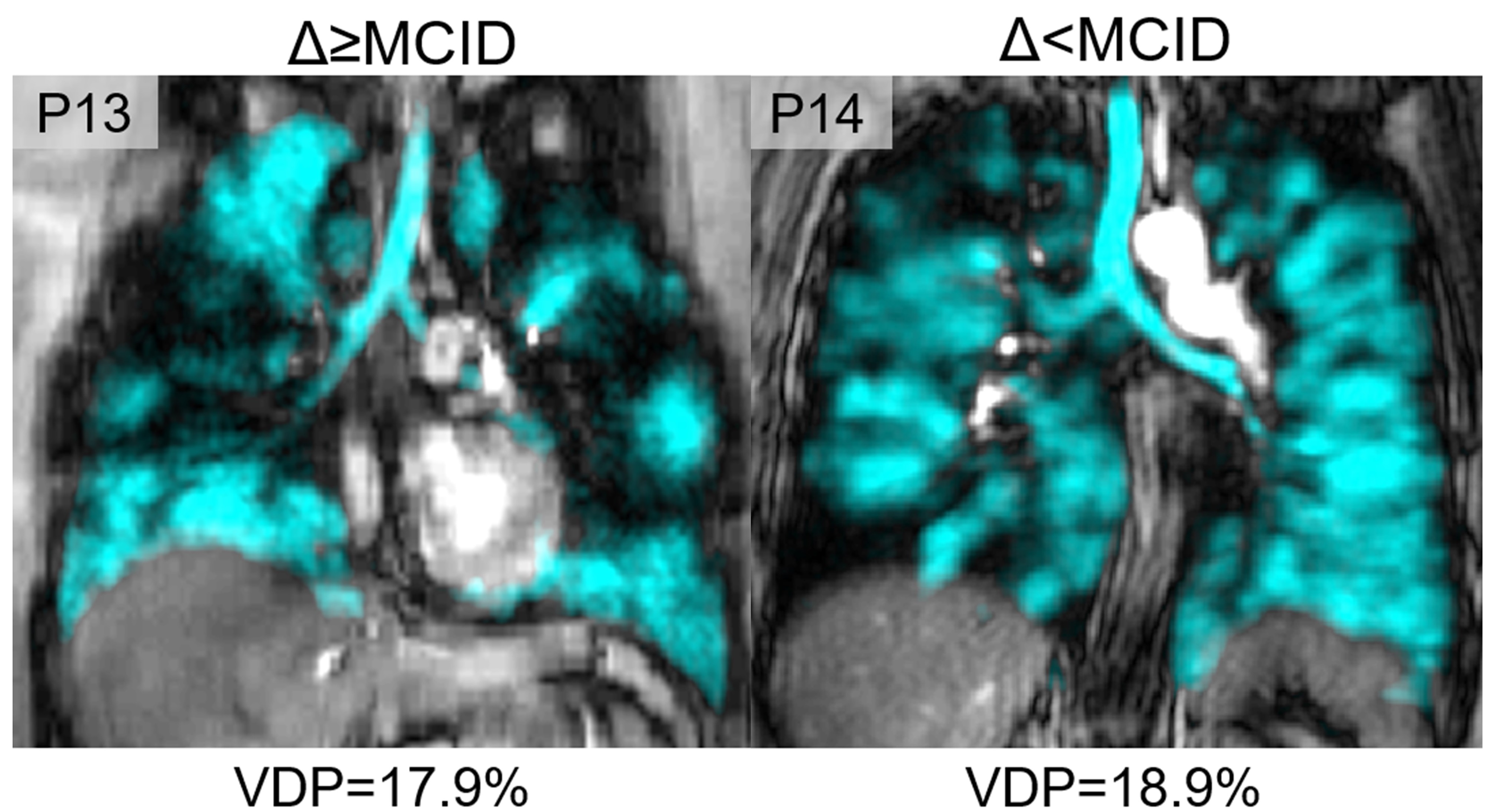
Figure 1. 1H and 129Xe MRI at week 0 for participants from the Δ≥MCID and Δ<MCID groups. P13 is a 26-year-old female with FEV1=65%, VDP=18%, OS Maximum 3D Diameter=107, FO Kurtosis=3.3, FO Median=1925, NGTDM Coarseness=0.0006 and NGTDM Strength=340. P14 is a 62-year-old female with FEV1=62%, VDP=19%, OS Maximum 3D Diameter=94, FO Kurtosis=2.5, FO Median=2902, NGTDM Coarseness=0.0011 and NGTDM Strength=408.
MCID=minimal clinically important difference; VDP=ventilation defect percent; OS=original shape; FO=first order; NGTDM=neighbourhood gray-tone difference matrix.
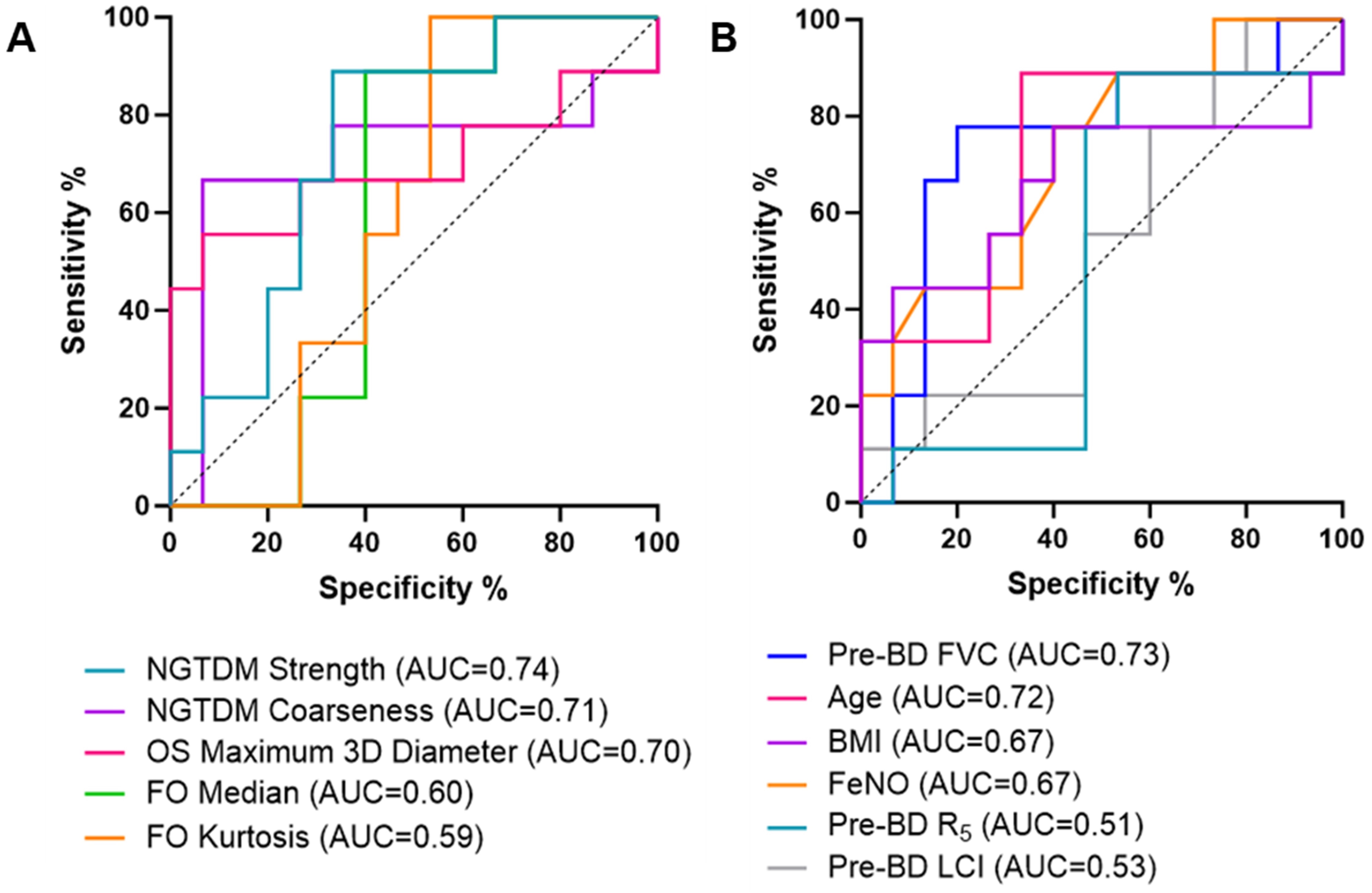
Figure 2. Receiver-operator-curves of individual components in A) MRI texture and B) clinical models. The graphs represent the strength of each individual feature in predicting change in FEV1.
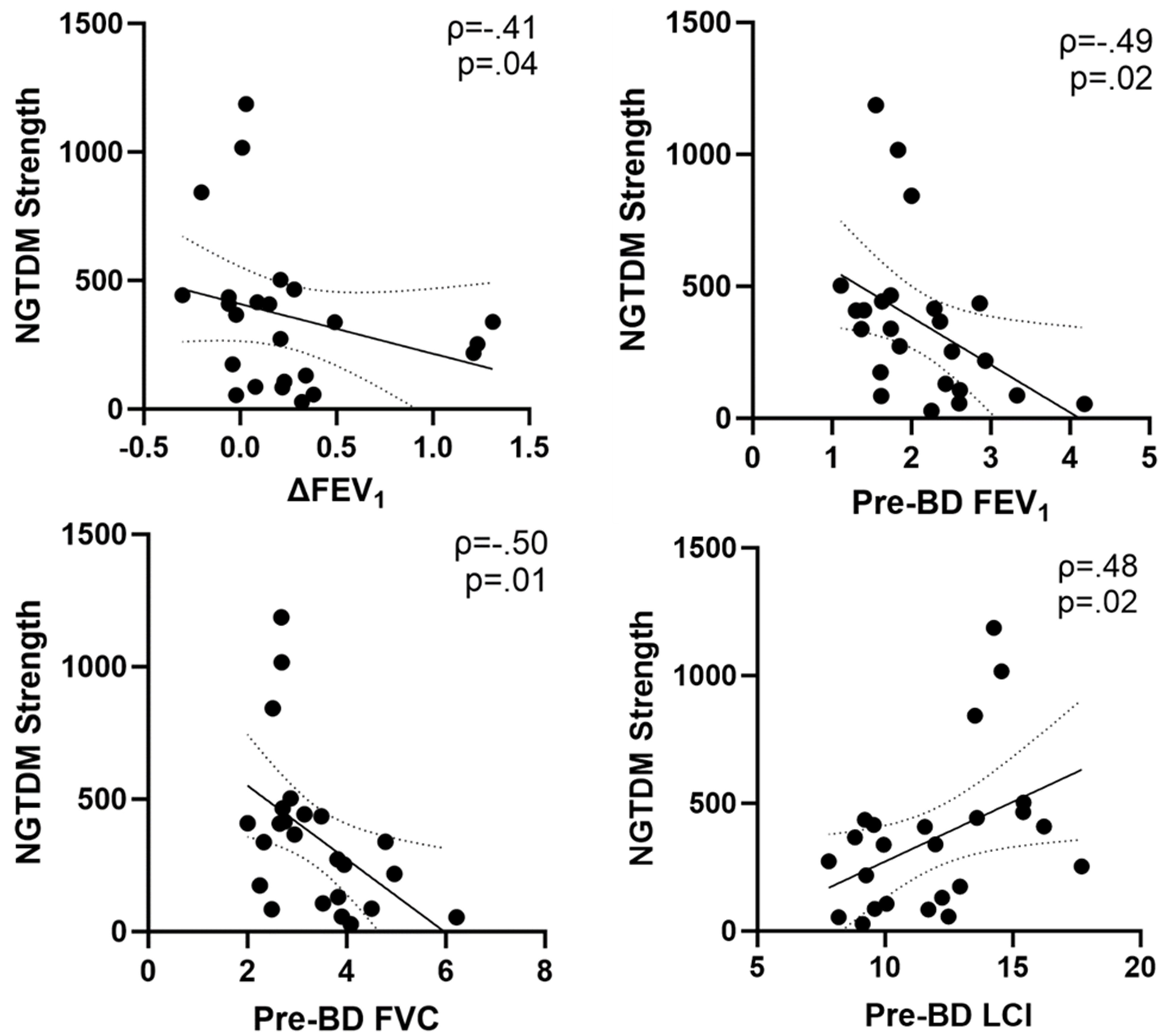
Figure 3. Scatter plots showing the correlations between MRI texture features and the change in FEV1 from Week 0 to Week 6. NGTDM strength significantly correlated with ΔFEV1 (ρ=-.41, p=.04), FEV1 (ρ=-.49, p=.02), FVC (ρ=-.50, p=.01) and LCI (ρ=.48, p=.02).
ρ=Spearman correlation coefficient; NGTDM=neighbourhood gray-tone difference matrix; FEV1=forced expiratory volume in 1-second; BD=bronchodilator; FVC=forced vital capacity; LCI=lung clearance index.