3194
The Successful Use of Unenriched Xenon in Dissolved Gas MRI: A Case Report1Division of General Medicine, Columbia University Medical Center, New York, NY, United States, 2POLARIS, Imaging Section, Division of Clinical Medicine, School of Medicine and Population Health, University of Sheffield, Sheffield, United Kingdom, 3Weill Cornell Medicine, New York, NY, United States, 4GE Healthcare, Munich, Germany, 5Department of Biomedical Engineering, Columbia University, New York, NY, United States, 6Department of Physics, Columbia University, New York, NY, United States
Synopsis
Keywords: Hyperpolarized MR (Gas), COVID-19, Xenon, Blood
Motivation: We sought to demonstrate the safety and efficacy of a hyperpolarized gas MRI protocol for assessing the pulmonary physiology of long-COVID.
Goal(s): Our goal was to gather preliminary dissolved phase images utilizing unenriched xenon.
Approach: Volumes of 300mL and 500mL of unenriched xenon were hyperpolarized using a 180W diode laser at 795nm and administered to two healthy subjects. Scans were obtained using a 3T SIGNATM Premier scanner and a 129Xe Quadrature T/R lung coil. Images were masked using signal-to-noise thresholding on tissue/plasma signal.
Results: We found strong red blood cell and tissue/plasma xenon signal in both subjects utilizing 300mL of unenriched xenon.
Impact: At present time, successful dissolved xenon imaging has only been described with the use of enriched xenon-129. Our findings suggest that less expensive and more widely available unenriched xenon may be used in dissolved gas MRI.
Introduction
Dissolved hyperpolarized xenon-129 (129Xe) imaging has been used in examining blood flow and gas exchange in various organ systems. 129Xe provides the unique property of resonating at different frequencies when dissolved in red blood cells (RBC), tissue/plasma (M), and when in its gaseous phase (Gas), allowing for their differentiation upon MR imaging. We set out to perform preliminary imaging on two healthy subjects using natural abundance (unenriched) xenon with limited expectations for signal.Xenon-129 has a natural abundance of 26.4%, whereas enriched 129Xe typically used in dissolved xenon MR has a mole fraction of approximately 86%1. Enriched 129Xe is significantly more expensive and less widely available than unenriched xenon.
To date, there has been no report in the literature of dissolved xenon MR imaging using an unenriched gas source however encouraging results for ventilation imaging have been demonstrated2.
Here we report the successful use of natural abundance xenon in the dissolved MR imaging of two healthy subjects.
Methods
Xenon underwent spin exchange optical pumping to achieve hyperpolarization using a high yield system (POLARIS, Sheffield, UK). The specified volume of the hyperpolarized xenon was then mixed with nitrogen to a yield total volume of one liter. MRI imaging was conducted on 3T SIGNATM Premier (GE Healthcare, Waukesha, WI) scanner with multi-nuclei spectroscopy (MNS) capability. A 3T 129Xe Quadrature T/R lung coil (Clinical MR Solutions, LLC) was used. Two dissolved phase xenon images were collected in each healthy participant and images were reconstructed with proprietary software and the scans were masked using signal-to-noise thresholding on tissue/plasma signal. All further analysis was performed in Python.Results
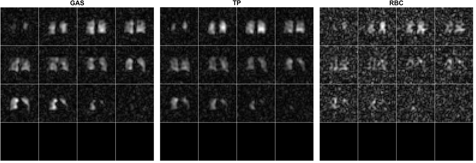
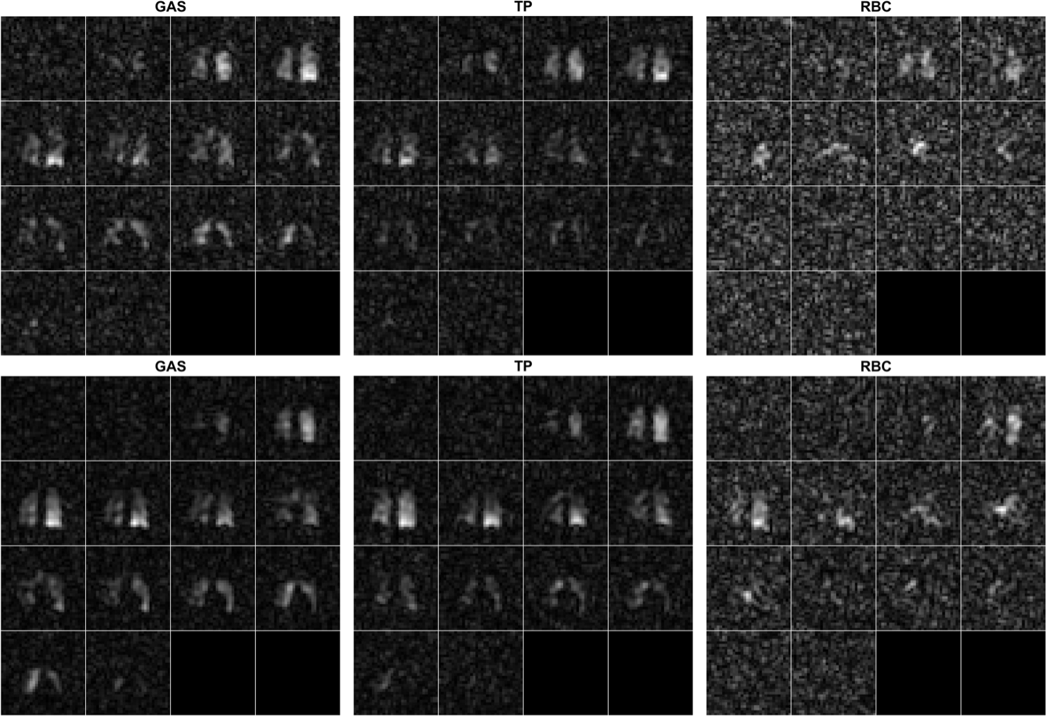
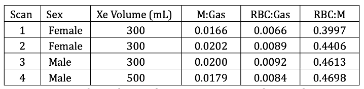
All four scans produced strong signal in all three frequency ranges (Gas, M, and RBC) [Figures 1, 2]. Intra-subject differences in Gas:M and Gas:RBC signal ratios were present, even at the same xenon volume [Figure 3].Net signal ratios were well-matched to those of dissolved phase scans in the literature, which utilized enriched xenon3.
An increase in Xenon volume from 300mL to 500mL revealed only minor improvements in signal-to-noise ratio (Gas: 0.35%, M: 5.24%, RBC: 14.13%) and a decrease in the ratio of dissolved to gaseous 129Xe.
Discussion
Unenriched xenon is far less expensive than enriched 129Xe. However, until now, there has been no report of its use in inhaled-gas MRI. Our findings suggest that future inhaled-gas study using unenriched xenon is possible.As for the intra-subject differences in Gas:M and Gas:RBC signal ratios, there exist multiple potential explanations such as a difference in acquisition time with regard to the respiratory cycle. Further examination is required to better understand the mechanisms underlying this, though such variability is likely not unique to the use of unenriched xenon.
Acknowledgements
Research reported in this abstract was supported by the National Heart, Lung, and Blood Institute of the National Institutes of Health under award number R01HL121270.References
1. Qing, K., Ruppert, K., Jiang, Y., Mata, J. F., Miller, G. W., Shim, Y. M., Wang, C., Ruset, I. C., Hersman, F. W., Altes, T. A., & Mugler, J. P., 3rd. (2014). Regional mapping of gas uptake by blood and tissue in the human lung using hyperpolarized xenon-129 MRI. J Magn Reson Imaging, 39(2), 346-359. https://doi.org/10.1002/jmri.24181
2. Collier, G. J., Schulte, R. F., Rao, M., Norquay, G., Ball, J., & Wild, J. M. (2023). Imaging gas-exchange lung function and brain tissue uptake of hyperpolarized (129) Xe using sampling density-weighted MRSI. Magn Reson Med, 89(6), 2217-2226. https://doi.org/10.1002/mrm.29602
3. Stewart, N. J., Norquay, G., Griffiths, P. D., & Wild, J. M. (2015). Feasibility of human lung ventilation imaging using highly polarized naturally abundant xenon and optimized three-dimensional steady-state free precession. Magn Reson Med, 74(2), 346-352. https://doi.org/10.1002/mrm.25732
Figures