3193
Development of a transmit gain calibration method using the Bloch-Siegert shift for nonproton hyperpolarized MRI on a preclinical 7T scanner1The University of Texas MD Anderson Cancer Center UTHealth Graduate School of Biomedical Sciences, Houston, TX, United States, 2Department of Imaging Physics, The University of Texas MD Anderson Cancer Center, Houston, TX, United States, 3Imaging Physics Residency Program, The University of Texas MD Anderson Cancer Center, Houston, TX, United States
Synopsis
Keywords: Hyperpolarized MR (Gas), Hyperpolarized MR (Gas), Transmit Gain Calibration, Bloch-Siegert Shift
Motivation: Transmit gain calibration is necessary for nonproton hyperpolarized MRI to accurately calibrate excitation angles.
Goal(s): To implement a phase-based TG calibration method using the Bloch-Siegert shift for nonproton HP MRI on a preclinical 7T scanner.
Approach: An off-resonance Fermi-shaped Bloch-Siegert pulse follows directly an excitation pulse. Percent difference between our measured Bloch-Siegert TG calibration factors and the validation TG calibration factors for 1H and 13C were calculated.
Results: Our method allows for fast, accurate, and robust TG calibration of preclinical nonproton HP MRI studies with minimal error (<5%) for 1H and 13C.
Impact: This method will facilitate efficient transmit calibration for 13C and HP 129Xe MRI on a preclinical 7T scanner, where limited bore size and the inability to easily implement a thermal calibration phantom can make 129Xe TG calibration a challenge.
Introduction
Transmit gain (TG) calibration is performed prior to MR image acquisition to determine the radiofrequency (RF) output necessary to produce desired excitation angles. Accurate TG calibration is crucial for reproducible imaging of nonproton hyperpolarized (HP) agents because each HP signal excitation attenuates the total available HP signal pool, which directly effects future measurements.We have previously implemented an automated script to perform saturation-recovery 13C TG calibration on a preclinical 7T MRI for metabolic imaging of HP [1-13C]-pyruvate in small animals. This approach leverages strong signal from a heavily doped 8M 13C-urea phantom. While a thermal 129Xe gas phantom was recently introduced for HP gas MRI1, this approach may not easily translate into a small calibration phantom that can fit alongside small animals in a 7T MRI.
We developed a TG calibration sequence for nonproton HP MRI using the Bloch-Siegert shift2,3 in preparation for upcoming HP 129Xe gas MRI small animal studies on a 7T MRI. Our method allows for fast, accurate, and robust TG calibration of nonproton HP MRI studies with minimal error (<5%) for 1H and 13C.
Methods
Bloch-Siegert Effect: The Bloch-Siegert shift describes the effect by which an off-resonant RF pulse induces a phase shift without altering spin nutation of the target nuclei:$$$\phi_{BS}=\int_{0}^{T}\frac{(\gamma B_{1}(t_{BS}))^{2}}{2(2\pi f_{BS})(t_{BS})}dt_{BS}\tag1$$$
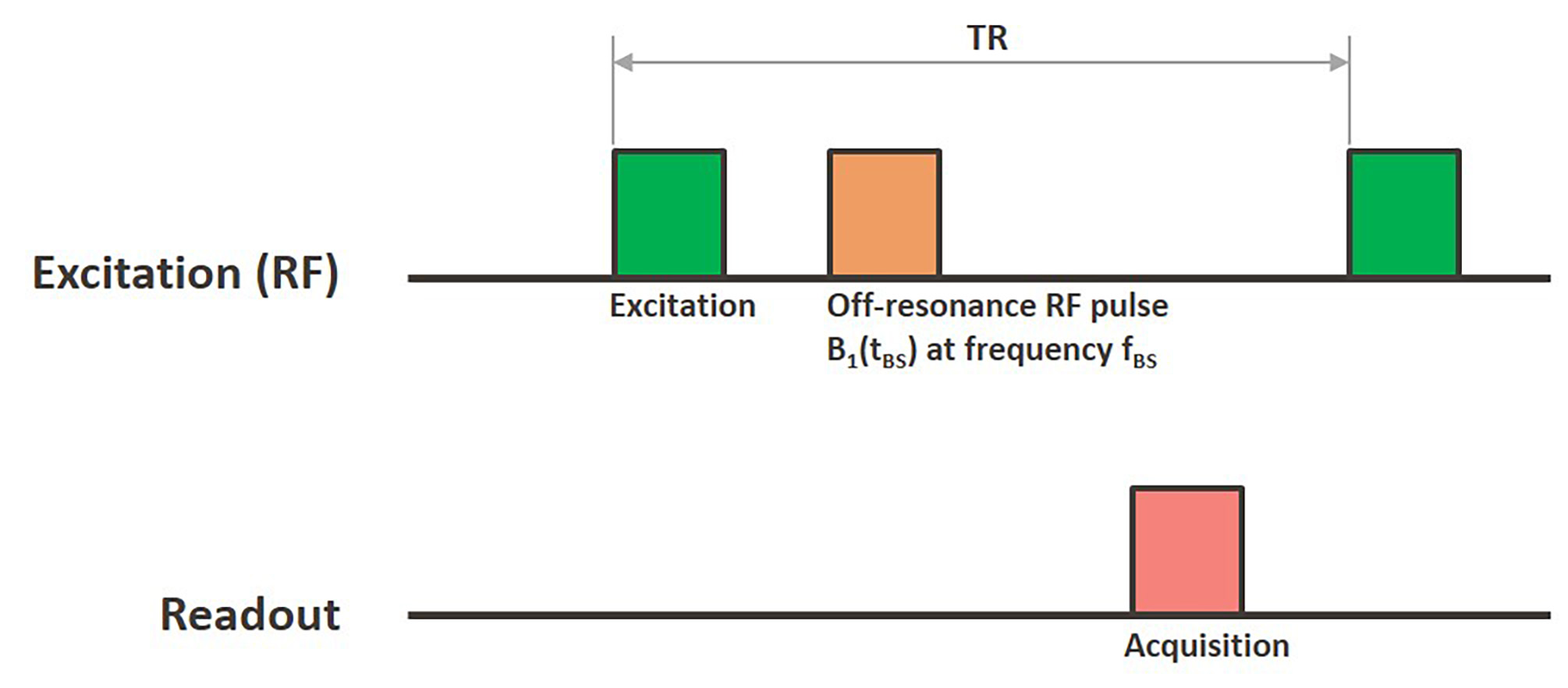
Briefly, an off-resonant RF pulse of duration tBS at offset frequency fBS is applied following a single excitation pulse (Figure 1). Two repeated measurements with ±fBS allow for the quantification of the Bloch-Siegert phase difference, φBS, which is proportional to the square of the amplitude of the RF field.
Imaging: All imaging and spectroscopy were carried out using a 7T Biospec USR7030 small animal MR scanner (Bruker Preclinical Imaging, Billerica, MA) and a dual-tuned 13C/1H volume coil with a 35 mm inner diameter. An optimized Fermi shaped RF pulse4 was used for off-resonant Bloch-Siegert pulse generation. An analysis script was implemented as a standalone MATLAB (The MathWorks, Natick, MA) executable function that can be called automatically following data acquisition.
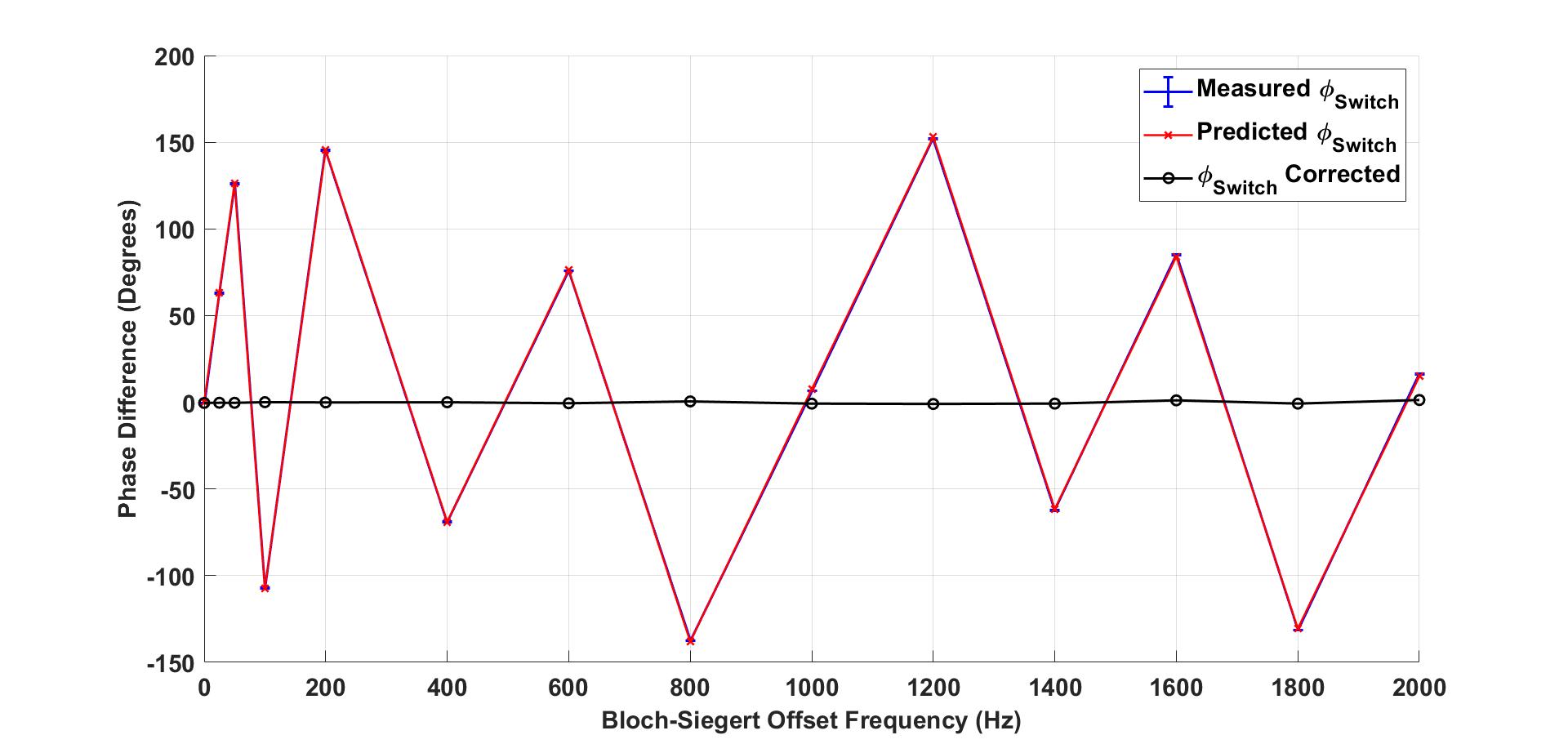
Phase Error Correction: During sequence development we discovered a persistent phase error (φSwitch) as a result of switching the frequency of the synthesizer to fBS, for the duration of the Bloch-Siegert pulse (tBS), between signal excitation and readout. In order to isolate φSwitch, the Bloch-Siegert pulse was driven with 0 W (φBS = 0°) and tested at several fBS values ranging 0 to 2000 Hz at 1H (Figure 2). Other important pulse parameters included tBS = 3.5 ms, TR = 10,000 ms, two repetitions, and two dummy scans.
Phase induced by switching the synthesizer frequency can be written as a function of the offset frequency, pulse duration, and a minor delay associated with the frequency switch:
$$$\phi_{Switch}=2\pi f_{BS}(t_{BS}+t_{Switch})\tag2$$$
The total phase (Δφ) observed following the Bloch-Siegert pulse reflects a combination of these two effects:
$$$\triangle\phi=\phi_{BS}+\phi_{Switch}\tag3$$$
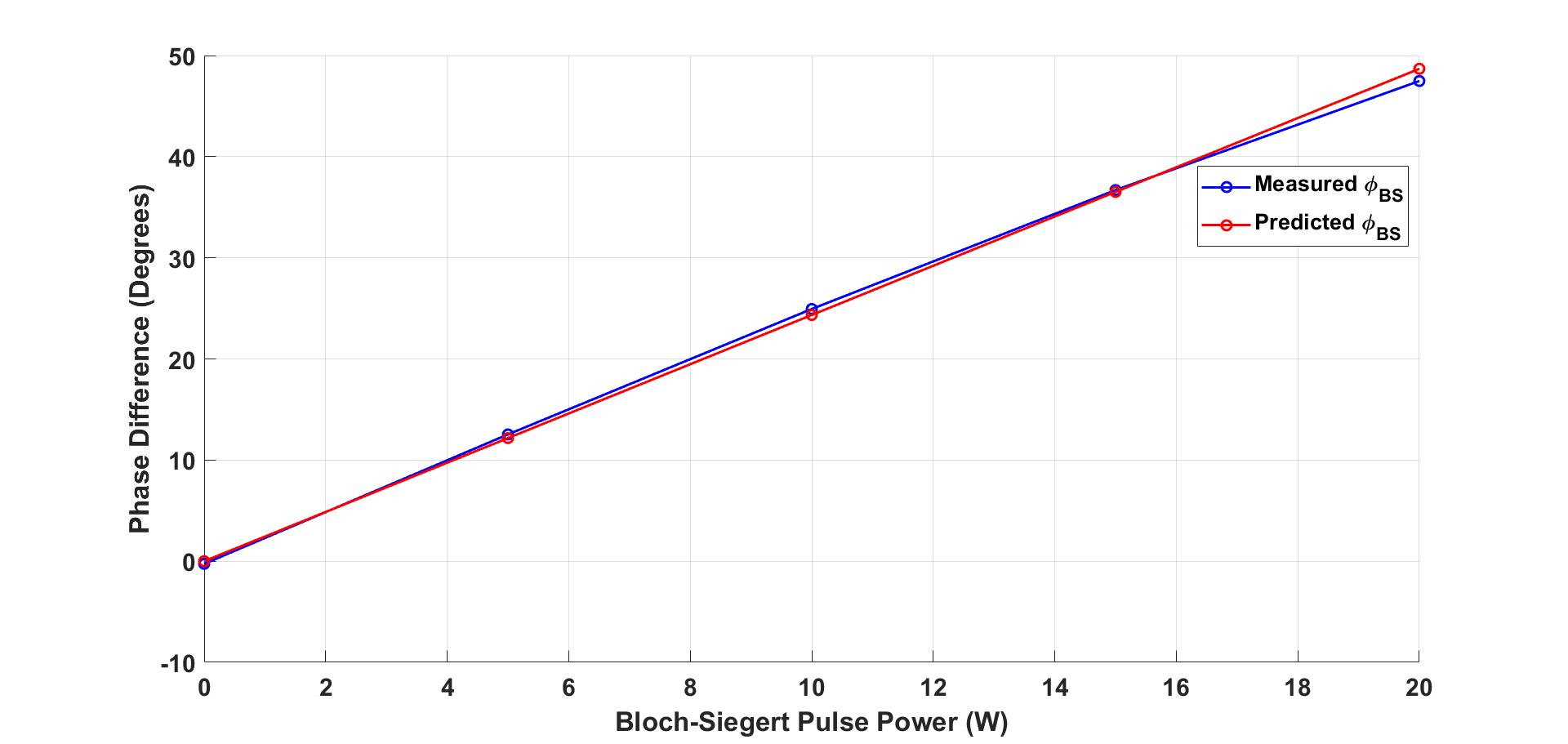
Bloch-Siegert Validation: Following characterization of φSwitch, the Bloch-Siegert pulse was driven with power values ranging 0 to 20 W at 1H (Figure 3) and 13C. Measured Δφ values were corrected for phase induced by the frequency switch. Next, φBS was used to calculate TG calibration factors at each Bloch-Siegert power level. Finally, percent difference between our measured Bloch-Siegert TG calibration factors and the validation TG calibration factors for 1H and 13C, measured automatically by the scanner and using our existing saturation-recovery 13C TG calibration method, were calculated.
Results
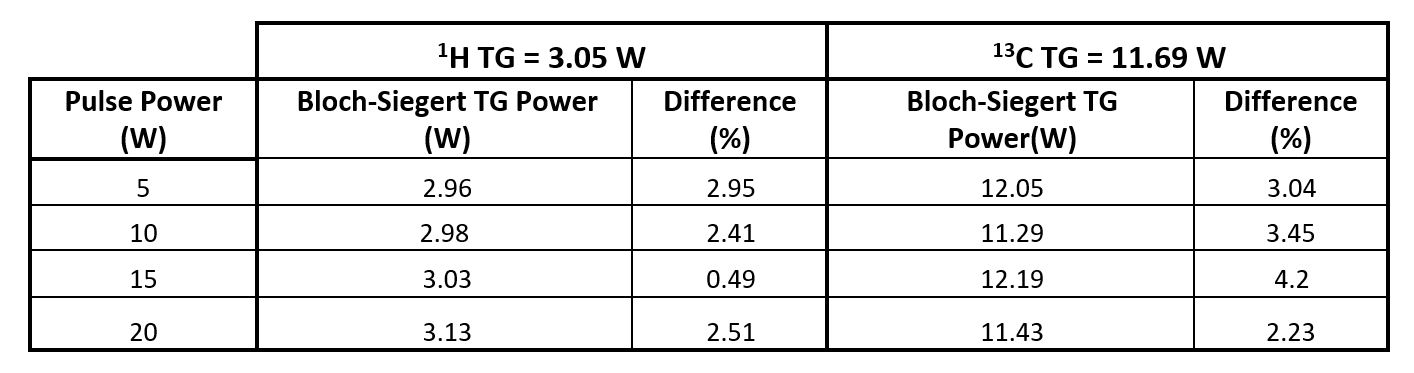
φSwitch was measured to be ~7° when tBS = 3.5 ms, fBS = 2000 Hz and tSwitch was calculated to be ~10 µS.Figure 4 provides a summary of percent difference between our measured Bloch-Siegert TG calibration factors and the validation TG calibration factors for 1H and 13C at power levels ranging 0 to 20 W. The reference power required for the scanner to generate a 90°, 1 ms hard excitation pulse was measured to be 3.05 W and 11.69 W for 1H and 13C, respectively.
Discussion and Conclusion
We successfully utilized the Bloch-Siegert effect to develop a robust pulse sequence that can be used for fast and automated TG calibration of nonproton nuclei on a 7T small animal MRI with minimal error (<5%). Our method performs well for 1H and 13C. We are currently in the process of testing our method using HP 129Xe gas and a dual-tuned 129Xe/1H volume coil. Our Bloch-Siegert pulse and supporting developmental tools can be found at: https://github.com/mda-mrsl/PV-BSXmitAcknowledgements
This work was supported by funding from the National Cancer Institute of the National Institutes of Health (R21CA280799, T32CA196561). The content is solely the responsibility of the authors and does not necessarily represent the official views of the sponsors.References
1. Bier EA, Nouls JC, Wang Z, et al. A thermally polarized 129 Xe phantom for quality assurance in multi-center hyperpolarized gas MRI studies. Magn Reson Med. 2019;82(5):1961-1968.
2. Sacolick LI, Wiesinger F, Hancu I, Vogel MW. B1 mapping by Bloch-Siegert shift. Magn Reson Med. 2010;63(5):1315-1322.
3. Schulte RF, Sacolick L, Deppe MH, et al. Transmit gain calibration for nonproton MR using the Bloch-Siegert shift. NMR Biomed. 2011;24(9):1068-1072.
4. Rodgers CT, Clarke WT, Snyder C, Vaughan JT, Neubauer S, Robson MD. Human cardiac 31P magnetic resonance spectroscopy at 7 Tesla. Magn Reson Med. 2014;72(2):304-315.
Figures