3191
Mapping Hyperpolarised 129Xe Gas Exchange with CSSR – CSI in a Model of Radiation-Induced Lung Injury1University College London, London, United Kingdom, 2University of Pennsylvania, Philadelphia, PA, United States, 3GlaxoSmithKline, Stevenege, United Kingdom, 4Bioxydyn Limited, Manchester, United Kingdom
Synopsis
Keywords: Hyperpolarized MR (Gas), Lung, Radiation induced lung injury, Modelling
Motivation: Regional assessment of functional decline due to radiation-induced lung injury (RILI) remains challenging
Goal(s): We aimed to assess the accuracy of the recently introduced kinetic model of xenon exchange (kMXE) by comparison with the established MOXE model using hyperpolarised 129Xe MRI
Approach: Employing a RILI rat model, we implemented gas exchange mapping of hyperpolarised 129Xe MRI chemical shift imaging data using 1D compartmental diffusion models.
Results: The kMXE results matched the MOXE model well. Both models demonstrated asymmetric outcomes in the RILI cohort, diverging from the homogeneous results in the healthy group.
Impact: Using hyperpolarised 129Xe MRI, we evaluated the effectiveness of kMXE and MOXE models and demonstrate their ability to uncover regional functional variations, providing potential biomarkers for assessing the longitudinal progression of radiation-induced lung injury.
Introduction
Radiation-induced lung injury (RILI) is a common side effect in the treatment of lung cancer. Given the varied onset of RILI, quantifying the pathophysiological alterations remains crucial. Although pulmonary function tests can evaluate lung function pre- and post-treatment, these assessments offer a global measure of lung function, lacking regional insights. Hyperpolarised 129Xe (HXe) MRI enables both regional and global evaluation of lung function permitting gas exchange mechanisms within the alveoli, tissue, and blood compartments to be modelled utilising characteristic spectroscopic peaks generated by the presence of 129Xe. The kinetic model of xenon exchange (kMXE) [1] was initially developed to parameterise dissolved compartments within animal models; aiming to resolve the overlapping membrane and RBC peak problem in certain species such as mice [2]. Before using it in a mouse model, it's essential to validate its accuracy against current diffusion models. To this end, we employ gas exchange modelling with kMXE and MOXE [3] in a RILI preclinical model.Methods
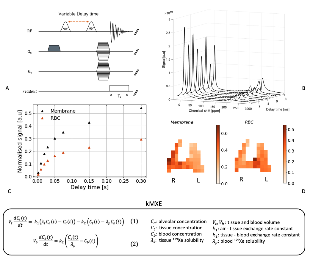
Sprague Dawley rats (approximately 500 g) were exposed to RILI over a six-week period, receiving 20 Gy of radiation targeting the entire right lung. At the end of the six weeks, the group, comprising two rats with RILI and one healthy Sprague Dawley rat, underwent imaging on a Bruker 3T scanner using a custom CSSR-CSI sequence. Figure 1A) illustrates the CSSR CSI pulse sequence diagram, showcasing complete lung spectra for the individual lung compartments, namely the alveoli (gas phase), tissue and blood plasma (membrane) and red blood cells (RBC) B). The rats were sedated and ventilated with room air until the start of imaging. All studies were approved by the Institutional Animal Care and Use Committee. Each image required the completion of every line of k-space during a breath-hold interval, maintaining a consistent delay period between the saturation and the excitation pulse. Subsequent images involved extended delay durations of up to 300 ms. Both the MOXE model and the kinetic model of xenon exchange (KMXE) – described in Figure 1 (Equations 1 and 2) – were fitted to the CSSR CSI images at the voxel level while adhering to user-defined, realistic parameter constraints.Results
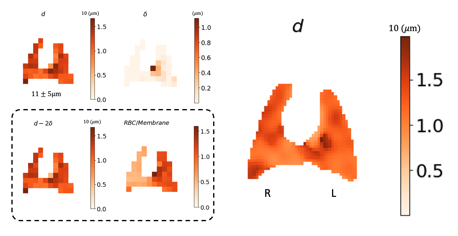
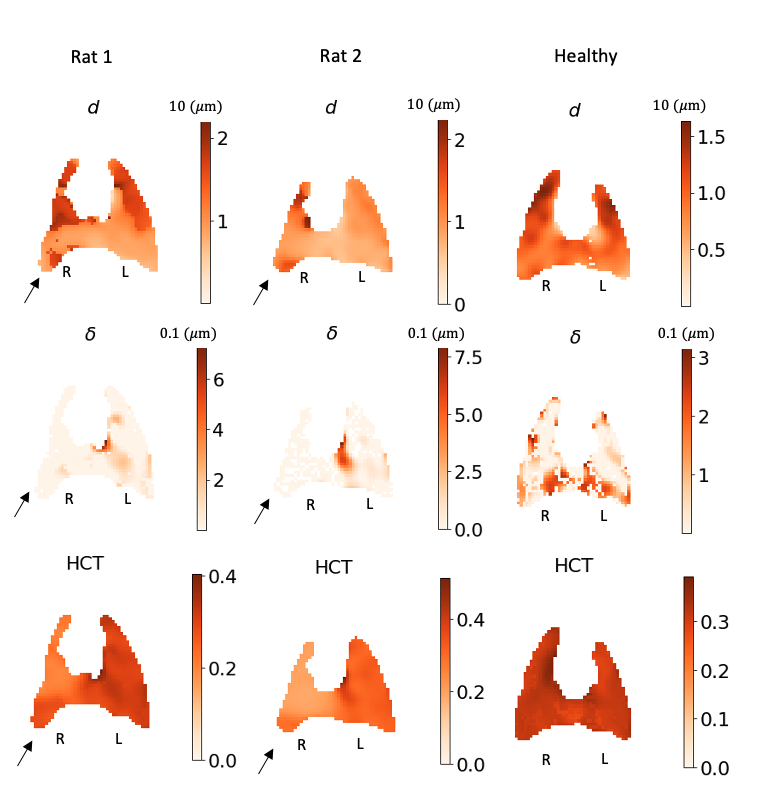
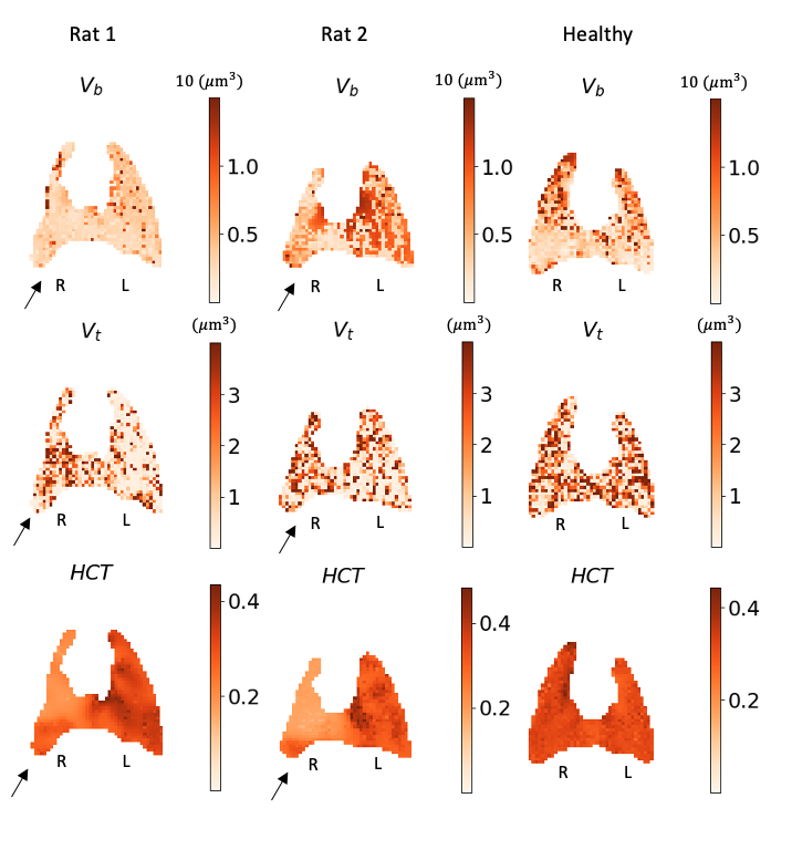
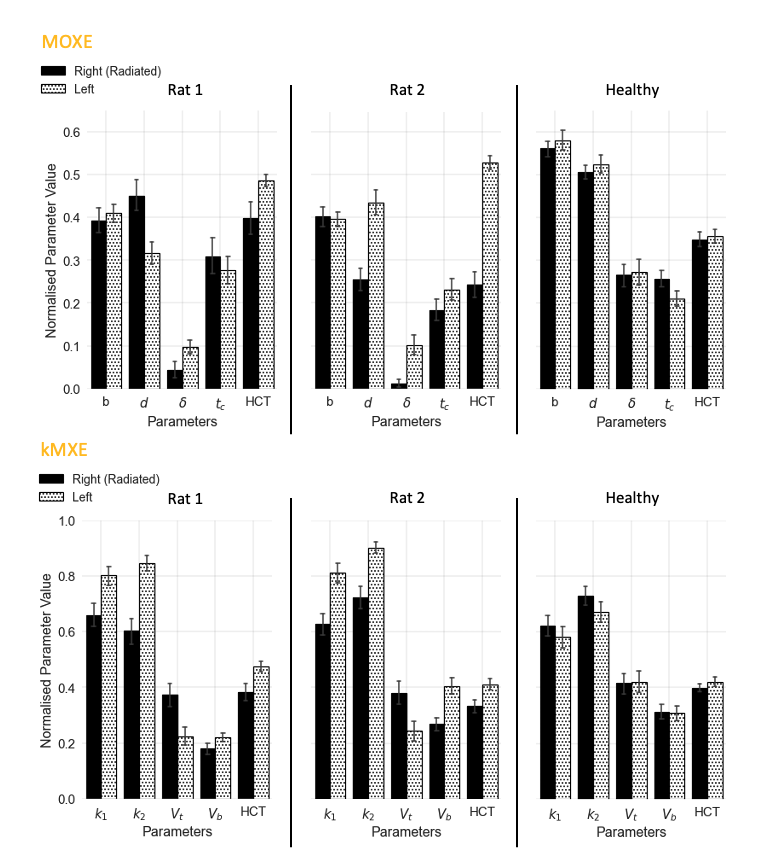
Figure 1 highlights normalised dissolved phase uptake signals and corresponding images D) derived from integrating the area under the spectra, depicting a distinct increase in membrane and RBC signal in the irradiated and control lung regions respectively. Figure 2 presents MOXE maps of the septal wall thickness (d), air-capillary barrier thickness (δ), a representative blood compartment (d-2δ), and a normalised RBC/membrane image of a radiation-induced lung injury (RILI) rat. Septal wall thickness was calculated to be 11 ± 5 μm, demonstrating considerable variability. Parameter maps from the MOXE and kMXE models are featured in Figures 3 and 4 for both RILI and healthy rats, accompanied by corresponding charts in Figure 5.Discussion
Both models attribute the tissue phase signal to a combination of tissue and blood plasma. Previous simulation studies [4] indicated that increasing barrier thickness led to MOXE solutions driving fitted parameters away from simulated anatomy, resulting in elevated plasma contributions and reduced haematocrit values [5, 6]. Figures 2, 3, and 4 demonstrate this trend, emphasising increased blood and tissue contributions in the irradiated and control areas of the lung, contradicting expected physiology [7]. Despite encountering similar limitations, the kMXE model assesses different physiological measures such as tissue and blood gas exchange rate constants (k1, k2) and volumes (Vt, Vb) that consider the corresponding tissue and blood surface areas and diffusion coefficients [8]. However, the constraints on the kMXE model parameters were stricter with greater ambiguity surrounding the limits for and potentially contributing to the observed noise in the image. Although kMXE maps (Figure 4) demonstrate closer alignment with expected pathophysiology [7], both models illustrate asymmetric outcomes in the RILI cohort, deviating from the homogeneous results observed in the healthy group, as depicted in the HCT maps shown in Figures 3 and 4 consistent with previous studies [9]. One limitation in this study pertains to the absence of information on compartmental flip angles, resulting in suboptimal signal strength scaling and compromised quantitative accuracy, which is anticipated to be resolved in subsequent studies. The qualitative asymmetry observed, however, is expected to remain consistent and is not an intrinsic limitation of the technique.Conclusion
By comparing the data derived from the kMXE and MOXE diffusion models, this research highlights the potential of both models for the preliminary analysis of RILI using HXe MRI data; validating KMXE’s ability to reveal significant distinctions between RILI-affected rats and the control group.Acknowledgements
This work is co-funded by an EPSRC Industrial CASE award (Voucher No. V20000074)aligned to the EPSRC UCL Centre for Doctoral Training in Medical Imaging(EP/S021930/1) and GlaxoSmithKline Research and Development Ltd (BIDS3000035683).References
- Taylor, Y., Kim, M., Jimenez-Royo, P., & J.M Parker, G. (2023). A novel compartmental model of 129Xe gas exchange using blind estimation of blood concentration. ISMRM. https://cds.ismrm.org/protected/23MPresentations/abstracts/4496.html
- Freeman, M. S., Cleveland, Z. I., Qi, Y., & Driehuys, B. (2013). Enabling hyperpolarized 129Xe MR spectroscopy and imaging of pulmonary gas transfer to the red blood cells in transgenic mice expressing human haemoglobin. Magnetic Resonance in Medicine, 70(5), 1192-1199
- Chang, Y. V. (2013). MOXE: a model of gas exchange for hyperpolarized 129Xe magnetic resonance of the lung. Magnetic resonance in medicine, 69(3), 884-890.
- Qian, Y., Ruppert, K., Amzajerdian, F., Xin, Y., Hamedani, H., Loza, L., ... & Rizi, R. R. Simulating the impact of asymmetric geometries on apparent alveolar septal wall thickness measurements with hyperpolarized xenon-129 MRI
- Stewart, N. J., Leung, G., Norquay, G., Marshall, H., Parra‐Robles, J., Murphy, P. S., ... & Wild, J. M. (2015). Experimental validation of the hyperpolarized 129Xe chemical shift saturation recovery technique in healthy volunteers and subjects with interstitial lung disease. Magnetic resonance in medicine, 74(1), 196-207.
- Chang, Y. V., Quirk, J. D., Ruset, I. C., Atkinson, J. J., Hersman, F. W., & Woods, J. C. (2014). Quantification of human lung structure and physiology using hyperpolarized 129Xe. Magnetic Resonance in Medicine, 71(1), 339-344.
- Gross, N. J. (1981). The pathogenesis of radiation-induced lung damage. Lung, 159, 115-125.
- Wolber, J., Santoro, D., Leach, M. O., & Bifone, A. (2000). Diffusion of hyperpolarized 129Xe in biological systems: effect of chemical exchange. In Proc 8th Annual Meeting ISMRM (p. 754).
- Zanette, B., Stirrat, E., Jelveh, S., Hope, A., & Santyr, G. (2018). Physiological gas exchange mapping of hyperpolarized 129Xe using spiral‐IDEAL and MOXE in a model of regional radiation‐induced lung injury. Medical Physics, 45(2), 803-816.
Figures