3188
An Unsupervised Deep Learning-Based Approach to Denoise Hyperpolarized 129Xe MR Images1Division of Pulmonary Medicine, Cincinnati Children's Hospital Medical Center, Cincinnati, OH, United States, 2Center for Pulmonary Imaging Research, Cincinnati Children's Hospital Medical Center, Cincinnati, OH, United States, 3Department of Biomedical Engineering, University of Cincinnati, Cincinnati, OH, United States, 4Department of Pediatrics, University of Cincinnati, Cincinnati, OH, United States, 5Imaging Research Center, Cincinnati Children's Hospital Medical Center, Cincinnati, OH, United States, 6Department of Radiology, Cincinnati Children's Hospital Medical Center, Cincinnati, OH, United States
Synopsis
Keywords: Hyperpolarized MR (Gas), Lung, Denoise, 129Xe MRI
Motivation: Hyperpolarized 129Xe (HXe) MRI is a powerful, FDA-approved modality to assess lung function. While improvements in 129Xe technology enable polarizations of ~50%, low SNR images still hinder image interpretation and quantification. With only modest improvements in polarization levels still possible, other means must be developed to improve HXe SNR.
Goal(s): Developed a denoising method to improve HXe SNR.
Approach: This study adapts Noise2Void (N2V) denoising for HXe imaging and evaluates its performance on ventilation, diffusion, and gas exchange images.
Results: Comparison with Block Matching 3D indicates the effectiveness of N2V in reducing noise and enhancing image quality.
Impact: Elevated noise levels in hyperpolarized 129Xe MR images lower image quality and quantitative accuracy and are a confounding factor for clinical interpretation. The objective of this work is to develop a 129Xe-MR image denoising technique based on unsupervised deep learning.
PURPOSE
Hyperpolarized 129Xe (HXe) MRI is an in vivo imaging technique that enables 3D visualization of lung function and structure1. With the recent U.S. FDA approval of HXe for clinical use, HXe will emerge as a vital player in clinical settings for assessing lung ventilation, air-space dimension, and gas exchange. Accurate interpretation of HXe images is of high clinical significance both in the context of diagnostics and therapeutic assessment. Elevated noise levels in HXe images, however, pose a challenge to quantify and adversely impact clinical assessment. Prior research has indicated that HXe MRI exhibits bias when Signal-to-noise (SNR) ratio is low for Ventilation Defect Percentage (VDP)2 and apparent diffusion coefficient (ADC)3. In recent years, a wide array of novel deep learning denoising techniques has emerged in image processing4. However, most of these approaches are supervised learning methods, necessitating paired training data (noisy and clear image pairs) for network training. Recently, a new technique known as Noise2void5 (N2V) has been developed, which does not require paired training samples or multiple noise realizations. In this work, we present an adaptation of N2V denoising for HXe imaging and assess the performance of this approach on ventilation, diffusion, and gas exchange images. We also evaluated the performance of the N2V denoising framework by comparing it to the Block-Matching 3D (BM3D)6 denoising technique.METHODS
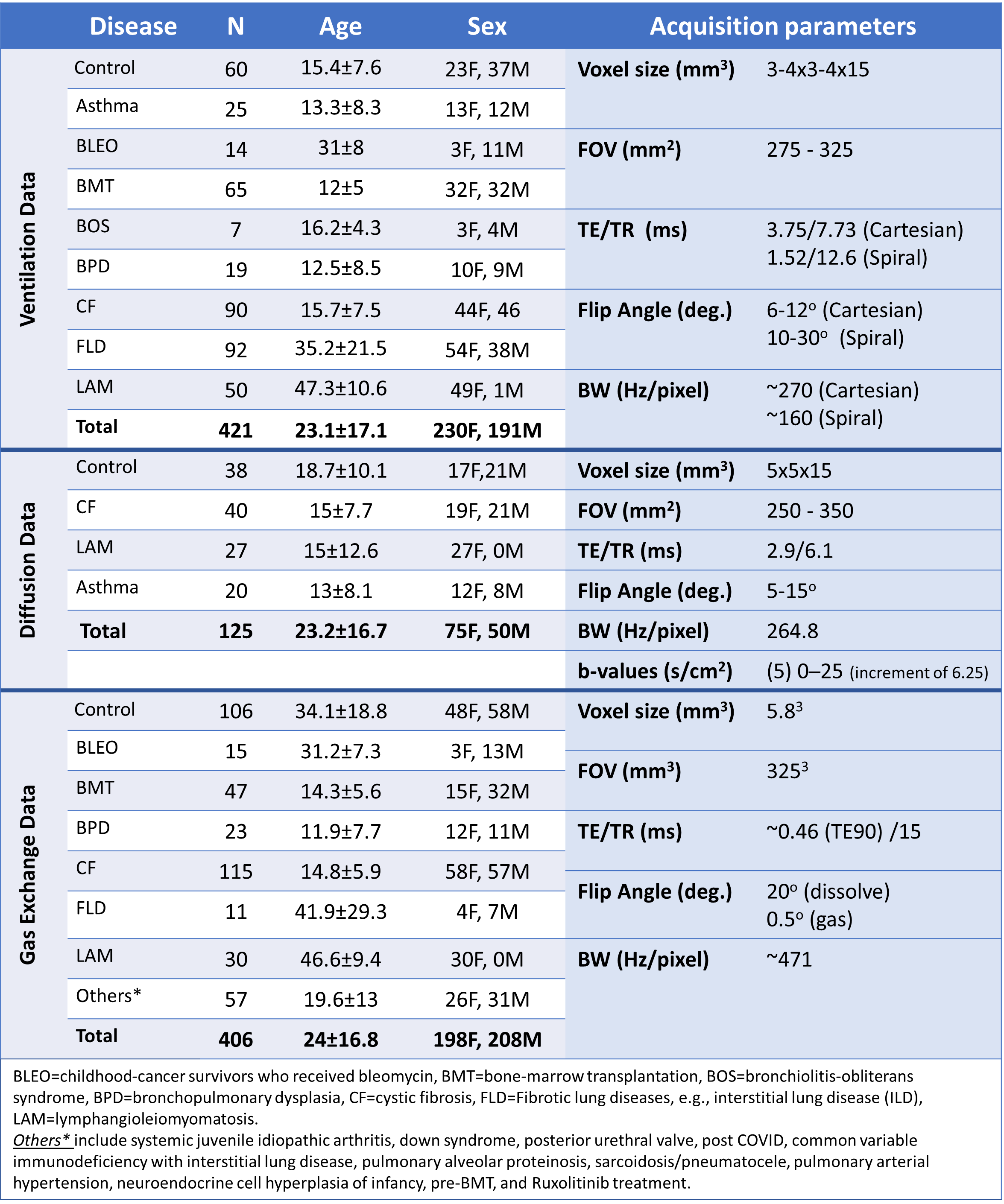
Participants: 952 HXe MRI datasets (ventilation: N=421, diffusion: N=125, and gas exchange: N=406) were retrospectively compiled from subjects who underwent imaging at Cincinnati Children's Hospital, including adults and children, healthy subjects, and a diverse array of individuals with cardiopulmonary conditions (Figure 1, footnote).Image Acquisition: Images were obtained using 3T-Philips scanners with either a home-built dual-loop, single-channel 129Xe transmit-receive7 or a commercial 129Xe chest coil (Clinical MR Solutions). We utilized 2D multi-slice gradient echo sequences (cartesian or spiral) for HXe ventilation and diffusion image acquisition (Table 1). Gas exchange data were collected using 3D radial with interleaved gaseous and dissolved excitation. Subjects inhaled a volume of HXe based on their lung capacity (1/6th for pediatric, up to 1L for adults) during a <16-second breath-hold. Hyperpolarization was performed on Polarean 9810/9820 hyperpolarizers (Polarean, Durham NC).
Network: N2V network architecture was adapted for this work5. Inputs are 64x64 patches for 2D images (ventilation and diffusion) and 64x64x8 for the 3D image (gas exchange: gas and dissolved). Images are normalized between [0,1] before being split into training, validation, and testing sets, with a 70/15/15% split for each disease type. Separate models were trained for each imaging type (e.g., ventilation). All models underwent 200 epochs on a 16-GB NVIDIA-RTX A4000. The performance of the N2V denoising framework was compared to BM3D6, which is based on an enhanced sparse representation in a transform domain.
Post-processing: SNR was calculated for all test images and denoising methods. VDP was determined as the proportion of lung volume with signal below 60% of the whole-lung mean on ventilation images. Log-linear fitting was used to estimate ADC from diffusion images. Denoising was applied to membrane and red blood cell (RBC) images after 1-point Dixon separation of the dissolved images. A one-way ANOVA test assessed intergroup differences in mean signal, SD noise, SNR, VDP, and ADC.
RESULTS
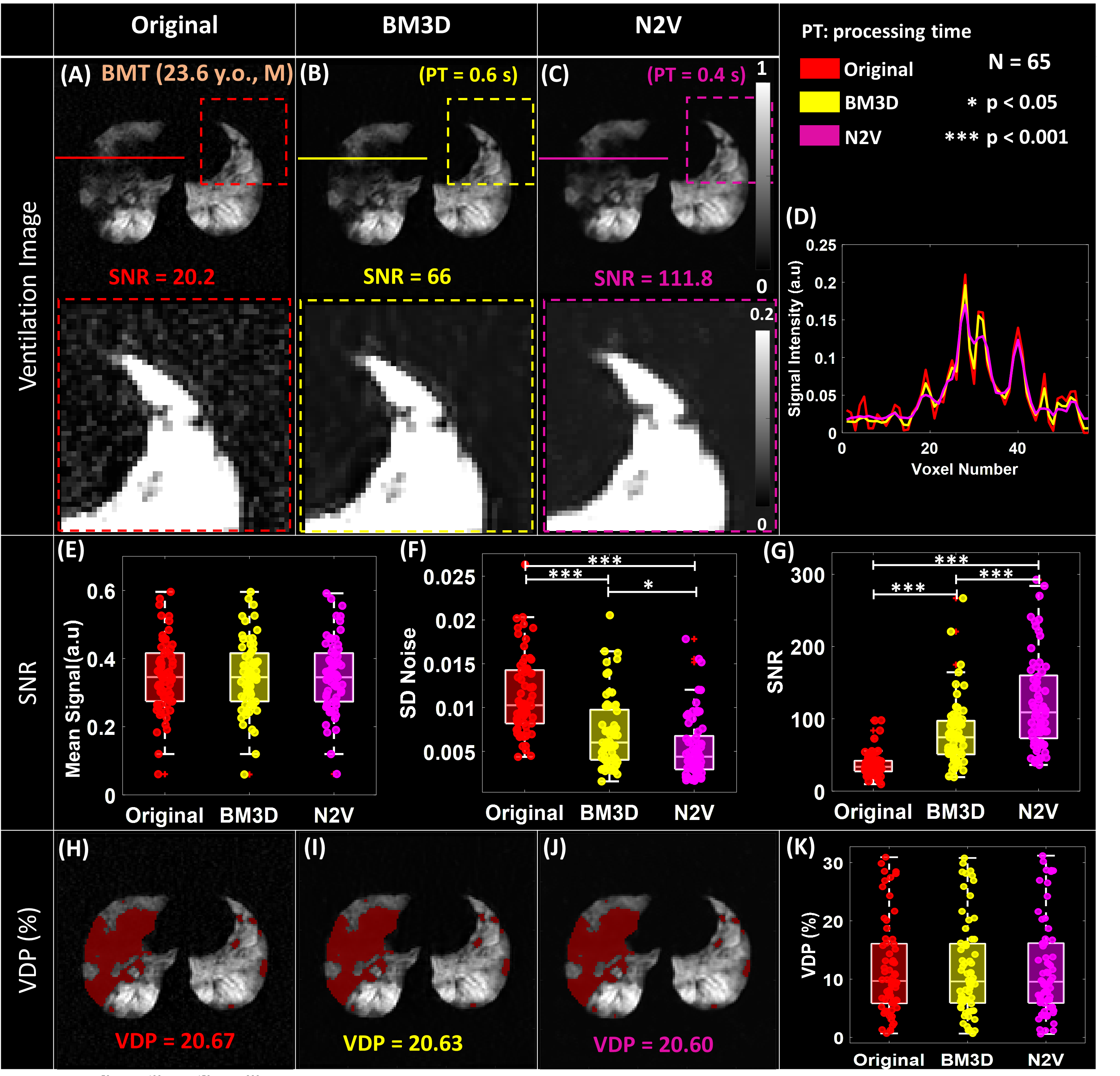
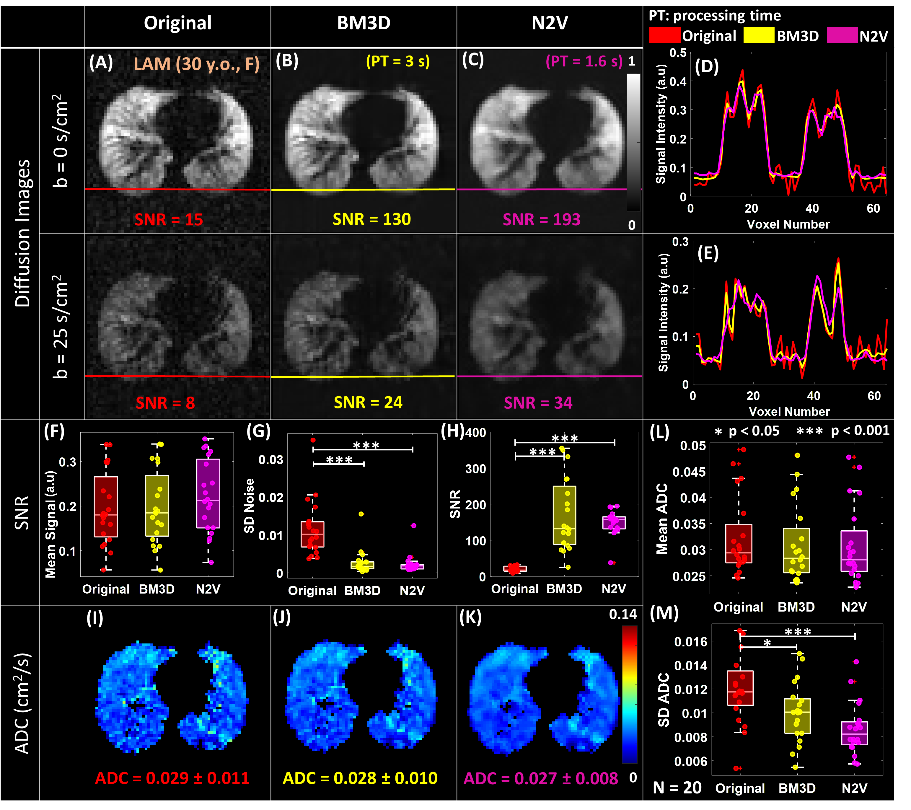
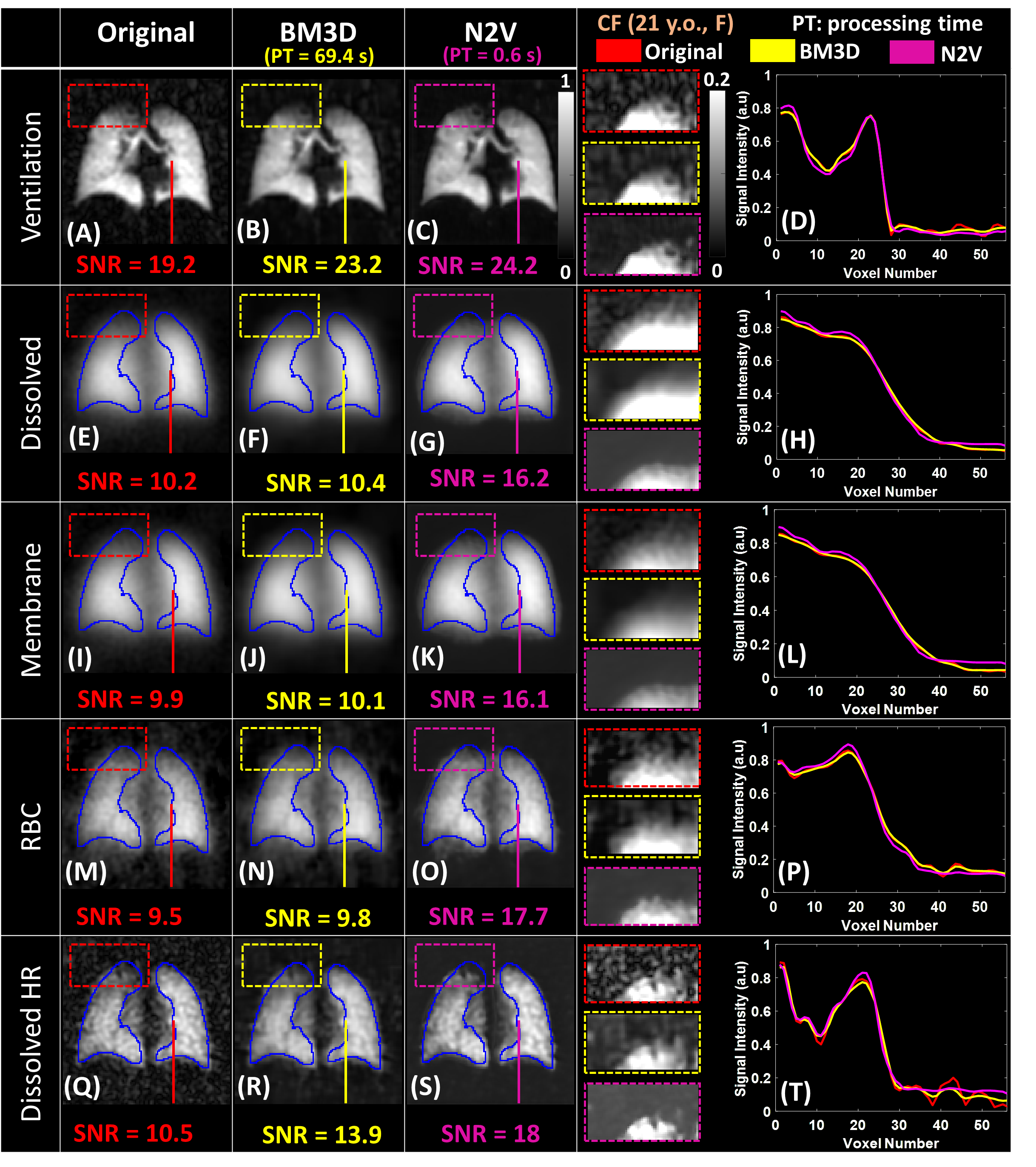
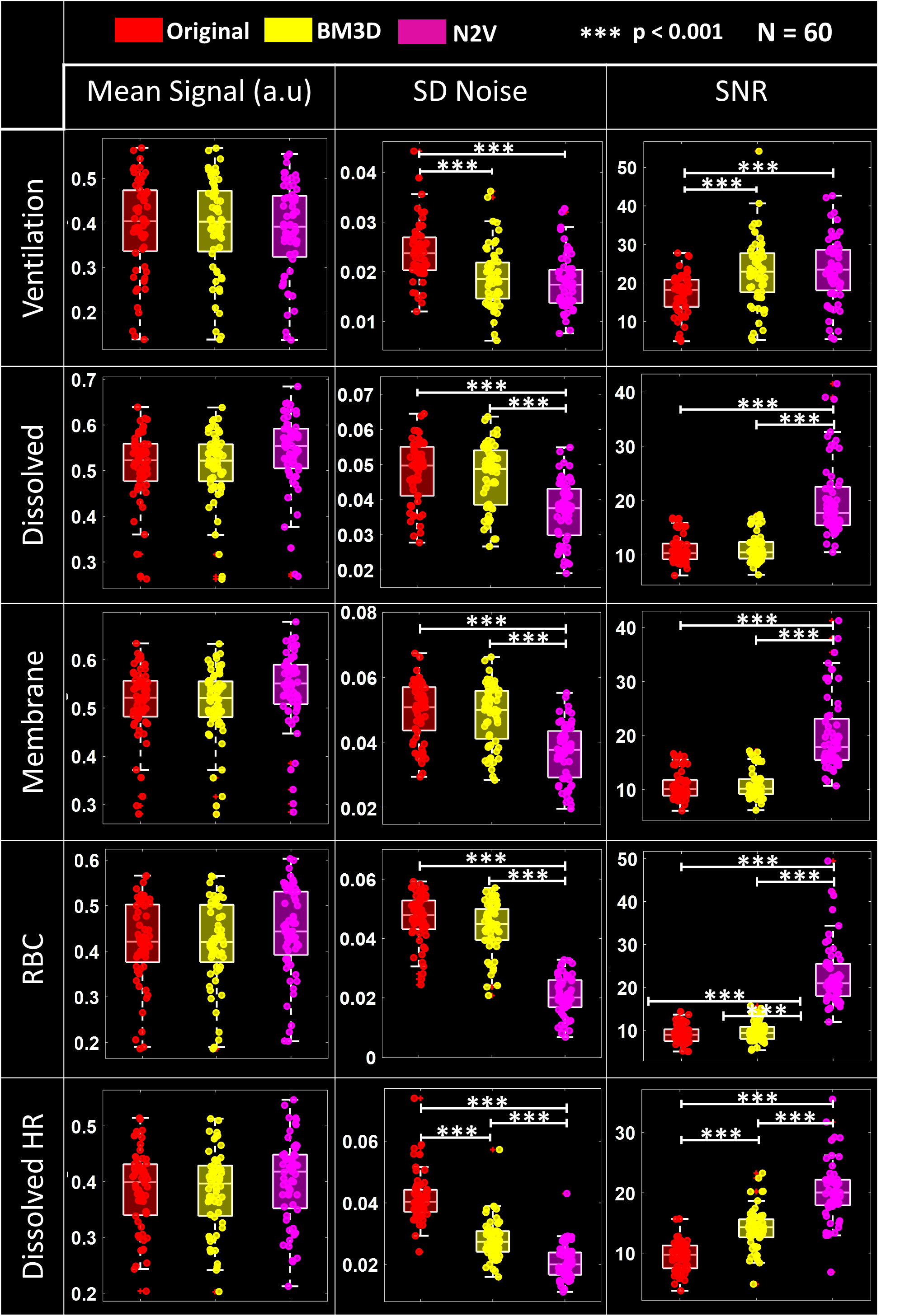
Figure 2 displays ventilation images for a BMT patient before and after denoising. SNR significantly improved (p<0.001), with a 3-fold increase using BM3D and a 5-fold increase using N2V compared to the originals. There was a substantial decrease in SD noise (p<0.001) without a significant change in mean signal. VDP remained unaffected by denoising. Figure 3 reveals similar outcomes for diffusion images, with significant SNR enhancement (p<0.001) post-denoising. Mean ADC remained unchanged, but there was a significant reduction in SD ADC. Figure 4 exhibits denoised gas exchange images, highlighting N2V's superior performance over BM3D. N2V yielded a substantial increase in SNR (p<0.001) and a decrease in SD noise (p<0.001) compared to the original and BM3D (Figure 5).DISCUSSION
We have applied the Noise2Void method to denoise HXe images. The significance of this approach is its unsupervised nature, dependency on a single noisy image, making it suitable for low-SNR HXe images. Our results demonstrate that N2V outperforms BM3D in denoising without affecting the quantitative outcomes. This ensures accurate interpretation of HXe images for diagnostics and therapeutic assessment, free from the interference of noise.CONCLUSIONS
The recent approval of HXe MRI for clinical use holds great diagnostic and therapeutic potential in assessing lung function. However, high noise levels in HXe images challenge accurate quantitation. This study adapts an unsupervised denoising approach, N2V, to enhance HXe imaging.Acknowledgements
The study was supported by NIH (R00HL111217, R01HL131012, R01HL166335, 2R01HL126771, R01HL151588, R01HL143011, R01HL166335, and R44HL123299), University of Cincinnati Cancer Center, Cystic Fibrosis Foundation (CLEVEL16A0), and the National Organization for Rare Disorders (20003).References
1. Niedbalski PJ, Hall CS, Castro M, et al. Protocols for multi-site trials using hyperpolarized 129Xe MRI for imaging of ventilation, alveolar-airspace size, and gas exchange: A position paper from the 129Xe MRI clinical trials consortium. Magnetic Resonance in Medicine. 2021;86(6):2966-2986.
2. He M, Zha W, Tan F, Rankine L, Fain S, Driehuys B. A comparison of two hyperpolarized 129Xe MRI ventilation quantification pipelines: the effect of signal to noise ratio. Academic radiology. 2019;26(7):949-959.
3. Bdaiwi AS, Niedbalski PJ, Hossain MM, et al. Improving hyperpolarized (129) Xe ADC mapping in pediatric and adult lungs with uncertainty propagation. NMR Biomed. 2021;35(3):e4639.
4. Lehtinen J, Munkberg J, Hasselgren J, et al. Noise2Noise: Learning image restoration without clean data. arXiv preprint arXiv:180304189. 2018.
5. Krull A, Buchholz T-O, Jug F. Noise2void-learning denoising from single noisy images. Paper presented at: Proceedings of the IEEE/CVF conference on computer vision and pattern recognition2019.
6. Dabov K, Foi A, Katkovnik V, Egiazarian K. Image denoising by sparse 3-D transform-domain collaborative filtering. IEEE Transactions on image processing. 2007;16(8):2080-2095.
7. Loew W, Thomen R, Pratt R, et al. A dual loop T/R -Xenon coil for homogenous excitation with improved comfort and size ISMRM; 2016.
Figures