3186
Manganese-enhanced gene expression MRI using Zip141NINDS, NIH, Bethesda, MD, United States
Synopsis
Keywords: Molecular Imaging, Cell Tracking & Reporter Genes, Zip14, Manganese-enhanced MRI, Neurons, Mesoscale connectivity
Motivation: Zip14 is an in vivo MRI-visible gene expression reporter system capable of producing focal signal changes without administration of additional contrast agents. However, it is not yet known how much contrast-enhanced MRI will improve sensitivity for Zip14 expression.
Goal(s): Our goal was to understand how systemically delivered manganese (Mn2+)-enhanced MRI (MEMRI) would improve Zip14 contrast.
Approach: Zip14 was expressed in the mouse brain and MRI was performed as previously described. MnCl2 was injected intraperitoneally and MEMRI was performed.
Results: MEMRI improved Zip14 contrast by approximately four-fold at the injection site, by two-fold at sites of anterograde tracing, and revealed previously undetectable tracing sites.
Impact: Although supplemental Mn2+ is not required to observe T1-weighted MRI signal enhancement produced by Zip14 expression, it can significantly improve contrast. Increased sensitivity for Zip14 expression with MEMRI will improve measurement of neural connectivity, degeneration, and plasticity in vivo.
Introduction
Recently, the MRI-visible gene expression reporter potential for the metal ion transporter Zip14 delivered by adeno-associated viruses (AAV-Zip14) was demonstrated1. Focal signal enhancement in T1 weighted MRI was observed in neuron specific Zip14 expressing areas of the mouse brain, including at the injection site and at anterograde projection sites. Excitingly, these results were obtained without needing to supplement the animal with manganese (Mn2+) which Zip14 is known to transport effectively2,3. However, performing Mn2+-enhanced MRI (MEMRI) by supplementing with systemic Mn2+ may increase sensitivity as well as add to evidence that the contrast is produced by Mn2+ transport into Zip14 expressing cells.Methods
Schematic experimental design is shown in Figure 1a. The Synapsin promoter, the gene coding for Zip14, and a histology-visible tag were packaged into AAVs and injected into the primary somatosensory area, barrel cortex (S1BC, n = 6). MRI were performed using conventional T1 weighted MPRAGE protocol immediately after and 1 week after injection of AAV-Zip14. Two weeks after AAV-Zip14 injection, 100 mM MnCl2•4H2O was delivered intraperitoneally at least 20 hours before MEMRI experiments (64 mg/kg) (dashed vertical lines in all figures). Due to global relaxation rate changes in the brain due to systemic Mn2+ uptake, a MEMRI-optimized MPRAGE protocol was implemented.For MEMRI timecourse experiments, a separate cohort of mice were injected with AAV-Zip14 (n = 3) and a control cohort were injected with AAV expressing channelrhodopsin (AAV-ChR2, n = 3) into the S1BC. MnCl2 was injected intraperitoneally, and imaging was performed at least two weeks after AAV injection using a T1-weighted GRE protocol at various timepoints up to one week after MnCl2 injection.
Results
Injection of AAV-Zip14 into the S1BC produced signal enhancement in the S1BC and VPM without needing to supplement with MnCl2 as previously described1. No enhancement was apparent in the S1BC (magenta arrow) and the VPM (cyan arrow) immediately after injection. Cortical lamina-specific enhancement was apparent in the S1BC and moderate enhancement was apparent in the VPM due to anterograde transport as soon as one week after AAV-Zip14 injection.Imaging performed one day after MnCl2 injection, using the same imaging protocol, showed improved contrast between normal appearing gray matter and the Zip14 expressing S1BC and VPM (Figure 1c). No enhancement of M1 immediately after and one week after injection of AAV-Zip14 into the S1BC was apparent (yellow arrow). MEMRI performed two weeks after AAV-Zip14 and one day after systemic MnCl2 injections revealed previously undetected, moderate M1 enhancement (Figure 1d).
Optimization of the MPRAGE protocol to account for brain-wide longitudinal relaxation rate changes due to MnCl2 supplementation substantially improved contrast between Zip14 expressing areas and surrounding normal tissue (Figure 2a). Relative enhancement of injected (white bars) with respect to contralateral control (gray bars) S1BC is shown in Figure 2b. There were no significant differences in signal intensity apparent immediately after injection of AAV-Zip14. The injected cortex was significantly hyperintense one week after AAV-Zip14 injection and at the two week post injection timepoint using MEMRI. Optimization of the MEMRI protocol led to improved contrast compared to the routine protocol. There was an approximately four-fold improvement. The same trends were observed for the enhancement of the VPM ipsilateral to the injected S1 due to anterograde transport of Zip14 from the injected S1BC (Figure 2c). No enhancement was apparent immediately after injection of AAV-Zip14 into the S1BC. Significant enhancement was apparent in the ipsilateral VPM one week after AAV-Zip14 injection. Similar magnitude, significant enhancement was apparent using MEMRI at the two week injection timepoint. Optimization of the MEMRI protocol approximately doubled the apparent enhancement of the ipsilateral VPM.
Zip14 expression alters Mn2+ uptake and sequestration dynamics (Figure 3). Enhancement due to Zip14 expression was detected using T1-weighted GRE two weeks after AAV-Zip14 into the S1BC before administration of MnCl2 (Figure 3b, Pre). No enhancement over baseline was apparent two weeks after AAV-ChR2 injection. After administration of MnCl2, the Zip14 expressing S1BC enhanced above neighboring normal-appearing gray matter significantly more than ChR2 expressing areas. It was apparent both visually and quantitatively that the Zip14 expressing areas remained hyperintense for at least a week after MnCl2 administration.
Discussion and Conclusion
MEMRI can improve in vivo sensitivity for Zip14 expression. It is not necessary to introduce systemic Mn2+ to robustly detect AAV-Zip14 enhancement at the injection site. However, improved contrast-to-noise afforded by MEMRI improves detection of anterograde sites. Considering that these sites are enhancing due to Zip14 expression in only a small volume fraction occupied by the processes of the expressing neuron, the extra sensitivity should enable measurement of connectivity, structural plasticity, and neurodegeneration in vivo.Acknowledgements
This work was supported by the Intramural Research program of the National Institutes of Neurological Disorders and Stroke (NINDS), National Institutes of Health. We thank Raymond Fields in the NINDS Viral Production Core Facility for virus productionReferences
1. Rallapalli, H., McCall, E. C., Bouraoud, N. & Koretsky, A. P. MRI detection of gene expression in the mammalian brain using the Manganese transporter Zip14. in Proceedings of the ISMRM (2023).
2. Steimle, B. L., Smith, F. M. & Kosman, D. J. The solute carriers ZIP8 and ZIP14 regulate manganese accumulation in brain microvascular endothelial cells and control brain manganese levels. Journal of Biological Chemistry 294, 19197–19208 (2019).
3. McCabe, S., Limesand, K. & Zhao, N. Recent progress toward understanding the role of ZIP14 in regulating systemic manganese homeostasis. Comput Struct Biotechnol J 21, 2332–2338 (2023).
Figures
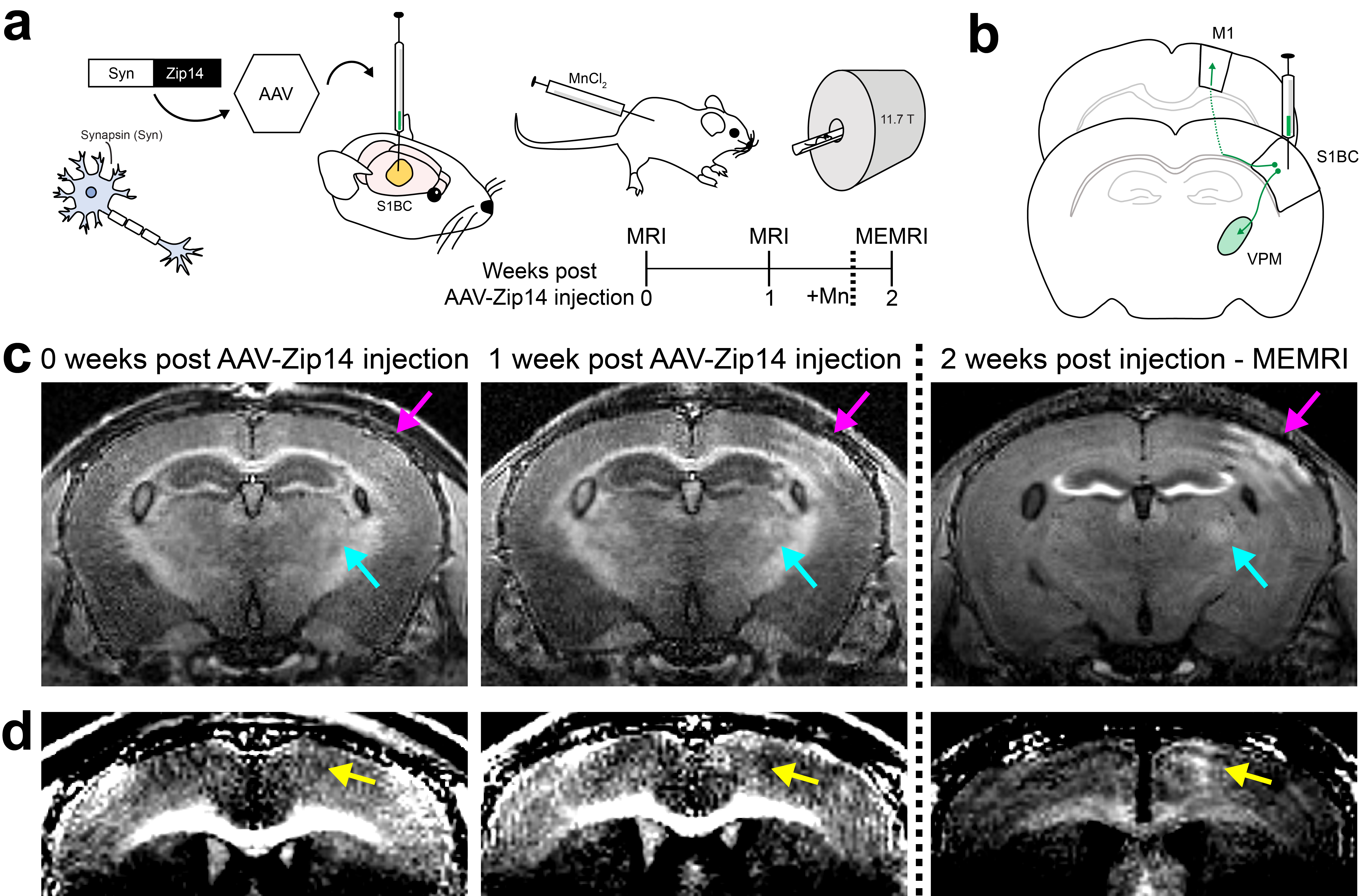
Mn2+ supplementation improves contrast produced by Zip14 expression in neurons and reveals previously undetected tracing sites.
(a) Schematic experimental design. Zip14 was expressed in the S1BC of the mouse brain. MRI was performed immediately and one week after injection. MnCl2 was injected intraperitoneally one day before MEMRI (+Mn, dashed line) (b) Tracing diagram from S1BC to VPM and M1 (c) Imaging results before and after MnCl2 injection (dashed line) show enhancement of S1BC (magenta) and VPM (cyan) (d) Enhancement of M1 (yellow) due to anterograde tracing visible on MEMRI
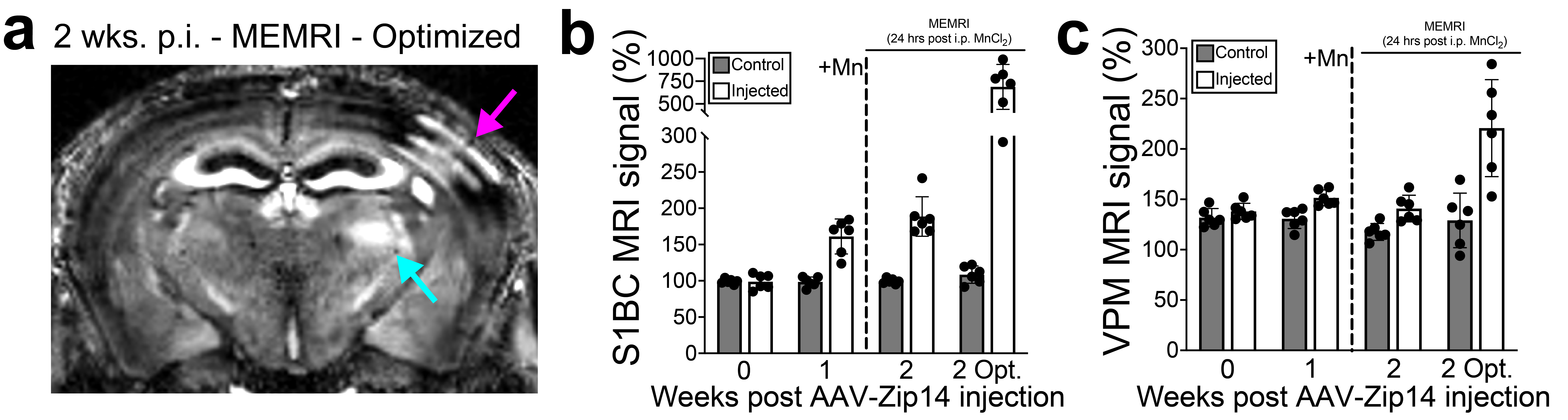
Optimized MEMRI protocol further improves contrast produced by Zip14 expression.
(a) Optimized MEMRI reveals improved contrast at the S1BC injection site (magenta) and the VPM (cyan) (b) Quantification of signal in the Zip14 expressing S1BC (white) compared to contralateral control S1BC (gray) revealed a greater than four-fold increase in enhancement when using the optimized MEMRI protocol (c) Signal quantification in the Zip14 expressing (white) and control (gray) VPM revealed that the optimized protocol doubled the contrast compared to the routine protocol.
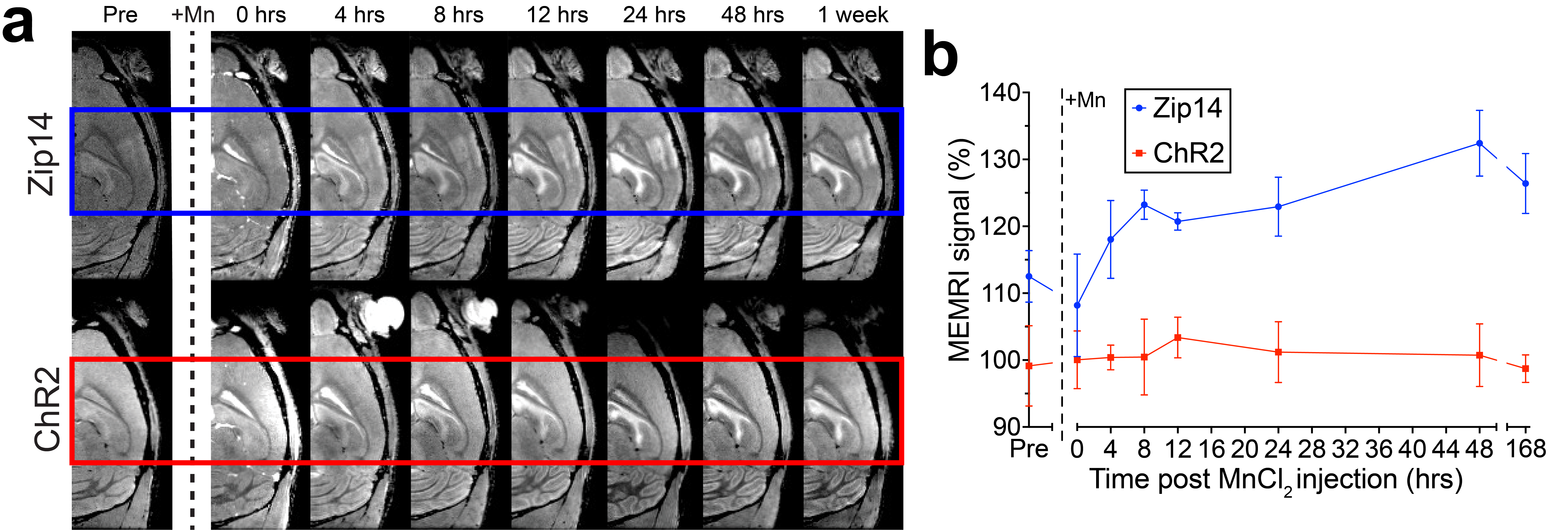
MEMRI timecourse analysis demonstrates Zip14 expression alters Mn2+ uptake and sequestration dynamics.
(a) MRI performed two weeks after injection of AAV-Zip14 (blue) showed moderate contrast in the S1BC compared to none in AAV-Channelrhodopsin (ChR2, red) before i.p. MnCl2 injection (dashed line). Enhancement was apparent in the AAV-Zip14 injected S1BC as soon as four hours after Mn2+ but not in the AAV-ChR2 injected S1BC. This effect lasted at least one week (b) Quantification of these effects confirmed significant increases in MEMRI enhancement rate due to Zip14 expression.